Case Report Open Access
Gastric Synchronous Stromal Tumor and Adenocarcinoma: A Fortuitous Association?
Haddad Anis*, Kacem Selma, Sebai Amine, Maghrebi Houcine, Makni Amine and Kacem Montassar
Department of Surgery “A”, La Rabta Hospital, Tunis, Tunisia
- *Corresponding Author:
- Haddad Anis
Department of Surgery “A”, La Rabta Hospital, Tunis, Tunisia
Tel: +21650928386
E-mail: anis.haddad2015@icloud.com
Received date: March 23, 2017; Accepted date: August 04, 2017; Published date: August 09, 2017
Citation: Anis H, Selma K, Amine S, Houcine M, Amine M, et al. (2017) Gastric Synchronous Stromal Tumor and Adenocarcinoma: A Fortuitous Association?. J Clin Exp Pathol 7:320. doi: 10.4172/2161-0681.1000320
Copyright: © 2017 Anis H, et al. This is an open-access article distributed under the terms of the Creative Commons Attribution License, which permits unrestricted use, distribution, and reproduction in any medium, provided the original author and source are credited.
Visit for more related articles at Journal of Clinical & Experimental Pathology
Abstract
The association of an adenocarcinoma with a stromal tumor in the stomach is rarely observed. We report the observation of a patient operated for a synchronous tumor associating subcardial adenocarcinoma with a stromal tumor of the gastric body. It is a 74 year-old patient, diabetic, who was admitted, in December 2016, for epigastralgia associated with vomiting evolving for 2 months. Physical examination was strictly normal. An oesogastroduodenal fibroscopy was performed showing a subcardial ulcerative budding formation with a 3 cm diameter and the presence, in the gastric body, of a second sub mucosal nodular formation with a 4 cm diameter. The anatomopathological examination of the biopsies concluded to an infiltrating and slightly differentiated adenocarcinoma at the subcardial level associated with chronic HP+ gastritis without signs of malignancy. The computed tomography, performed as part of the extension study, revealed a macro-lobulated tissue mass, posterior, pre-pyloric, with submucosal development, prolapsed in the peritoneum whose enhancement characteristics evoke a stromal tumor but also an irregular gastric cardio-tuberosal thickening predominant on the small gastric curvature and associated with necrotic pre-gastric centimetric satellite ganglia. The patient had a total gastrectomy with lymph node dissection type D1, 5 and oesojejunal anastomosis on a Y-shaped loop. The anatomo-pathological examination of the surgical specimen concluded to a slightly differentiated adenocarcinoma of the cardia classified as pT3N2 associated with a stromal tumor of the gastric body Synchronous development of gastric adenocarcinoma and GIST is rare. During this association, the stromal tumor is often characterized by low risk of recidivism.
Keywords
Adenocarcinoma; Interstitial cells of cajal; Gastrectomy
Introduction
Stromal tumors (GIST) are the most frequent mesenchymal tumors of the digestive tract [1,2] with an incidence of 10 to 20 cases per 10 million people. They sit mainly in the stomach and represent 1 to 2.2% of malignant digestive tumors. 95% of malignant tumors of the digestive tract are adenocarcinomas.
The association of two types of tumors in the stomach is possible. These are mainly adenocarcinomas associated with lymphoma or a carcinoid tumor. The association of an adenocarcinoma with a stromal tumor in the stomach is rarely observed.
We report the observation of a patient operated for a synchronous tumor associating subcardial adenocarcinoma with a stromal tumor of the gastric body.
Case Report
It is a 74 year-old patient, diabetic, who was admitted in December 2016 with epigastralgia associated with vomiting evolving for 2 months. Physical examination was strictly normal.
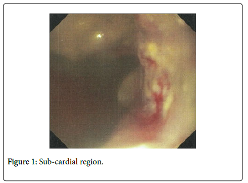
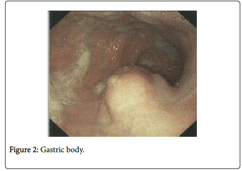
An oesogastroduodenal fibroscopy was performed showing a subcardial ulcerative-budding formation with a 3 cm diameter (Figure 1) and the presence, in the gastric body, of a second submucosal nodular formation with a 4 cm diameter (Figure 2).
The anatomopathological examination of the biopsies showed an infiltrating and slightly differentiated adenocarcinoma at the subcardial level associated with chronic HP+ gastritis without signs of malignancy in the gastric body.
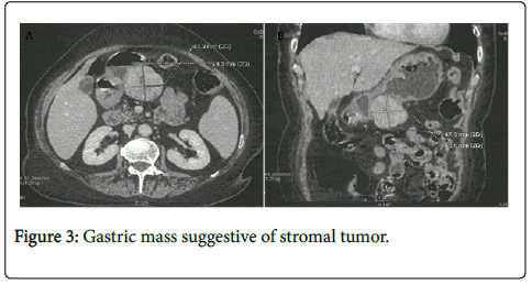
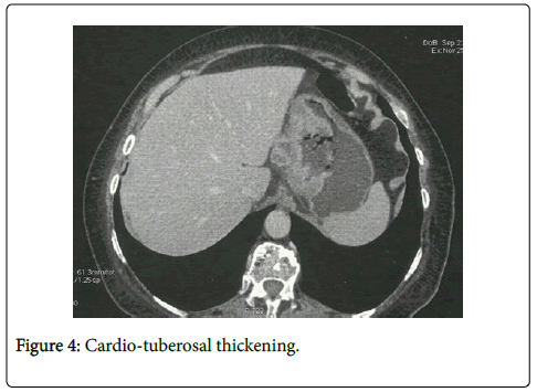
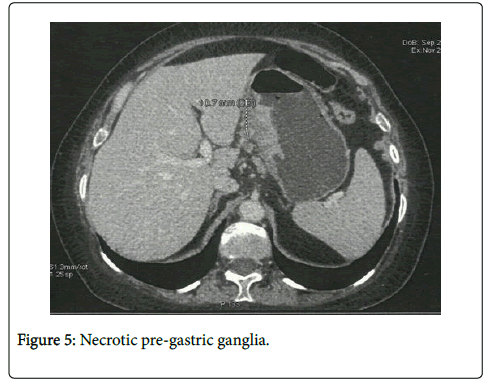
The computed tomography, performed as part of the extension study, revealed a macro-lobulated tissue mass, posterior, pre-pyloric, with submucosal development, prolapsed in the peritoneum, measuring 50 × 48 × 45 mm and whose enhancement characteristics evoked a stromal tumor (Figure 3A and 3B) but also an irregular gastric cardio-tuberosal thickening predominant on the small gastric curvature (Figure 4) and associated with necrotic pre-gastric centimetric satellite ganglia (Figure 5).
The patient had a total gastrectomy with lymph node dissection type D1, 5 and oeso-jejunal anastomosis on a Y-shaped loop.
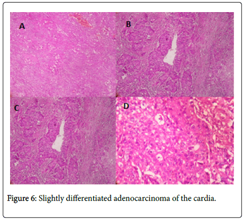
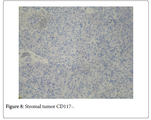
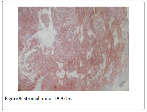
The anatomo-pathological examination of the surgical specimen showed a slightly differenciated adenocarcinoma of the cardia classified as pT3N2, 3N+/8N (Figure 6A-D) associated with a stromal tumor of the gastric body CD34-, CD117-, DOG1+, with rare mitoses, a mitotic index <5 and without nuclear atypia (Figure 7A and 7B) considered to be at low risk of recidivism (Figures 8 and 9).
Discussion
Stromal tumors (GIST) are mesenchymal tumors developed from the interstitial cells of Cajal. They, therefore, express a specific marker to these cells, the c-Kit (CD117), and this in 95% of cases, which makes it possible to differentiate them from the other digestive mesenchymal tumors [3]. They may also express other markers such as the BCL-2 (80%), the CD34 (70%), the SMA (35%), the DOG-1, the S100 protein (10%) and the desmine (5%) [3,4].
The preferred location of these tumors is the stomach (60 to 70%) followed by the small intestine (20 to 30%), the colon and rectum (5 to 10%) and finally the oesophagus (5%) [2].
The most common clinical signs of GIST are abdominal pain, digestive haemorrhage and abdominal mass. The symptomatology depends on the location and size of the tumor.
Preoperative diagnosis of the stromal tumor, if associated with adenocarcinoma, is difficult and rarely done. Generally, adenocarcinoma masks clinically and endoscopically the stromal tumor but also at imaging, in which it is confused with adenopathies [5].
In addition, the tumor is often submucosally developed, making in most cases endoscopic biopsies negative. Thus, in the case of an association of adenocarcinoma and a stromal tumor, the diagnosis of the GIST is usually performed peroperatively and/or by the anatomopathological examination of the surgical specimen [5]. Lin et al. estimated the preoperative diagnosis rate of such an association to be only 2.4%.
The association of a stromal tumor with another malignant tumor is estimated from 4.5% to 35% depending on the series [4]. Generally, they are gastrointestinal adenocarcinoma (47%), prostatic adenocarcinoma (9%), lymphoma and leucemia (7%) and breast cancer (7%) [4]. A few cases of association of gastric stromal tumors and gastric adenocarcinomas have been reported in the literature.
Liszka et al. [6] reported that during this association, the stromal tumor is generally small in size, often less than 2 cm and presents a low risk of invasion and recidivism compared to isolated GIST, with a mitotic index often less than 5 and a Ki-67 less than 5% [4], suggesting a probable inhibition of the malignancy of the stromal tumor by adenocarcinoma.
An immuno-histo-chemical feature of stromal tumors associated with adenocarcinoma is the low expression of CD117 and CD34 [7] with often an intense expression of the DOG-1 [8].
In the presence of such an association, an R0 resection of the adenocarcinoma and the stromal tumor is necessary. Postoperative chemotherapy may be proposed for one or both tumors according to indications.
Conclusion
Synchronous development of gastric adenocarcinoma and GIST is rare. During this association, the stromal tumor is often characterized by a small size, a low mitotic index, minimal or absent nuclear atypies, a low malignant potential but also a weak expression of CD117 and CD34 with significant expression of DOG-1.
References
- Nilsson B, Bümming P, Meis-Kindblom JM, Odén A, Dortok A, et al. (2005) Gastrointestinal stromal tumors: the incidence, prevalence, clinical course, and prognostication in the preimatinibmesylate era—a population-based study in western Sweden. Cancer 103: 821-829.
- Miettinen M, Lasota J (2003) Gastrointestinal stromal tumors (GISTs): definition, occurrence, pathology, differential diagnosis and molecular genetics. Pol J Pathol 54: 3-24.
- Miettinen M, Sarlomo-Rikala M, Lasota J (1999) Gastrointestinal stromal tumors: recent advances in understanding of their biology. Hum Pathol 30: 1213-1220.
- Yan Y, Li Z, Liu Y, Zhang L, Li J, et al. (2003) Coexistence of gastrointestinal stromal tumors and gastric adenocarcinomas. Tumor Biol 34: 919-927.
- Lin M, Lin JX, Huang CM, Zheng CH, Li P, et al. (2014) Prognostic analysis of gastric gastrointestinal stromal tumor with synchronous gastric cancer. World J Surg Oncol 12: 25.
- Liszka┼ü, Zieli┼?ska-Pajak E, Pajak J, Go┼?ka D, Huszno J (2007) Coexistence of gastrointestinal stromal tumors with other neoplasms. J Gastroenterol 42: 641-649.
- Agaimy A, Wünsch PH, Sobin LH, Lasota J, Miettinen M (2006) Occurrence of other malignancies in patients with gastrointestinal stromal tumors. Semin Diagn Pathol 23: 120-129.
- Zhen Liu, Liu S, Zheng G, Yang J, Hong L, et al. (2016) Clinicopathological features and prognosis of coexistence of gastric gastrointestinal stromal tumor and gastric cancer. Medicine 95: 45.
Relevant Topics
Recommended Journals
Article Tools
Article Usage
- Total views: 2546
- [From(publication date):
August-2017 - Dec 19, 2024] - Breakdown by view type
- HTML page views : 1921
- PDF downloads : 625

