Gallstone Ileus - A Case Report
Received: 02-Feb-2024 / Manuscript No. roa-24-128081 / Editor assigned: 05-Feb-2024 / PreQC No. roa-24-128081 / Reviewed: 19-Feb-2024 / QC No. roa-24-128081 / Revised: 23-Feb-2024 / Manuscript No. roa-24-128081 / Published Date: 29-Feb-2024
Abstract
Gallstone ileus is a rare complication of secondary gallstone formation, most often due to a cholecysto-duodenal fistula with the passage of a stone into the small intestine. Obstruction occurs when the stone obstructs a narrowed segment of the digestive tract. Gallstone ileus is a surgical emergency, and its management is focused on relieving the obstruction. Treatment of the biliary-digestive fistula is considered based on the local and general condition. The best approach remains prevention through the diagnosis and treatment of acute cholecystitis.
Keywords
Ileus; Obstruction; Surgical Emergency; Gallstone
Introduction
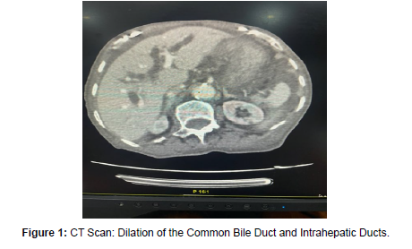
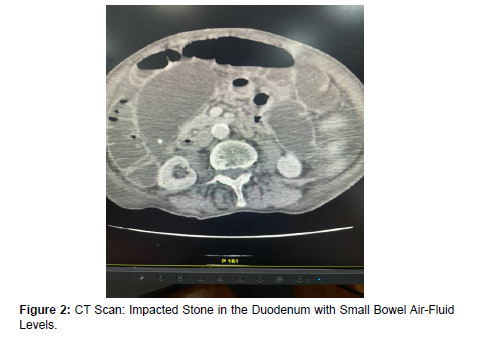
Gallstone ileus is an uncommon complication of gallstone disease, occurring in less than 0.5% of cases. It should be suspected in any patient with signs of bowel obstruction associated with aerobilia and an ectopic location of a stone, especially in elderly individuals. The obstruction typically occurs at the ileocecal region, with colonic locations being much rarer, representing 2.5% of cases. Treatment is generally surgical, except for rare cases of spontaneous stone passage [1] (Figure 1-3).
Figure 1: CT Scan: Dilation of the Common Bile Duct and Intrahepatic Ducts.
Figure 2: CT Scan: Impacted Stone in the Duodenum with Small Bowel Air-Fluid Levels.
Figure 3: CT Scan: Diffuse Small Bowel Air-Fluid Levels Associated with Gallstone Ileus.
Discussion
Gallstone ileus accounts for 2% of all small bowel obstructions, but in individuals over 70 years old, it may contribute to 25% of cases. It predominantly affects women (sex ratio reported between 4 to 16/1). Pathophysiologically, repeated episodes of gallstone cholecystitis lead to peri-vesicular inflammation, fistula formation, and migration of gallstones into the digestive tract. In 10-20% of cases, gallstones become impacted, typically in the small intestine, causing partial or complete mechanical obstruction. The obstruction most commonly involves the terminal ileum but can also affect the duodenum (Bouveret’s syndrome) and, less frequently, the colon. Contributing factors to obstruction include inflammatory or tumor-related strictures and postoperative adhesions. The most frequent site of impaction is the ileocecal valve (60% of cases), followed by the proximal ileum (25% of cases) and distal jejunum (9% of cases). In the majority of cases, gallstone migration into the digestive lumen occurs through a biliary-intestinal fistula complicating cholecystitis. Rigler’s triad, including aerobilia, small bowel obstruction, and identification of an ectopic gallstone, is often sought for diagnosis. In this case, two signs were considered significant: an ectopic gallstone and small bowel obstruction [2].
Conclusion
Gallstone ileus primarily affects elderly women, and its clinical presentation is often atypical. Abdominal CT scan plays a diagnostic role by visualizing the obstruction. Surgical treatment involves enterolithotomy, with or without repair of the cholecysto-intestinal fistula and cholecystectomy [3,4].
References
- Karim Ibnmajdoub Hassani, Julie Rode, Jane Poincenot et Jean-Manuel Gruss (2010) Gallstone ileus with spontaneous evacuation of a large stone: report of a case. Pan Afr Med J 4: 10.
- Narjis Y, Chelala E, Dessily M,Allé JL (2010) Biliary ileus: diagnostic and therapeutic aspects. Report of a case. Revue Medicale de Bruxelles, 31: 463-465.
- Course Sheet: Gall Ileus and Bouveret Syndrome.
- Hakima Abid, Fatima Babakhouya, Ahmed Zerhouni, Imane Toughrai, Hicham Sbai, et al. (2018) Biliary ileus: a rare cause of intestinal obstruction, about a case. Pan Afr Med J 29: 20
Citation: Kenza S, Rabileh M, Omar A, Fatimazahra L (2024) Gallstone Ileus - ACase Report. OMICS J Radiol 13: 543.
Copyright: © 2024 Kenza S, et al. This is an open-access article distributed underthe terms of the Creative Commons Attribution License, which permits unrestricteduse, distribution, and reproduction in any medium, provided the original author andsource are credited.
Select your language of interest to view the total content in your interested language
Share This Article
Open Access Journals
Article Usage
- Total views: 1382
- [From(publication date): 0-2024 - Feb 21, 2026]
- Breakdown by view type
- HTML page views: 1038
- PDF downloads: 344


