Make the best use of Scientific Research and information from our 700+ peer reviewed, Open Access Journals that operates with the help of 50,000+ Editorial Board Members and esteemed reviewers and 1000+ Scientific associations in Medical, Clinical, Pharmaceutical, Engineering, Technology and Management Fields.
Meet Inspiring Speakers and Experts at our 3000+ Global Conferenceseries Events with over 600+ Conferences, 1200+ Symposiums and 1200+ Workshops on Medical, Pharma, Engineering, Science, Technology and Business
Case Report Open Access
‘Flexor Hallucis Longus Loose Bodies- An Unusual Cause of Plantar Midfoot Pain’
| Shah A1*, Botchu R2 and Rennie WJ1 | |
| 1Leicester Royal Infirmary, Leicester, UK | |
| 2Kettering General Hospital, Kettering, UK | |
| Corresponding Author : | Dr. Amit Shah Leicester Royal Infirmary Infirmary Square, Leicester LE1 5WW, UK Tel: +44 300 303 1573 Fax: +44 116 258 8210 E-mail: doctigs@gmail.com |
| Received April 02, 2013; Accepted October 24, 2013; Published October 27, 2013 | |
| Citation: Shah A, Botchu R, Rennie WJ (2013) ‘Flexor Hallucis Longus Loose Bodies- An Unusual Cause of Plantar Midfoot Pain’. OMICS J Radiology 2:152. doi: 10.4172/2167-7964.1000152 | |
| Copyright: © 2013 Shah A, et al. This is an open-access article distributed under the terms of the Creative Commons Attribution License, which permits unrestricted use, distribution, and reproduction in any medium, provided the original author and source are credited. | |
Visit for more related articles at Journal of Radiology
| Keywords |
| Flexor hallucis longus; Joint loose bodies; Mid foot pain |
| Abbreviations |
| FHL: Flexor Hallucis Longus; FDAL: Flexor Digitorum Accessorius Longus |
| Introduction |
| Plantar midfoot pain is a frequently encountered complaint. It is most commonly seen in athletes and secondary to overuse trauma. The aetiology for plantar midfoot pain includes trauma-related conditions, infection (commonly secondary to diabetes), joint disorders, benign or neoplastic soft-tissue/bone masses and tendon disorders. The Flexor Hallucis Longus (FHL) tendon is the second strongest tendon in the body and among the least commonly injured tendons, which when injured usually occurs at the level of the medial malleolus [1]. We present a seventy-eight year-old male with midfoot pain on weightbearing due to loose bodies within the FHL tendon sheath in the midfoot and discuss the common aetiologies of plantar midfoot pain. |
| Case Report |
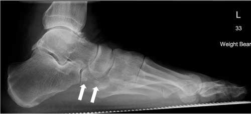
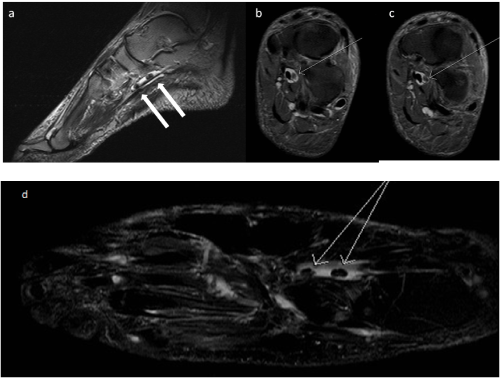
| Seventy- eight year old male, presented with a ten month history of insidious onset plantar midfoot pain which was exacerbated on weight bearing. There was no history of trauma, diabetes or rheumatological disease. He was not on any regular medications. There was no improvement with analgesia prescribed by his General Practitioner. Hence, was referred to a foot and ankle specialist. Clinical examination revealed tenderness at the plantar aspect of the midfoot with point tenderness in the region of the base of the second metatarsal. A clinical diagnosis of a midfoot ganglion or tenosynovitis was considered. Radiographs performed in clinic were assessed by the orthopedic team and were assumed to demonstrate mild osteoarthritis at the ankle joint with osteophytosis only. No osteochondral lesion involving the talar dome was noted. No other significant abnormality was noted to account for the symptoms of plantar midfoot pain. A MRI was performed to evaluate the ankle and midfoot. This demonstrated two calcific loose bodies within the FHL tendon sheath. A retrospective review of the radiographs revealed two 5 mm ossific loose bodies in relation to plantar aspect of the foot (Figure 1). One was noted plantar and distal to the navicular, in the region of the Master Knot of Henry and the other was just proximal to the base of the second metatarsal (Figure 2). FHL tenosynovitis with fluid along the tendon sheath distal to the talocrural joint was noted on MRI. The site of the loose bodies correlated clinically with the patient’s point tenderness and was inferred to be the cause of his symptoms. During his follow-up consultation the patient had reported an improvement in his symptoms with conservative orthotic treatment, off-loading the medial arch, hence opted for non-surgical management. |
| Discussion |
| Flexor hallucis longus is one of three muscles that make up the deep posterior compartment of the leg. It arises from the inferior twothirds of the posterior surface of the fibula, interosseous membrane and adjacent intermuscular septum. The tendon arises just above the level of the medial malleolus, courses obliquely through a fibroosseous tunnel and along a groove passing under the inferior aspect of the sustentaculum tali. As the tendon passes forward in the sole of the foot it crosses the tendon of the flexor digitorum longus, to which it is connected by a fibrous slip at the Knot of Henry. At the midfoot it runs forward between the two heads of the flexor hallucis brevis and the sesamoid bones of the greater hallux to insert into the base of the distal phalanx [2]. It is a flexor of the great toe and assists in plantar flexion of the foot at the ankle. Several studies have confirmed that the FHL tendon sheath communicates with the ankle joint [3-6]. These have a proximal and distal component, which together make up the synovial sheath of the FHL tendon. The distal extent of the proximal synovial sheath ends in the region of the cuneonavicular joint [7] but variation in its termination has been shown which include the region of the calcaneocuboid joint, the cuboid, the cuboid-metatarsal joint and the base of the first metatarsal [8]. A communication between the proximal and distal component is extremely rare. This may explain why the osseous bodies stopped at in the region of the navicular in our case. |
| The pathologies of the posterior tibialis, Achilles and peroneal tendons are relatively common and well described as a cause for hind and midfoot pain [6,9]. The other differential diagnoses of plantar midfoot pain or arch pain include plantar fasciitis [10,11] and plantar fibromatosis, tarsal tunnel syndrome, stress fractures and trauma. Abnormalities of the FHL tendon and its sheath must also be considered in the differential diagnosis. Pain or mechanical symptoms related to the FHL are often confused with plantar fasciitis or tarsal tunnel syndrome [12] and may be inadequately treated. Plain radiographs of the ankle are performed to evaluate for bone spurs, degenerative changes and osteochondral lesions. These may also be helpful to elicit ossific loose bodies as seen in our case. Imaging with MRI will help evaluate the soft tissues, ligaments and tendons in detail. The FHL is a major initiator of and is significantly responsible for weight bearing in the ‘toe-off’ phase of the gait cycle [13]. Therefore, a prompt diagnosis and management of FHL dysfunction is essential to decrease morbidity due to chronic pain, disability and to maintain normal gait. |
| Common causes of FHL dysfunction include stenosing tenosynovitis, pseudocyst or a tendon tear. Stenosing tenosynovitis of the FHL tendon may result as a sequalae of trauma such as medial malleolar or calcaneal facture causing a check-rein deformity or repetitive micro trauma which is typically seen in athletes and ballet dancers. In our case, pain was caused by loose bodies in the FHL tendon sheath causing tenosynovitis due to physical impingement and irritation. FHL tenosynovitis has also been reported to be caused by Flexor Digitorum Accessorius Longus (FDAL). FDAL is an anomalous muscle with a reported prevalence of 2-8% in cadaveric studies [14-16]. The FDAL is intimately related to the neurovascular bundle and may abut, compress, or impinge upon the posterior tibial and or lateral plantar nerves. In our case, no accessory muscles were present. |
| Loose bodies are commonly intra-articular with the knee joint most commonly affected. These are either chondral, osseous or osteochondral. These may arise from injury to the synovial lining, cartilage or bone. The aetiology of loose bodies includes infection (tuberculosis), inflammatory (Rheumatoid Arthritis) and degenerative conditions (osteoarthritis) and metaplasia of the synovium (synovial chondromatosis). In our case the osseous bodies were assumed to be due to talocrural osteoarthritis as there was no evidence of synovial proliferation, metaplasia or relevant inflammatory arthritic changes in the talocrural joint. |
| Loose bodies have been demonstrated within tendon sheaths and have been reported in the biceps tendon [17,18] and finger flexor tendon sheath [19]. There has been no previously reported case of FHL osseous bodies. Synovial chondromatosis is a benign condition where the synovial lining of joints, bursae or tendon sheaths undergoes metaplasia forming cartilaginous loose bodies. Synovial chondromatosis of the foot and ankle is rare and typically occur in young-middle aged adults and demonstrates evidence of hypertrophic and inflammatory changes on MRI [20,21]. Soft tissue calcific lesions seen on plain radiography affecting tendon sheaths include giant cell tumours of tendon sheaths and calcific tendonitis, which can mimic loose bodies within the tendon sheath. Our case had no such features to suggest any of these conditions. |
| In our case, loose bodies were seen in the FHL tendon sheath at the level of the navicular and base of the 2nd metatarsal. There was associated tenosynovitis of the FHL but no tear was noted. We hypothesise that as there was no evidence of synovial abnormality or past history of rheumatological disease, the loose bodies originated from the degenerative osteophytes at the ankle joint which traversed into the FHL tendon sheath through its communication with the ankle joint. The gradual onset of pain was most likely secondary to the loose bodies gradually tracking more distally to the midfoot where they caused a mechanical type of stenosing tenosynovitis during weightbearing. In the toe-off phase of the gait cycle, increased pressure within the talocrural joint due to plantar flexion may predispose intra-articular bodies to escape through the lax posterior capsular communication into the FHL tendon sheath. The osseous bodies, if smooth and of a small enough calibre may track down the tendon sheath to points of narrowing like the Knot of Henry and can cause physical impingement, as is suspected in our case, or stenosing tenosynovitis. Movement of these bodies, tracking up or down the tendon sheath dependant on the varying pressures generated during the gait cycle can result in a waxing and waning phenomenon of pain. MRI can decipher these loose bodies (Figure 2). |
| Conclusion |
| Our case highlights an unusual cause for plantar midfoot pain due to loose bodies within the FHL tendon sheath One should consider this rare entity during the evaluation of the FHL in patients with plantar midfoot pain. A clinical and radiological assessment is essential to clinch this diagnosis. |
References |
|
Figures at a glance
 |
 |
| Figure 1 | Figure 2 |
Post your comment
Relevant Topics
- Abdominal Radiology
- AI in Radiology
- Breast Imaging
- Cardiovascular Radiology
- Chest Radiology
- Clinical Radiology
- CT Imaging
- Diagnostic Radiology
- Emergency Radiology
- Fluoroscopy Radiology
- General Radiology
- Genitourinary Radiology
- Interventional Radiology Techniques
- Mammography
- Minimal Invasive surgery
- Musculoskeletal Radiology
- Neuroradiology
- Neuroradiology Advances
- Oral and Maxillofacial Radiology
- Radiography
- Radiology Imaging
- Surgical Radiology
- Tele Radiology
- Therapeutic Radiology
Recommended Journals
Article Tools
Article Usage
- Total views: 15345
- [From(publication date):
November-2013 - Jul 02, 2025] - Breakdown by view type
- HTML page views : 10741
- PDF downloads : 4604
