Make the best use of Scientific Research and information from our 700+ peer reviewed, Open Access Journals that operates with the help of 50,000+ Editorial Board Members and esteemed reviewers and 1000+ Scientific associations in Medical, Clinical, Pharmaceutical, Engineering, Technology and Management Fields.
Meet Inspiring Speakers and Experts at our 3000+ Global Conferenceseries Events with over 600+ Conferences, 1200+ Symposiums and 1200+ Workshops on Medical, Pharma, Engineering, Science, Technology and Business
Case Report Open Access
First Rib Osteochondroma
| Jhunjhunwala HR1* and Neel R Dalal2 | |
| 1Institute of Medical Science of Bombay Hospital, Mumbai, India | |
| 2Post Graduate Institute of Medical Science of Bombay Hospital, Mumbai, India | |
| *Corresponding Author : | Jhunjhunwala HR Senior Ortho Consultant Post Graduate Institute of Medical Science of Bombay Hospital, Mumbai, India Tel: 9821064068 E-mail: drhrj2@gmail.com |
| Received: February 26, 2016; Accepted: March 11, 2016; Published: March 14, 2016 | |
| Citation: Jhunjhunwala HR, Dalal NR (2016) First Rib Osteochondroma. J Pain Relief 5:237. doi:10.4172/2167-0846.1000237 | |
| Copyright: © 2016 Jhunjhunwala HR, et al. This is an open-access article distributed under the terms of the Creative Commons Attribution License, which permits unrestricted use, distribution, and reproduction in any medium, provided the original author and source are credited. | |
Visit for more related articles at Journal of Pain & Relief
| Introduction |
| Osteochondroma are most common bone neoplasm accounting for 30-40% of benign osseous tumours [1,2]. Osteochondroma is benign developmental growth defect in which there is focal herniation of lateral component of epiphysial plate with a cartilaginous cap [3]. Understanding and recognizing the spectrum of the appearance and complications of first rib osteochondroma is important because of its dreadful complications that may follow if not taken care. |
| Case Report |
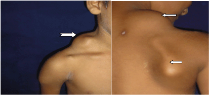
| A 10 year boy came with chief complaint of swelling in the right supraclavicular region disfiguring the area. There was another exostosis on inner side of right scapula which was rubbing on inner side of scapula on chest wall. CT scan has confirmed exostosis at the right supraclavicular area arising from right first rib and inner side of inferior end of right scapula. Multiple small exostosis were also found on lower end of right femur, upper end of right tibia and lower end of right radius which were not causing any pain and were not disfiguring (Figure 1). |
| Discussion |
| Tumours of the ribs compromise approximately 2% of all tumours of the body and may be benign or metastatic. Approximately 60% of the resected tumours are benign. Osteochondroma are the most common benign bone tumours [1,2] These tumours occurs more frequently in males. Male/Female Ratio of 3:1. |
| Osteochondroma begin in the childhood and grow until completion of skeletal maturity. They develop from an aberrant focus of growth plate beneath the ring of Ranvier, which continues to grow and undergo enchondral ossification in parallel with general skeletal growth. When skeletal maturity is reached, the cartilage cap thins and eventually completely ossifie [3]. Osteochondroma of first rib is frequently asymptomatic and the development of pain usually becomes an issue when an osteochondroma is repeatedly bumped on its prominence, or if a painful bursa develops or malignant changes occur. |
| First rib exostosis presents with swelling in the supraclavicular region, producing disfigurement and pain. |
| The complications of osteochondroma of first rib are often the result of mechanical interference with neighboring anatomic structures. The exostosis can injure the lung and pleura during respiration giving rise to pneumothorax, or haemothorax. Osteochondroma may cause compression of adjacent vascular or neural structures [4-9]. |
| There is an incidence of venous thoracic outlet syndrome associated with first rib osteochondroma which is a life threatening complication [7,8] Reports illustrate that rib exostosis can present acutely as a life- threatening bleeding or as chronic complication in the form of pneumonitis and empyema [4-6]. Chondrosarcoma is an exceedingly rare complication in 1-4% of osteochondroma which evolves very slowly, usually occurring in adult life [10]. |
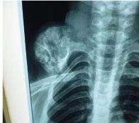
| There are certain path gnomic findings associated with osteoshondroma (Figure 2). |
| 1. The lesion protrudes from the host bone with a sessile (broad based) stalk. |
| 2. It occurs in the metaphysis |
| 3. The cortex and cancellous bone of the osteochondroma blend with the cortex and cancellous bone of the host. This is the main radiographic finding and any deviation from this feature should raise suspicion of a more serious lesion. |
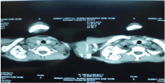
| 4. Coat Hanger Lession- Away from costo chondral joint (Figure 3). |
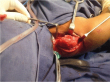
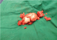
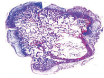
| Surgical resection can be expected to result in a successful outcome for symptomatic osteochondroma with low morbidity. We preferred a supra clavicular approach. A 7 cm incision was taken in the supra clavicular region. The base of the exostosis was approached by soft tissue dissection all around the exostosis and it was excised from the base with the cartilaginous cap in situ with it. The excised tissue was sent for histo pathology which confirmed the diagnosis of enchondroma. The scapular lesion was also removed. Post operatively patient is doing excellent (Figure 4a-4c). |
| Conclusion |
| In general all the osteochondroma are resected on development of complications. First rib exocytosis is a rare site and as it can cause life threatening complications and for this reason it should be resected as soon as diagnosed. |
| References |
References
- Cemil M, PurutMd (1997) Lesions ofthe Chest Wall. The Biological Basis of Modern Surgical Practice (15thedn.) Philadelphia, USA: Wb Saunders pp: 1896-1897.
- Pairolero PC (1994) Chest Wall Tumours(4th Edn.). General Thoracic Surgery.
- D’ambrosia R, Ferguson A (1968) The Formation of Osteochondroma by Epiphyseal Cartilage Transplantation. Clin Orthop61: 103-115.
- Uchida K, Kurihara Y, Sekiguchi S, Doi Y, Matsuda K, et al. (1997) Spontaneous Haemothorax Caused By Costal Exostosis. Eur Respir J 10: 735-736.
- Harrison Nk, Wilkinson J, O’donohue J, et al. (1994) Osteochondroma ofthe Rib: An Unusual Cause of Haemothorax. Thorax 49: 618-619.
- HajjarWm, El-MedanyYm, Essa Ma,Rafay MA, Ashour MH,et al. (2003) Unusual Presentation of Rib Exostosis. Ann Thorac Surg 75: 575-577.
- O'brien PJ, Ramasunder S, Cox Mw (2010) Venous Thoracic Outlet Syndrome Secondary to First Rib Osteochondroma in A Pediatric Patient.J Vasc Surg 53:811-813.
- Rosset P, Martinat H, Barsotti J, Gaisne E (1990) OsteogenicExostosis Of First Rib A Rare Etiology Of Thoracic Outlet Syndrome.Apropos of a case.Rev Chir Orthop Reparatrice Appar Mot 76:62-65.
- Ahmed Ar Tan Ts, UnniKk,Collins MS, Wenger DE,et al. (2003) Secondary Chondrosarcoma In Osteochondroma: Report Of 107 Patients. Clin Orthop Relat Res 411:193.
- Bottner F, Rodl R, Kordish I,Winklemann W, Gosheger G,et al. (2003) Surgical Treatment Of Symptomatic Osteochondroma. A Three To Eight Year Follow-Up Study. J Bone Joint Surg Br 85:1161.
Figures at a glance
 |
 |
 |
| Figure 1 | Figure 2 | Figure 3 |
 |
 |
 |
| Figure 4a | Figure 4b | Figure 4c |
Post your comment
Relevant Topics
- Acupuncture
- Acute Pain
- Analgesics
- Anesthesia
- Arthroscopy
- Chronic Back Pain
- Chronic Pain
- Hypnosis
- Low Back Pain
- Meditation
- Musculoskeletal pain
- Natural Pain Relievers
- Nociceptive Pain
- Opioid
- Orthopedics
- Pain and Mental Health
- Pain killer drugs
- Pain Mechanisms and Pathophysiology
- Pain Medication
- Pain Medicine
- Pain Relief and Traditional Medicine
- Pain Sensation
- Pain Tolerance
- Post-Operative Pain
- Reaction to Pain
Recommended Journals
Article Tools
Article Usage
- Total views: 12231
- [From(publication date):
March-2016 - Jul 01, 2025] - Breakdown by view type
- HTML page views : 11383
- PDF downloads : 848
