Case Report Open Access
First Report of Urolithiasis in a Donkey in Western Kordufan, Sudan
Shadia Ahmed M Lazim*, Khareef Abd Elrahman and Mohammed Yagoub MohammedCollege of Veterinary Science, University of West Kordofan, Sudan
- *Corresponding Author:
- Shadia Ahmed M Lazim
College of Veterinary Science
University of West Kordofan, Sudan
Tel: +2499 121055329
E-mail: shadia91@gmail.com; shadia333@hotmail.com
Received date: February 16, 2017; Accepted date: February 21, 2017; Published date: February 27, 2017
Citation: Lazim SAM, Elrahman KA, Mohammed MY (2017) First Report of Urolithiasis in a Donkey in Western Kordufan, Sudan. J Tradit Med Clin Natur 6:211.
Copyright: © 2017 Lazim SAM, et al. This is an open-access article distributed under the terms of the Creative Commons Attribution License, which permits unrestricted use, distribution, and reproduction in any medium, provided the original author and source are credited.
Visit for more related articles at Journal of Traditional Medicine & Clinical Naturopathy
Abstract
A 7 year old donkey was introduced to the clinic of Ghubaysh college of Veterinary Science showing haematuria, dysuria, stranguria, oliguria, and tenesmus, and colic, stiffness of the hindquarters, painful micturition and penile protrusion from the sheath. Sampled urine was yellow, very cloudy turbid and alkaline. The sediment showed medium numbers of epithelial cells, high numbers of leukocytes. There were high number of pus cells and RBCs. Calcium oxalate and Triple phosphate had been seen in high concentration. Furthermore, a remarkable quantity of calcium was present in the uroliths. By rectal palpation the bladder contain many uroliths with different size. By palpation there was urolith in urethral in the base of the penis. This final investigations result revealed urolithiasis and urinary tract inflammation.
Keywords
Donkey; Uroliths; Urine; Colic; Urethral
Introduction
Urolithiasis in bladder commonly leads to cystitis. Uroliths can be located anywhere within the urinary tract but they are most commonly found in the bladder. Males are affected more commonly than females. The prevalence of urolithiasis is however less common in equids [1]. Some authors believe that urinary system infections occur less frequently in the equine than in other species such as dogs, cats, swine, bovines, ovine and caprine [2,3]. The prevalence of urolithiasis in horses has been estimated at 0.11% over a 20-year period [4]. Out of 68 horses reported with urolithiasis, 59.7% had calculi in bladder and 24% had urethral calculi while 12.6% had renal calculi, and 3.7% had ureteral calculi [4]. The factors that help predispose a horse for urolith formation are prolonged urine retention, incomplete bladder emptying, increased mineral content of the water as well as decreased water intake. In addition, once the process is activated, the alkaline environment and high level of calcium carbonate mineral in normal equine urine favor crystallization. Urolithiasis can be diagnosed by rectal palpation and endoscopy is indicated to confirm urethral and cystic calculi [5]. Uroliths are composed primarily of calcium carbonate [4,6] and also contain magnesium and phosphate [7] reported that the prominent composition of the nephroliths was magnesium ammonium phosphate. Urolithiasis due to calcium oxalate calculi is relatively uncommon, with calcium carbonate calculi tending to develop more commonly in animals grazing on oxalate containing plants [8]. Recently, [9] reported that the most common component in canine uroliths is struvite (magnesium ammonium phosphate). High incidence of inflammations of the urinary bladder in equine, 19.55% reported in Perillo et al. [10]. The buffalo calves affected with urolithiasis showed anorexia or reduced appetite, restless and had a signs of colic, such as treading, kicking at the abdomen, tail switching and sinking of the back [11]. The case of a 7-year-old gelded donkey that sustained a type IV rectal prolapse secondary to a long-standing cystic calculus after several episodes of intermittent mucosal prolapse [12].
Treatment of uroliths includes removal of the stone either surgically (most common in male horses) [13-15] or manual removal via dilation of the urethra. The uroliths can easy fragmented by laser lithotripsy [16].
The surgical procedures include midline or paramedian laparotomy and cystotomy, pararectal cystotomy, subischial urethrostomy, urethral sphincterotomy, and laser or shock wave lithotripsy. The selection of a suitable procedure is depending on the size, location, and number of uroliths and the sex of the horse; and the availability of surgical facilities [17].
Case Report
Animal rearing
The donkey was fed a mixture of sorghum Straw and barley grain. Drinking wate
Water analysis
Drinking water of donkey was analyzed in the laboratory of College of Applied Sciences and Industrial, University of Bahri for some elements. The results are indicated in Table 1.
| Parameter ppm | Water sample |
|---|---|
| Na | 170 |
| K | 52.5 |
| Ca | 87.5 |
Table 1: Chemical values of water sample.
Urine analysis
The urine sample was analysis in the Khareef Laboratory in Ghubaysh. Sampled urine was yellow, very cloudy, turbid and alkaline. The sediment showed medium numbers of epithelial cells, high numbers of leukocytes. There were high number of pus cells and RBCs. Calcium oxalate had been seen in high concentration.
Urolith analysis
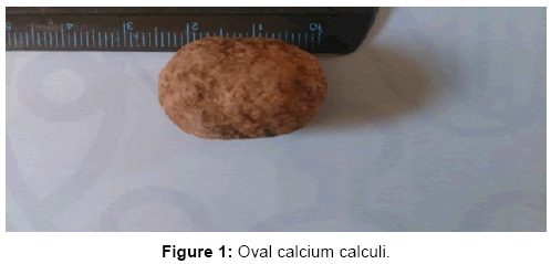
There was a calculi in urethral area of donkey. Grossly, urolith was oval shaped with a diameter of approximately 7 cm and a length of 2.5 cm. The majority of the surface area of the urolith was smooth textured with poles (Figure 1).
A fragment of urolith material from the case was submitted to the Forensic Laboratories/Ministry of interior Police Forces HQ for initial determination of mineral composition. Initial observation by the X-Ray Flurescence spectrometer/(EDX-8000/Japan) showed that the specimen was composed of 93.95% calcium. The result of that analysis was showed in Table 2.
| Element | % |
|---|---|
| Calcium | 93.95 |
| Potassium | 1.72 |
| Manganese | 1.32 |
| Silicon | 1.22 |
| Selenium | 0.92 |
Table 2: Constituents of analyzed uroliths.
Discussion
This is the first record of urethral calculus in donkeys in Sudan. The prevalence of equine urolithiasis is low (3.7%). The use of large quantities of grain and drinking salty water may cause an excessive ingestion of calcium. The affected donkey is mainly kept on grain foods al through which may suggest the finding of calcium stone formation. However, increased calcium content of the water in this report may also predispose to uroliths formation. Previous reports showed that, equine uroliths have a diameter of 0.5–21 cm and are found most often within the bladder [17]. Some cases are removed manually; others surgically through urethral sphincterotomy. In this case, the large size and lengthy of the calculi lead to it is removal by surgically using perineal urethrotomy under epidural anesthesia.
Conclusion
This is the first report of uroliths in donkey in Sudan. More investigation should be done about causes and prevention of the disease in donkeys in Westren Kourdofan State.
Acknowledgements
We are indebted to the laboratory of College of Applied Science and Industrial, University of Bahri for water analyzed. We are also grateful to Forensic Laboratories/Ministry of interior Police Forces HQ for uroliths analysis.
References
- Alicia F, Sabrina HB, Jan FH (2009).Urolithiasis. CompendiumEquine.CompendContinEduc Vet 4: 125-132.
- Cornelius CE (1963) Studies on ovine urinary biocolloids and phosphaticcalculosis. Ann NY AcadSci104: 638-657.
- Bailiff NL, Westropp JL, Jang SS, Ling GV (2005)Corynebacteriumurealyticum urinary tract infection in dogs and cats: 7 cases (1996-2003). J Am Vet Med Assoc 226: 1676-1680.
- Laverty S, Pascoe JR, Ling GV (1992) Urolithiasis in 68 horses.Vet Surg21:56-62.
- Saam D (2001) Urethrolithiasis and nephrolithiasis in a horse. Can Vet J42:880-883.
- Diaz-Espineira M, Escolar E,Bellanato J (1995). Crystallinecomposition of equine urinary sabulous deposits. Scanning Microsc9:1071-1077.
- Mohammadreza A, Amir HA (2009) Nephrolithiasis in two Arabian horses. Iranian J Vet SciTechnol1: 53-57.
- Abdel-HadyAAA(2014). Spontaneous repelling of a large urocystolith in a Working She-donkey. Sch J Agric Vet Sci1: 105-106
- TionMT, Dvorska J,SaganuwanSA (2015) A reveiw on urolithiasisin dogs and cats. Bulg J Vet Med 18: 1-8.
- Perillo A,Passantino G,Passantino L,Cianciotta A, Lo Prest i G,et al. (2008)Cystitises in the equine species: pathogenetic causes, histomorphological aspects and analogies with the human species.J Biomed Res 19: 2.
- Mohamed TH, Wael D (2015)Clinical, Biochemical and Ultrasonographic Findings in Buffalo Calves with Obstructive Urolithiasis. Global Veterinaria 14: 118-123.
- Robert MP, Main de B, Depecker MC,de Foumestraux, Touzot-Jourde G, et al. (2015) Type IV rectal prolapse secondary to a long-standing urinary bladder lithiasis in a donkey. Equine Veterinary Education.
- Lowe JE (1961)Surgical removal of equine uroliths via the laparocystotomy approach. J Am Vet Med Assoc139:345-348.
- Hanson RR, Poland HM (1995) Perinealurethrotomy for removal of cystic calculi in a gelding. J Am Vet Med Assoc207:418-419.
- Beard W (2004)Parainguinallaparocystotomy for urolith removal in geldings. Vet Surg 33: 386-390.
- Grant DC, Westropp JL, Shiraki R, Ruby AL (2009) Holmium:YAG Laser Lithotripsy for Urolithiasis in Horses. J Vet Intern Med 23:1079-1085.
- Thomas JD (2011). Urolithiasis in horses. In: Merck Veterinary Medicine, Last full revision.
Relevant Topics
- Acupuncture Therapy
- Advances in Naturopathic Treatment
- African Traditional Medicine
- Australian Traditional Medicine
- Chinese Acupuncture
- Chinese Medicine
- Clinical Naturopathic Medicine
- Clinical Naturopathy
- Herbal Medicines
- Holistic Cancer Treatment
- Holistic health
- Holistic Nutrition
- Homeopathic Medicine
- Homeopathic Remedies
- Japanese Traditional Medicine
- Korean Traditional Medicine
- Natural Remedies
- Naturopathic Medicine
- Naturopathic Practioner Communications
- Naturopathy
- Naturopathy Clinic Management
- Traditional Asian Medicine
- Traditional medicine
- Traditional Plant Medicine
- UK naturopathy
Recommended Journals
Article Tools
Article Usage
- Total views: 3809
- [From(publication date):
February-2017 - Nov 21, 2024] - Breakdown by view type
- HTML page views : 3147
- PDF downloads : 662

