Fahr's Disease in a Nigerian Pediatric Patient: A Rare Neurodegenerative Disorder
Received: 29-Jun-2023 / Manuscript No. roa-23-104201 / Editor assigned: 01-Jul-2023 / PreQC No. roa-23-104201 (PQ) / Reviewed: 21-Jul-2023 / QC No. roa-23-104201 / Revised: 24-Jul-2023 / Manuscript No. roa-23-104201 (R) / Published Date: 31-Jul-2023 DOI: 10.4172/2167-7964.1000465
Abstract
Introduction: Fahr’s disease is a rare neurodegenerative disorder characterized by abnormal calcium deposition in the basal ganglia, thalamus, dentate nucleus, subcortical white matter and hippocampus. The disease is characterized by impaired cognitive development, seizures, motor deficits and other neuropsychiatric symptoms. It is largely hereditary, with some cases occurring sporadically. Computed tomography and Magnetic resonance imaging are pivotal in its imaging, as they usually would help in identifying bilateral basal ganglia calcification and also identify other associations.
Objectives: To report a rare neurodegenerative disorder (Fahrs disease) in a Nigerian child. To review pertinent literature about this condition. To highlight the role of diagnostic imaging especially with Computed tomography, and also the findings on this modality.
Case Report: The case of a 7-year old male who was admitted in the Children emergency unit of the Irrua Specialist Teaching Hospital, on account of seizures, fever, dyspnoea and loss of consciousness. Serum electrolytes were deranged notably; hypokalemia, hyperuricemia and reduced bicarbonate. Computed tomographic scanner revealed bilateral symmetrical basal ganglia calcification, faint subcortical calcification in the frontal lobes and diffuse brain inflammatory changes. Chest radiograph done prior to the CT showed right sided pleural effusion.
Conclusion: Fahr’s disease is a neurodegenerative entity which is very rare in children. Radiologic imaging of the brain using cross sectional modalities remains a centrepiece in unravelling its diagnosis.
Keywords
Basal ganglia; Calcification; Fahr disease; Computed tomography; Irrua
Introduction
Fahr’s disease also called bilateral Striatopallidodentate calcinosis or Chavany-Brunches syndrome, is a rare neurodegenerative disorder characterized by symmetrical bilateral calcification in the basal ganglia, nucleus gyrus, and cerebral cortex [1-3]. The syndrome was first described in 1930 by a German Pathologist called Karl Theodor Fahr [2]. The disease is characterized by deposition of mineral elements, predominantly calcium, as well as variable amounts of Zinc, Iron, Aluminium, Silicon, Copper and Phosphorus within small capillaries in the areas of the brain which controls body movements. Such areas of the brain includes; the basal ganglia, thalamus, dentate nucleus, subcortical white matter, cerebral cortex and hippocampus [1,4].
There is paucity of recent published data on the incidence and prevalence of this condition. However, the overall incidence of basal ganglia calcification detected on computed tomography has previously been reported as between 0.24% and 1.64%, while the prevalence of Fahr disease has been reported as <1/1,000,000 [5-8]. Males have been reported to be more commonly affected than females. Typical age of onset is between the third and sixth decade of life [9-11]. Although, a few cases of the disease have been diagnosed and reported in the pediatric age group, in Nigeria cases of Fahr disease in children have been very rare [12].
The disease is most commonly inherited as an autosomal dominant trait. It may also be inherited in an autosomal recessive or sporadic fashion [13,14].
There has also been some scientific debates as to whether the term Fahr disease is synonymous with Fahr syndrome, although both terms have been used interchangeably. However, it has been advocated that the term Fahr disease be used to describe bilateral basal ganglia calcification with familial Autosomal dominant pattern of inheritance, also called primary familial brain calcification, whereas Fahr’s syndrome should be reserved for secondary causes of bilateral basal ganglia calcification such as; metabolic and endocrinopathies (e.g hypoparathyroidism, pseudohypoparathroidism, hyperthyroidism etc), vasculitis (e.g SLE), hypoxia, and infective causes [13-15].
Calcification of the basal ganglia with only a small amount of the calcification confined to the globus pallidus alone is commonly seen in the elderly occurring as an age dependent phenomenon(senile change) where it is commonly considered a normal finding [15].
Pathogenesis of this condition involves calcium deposition on the walls of capillaries and small arteries and veins in the regions of the brain which controls movement of the body. There is resultant generation of free radicals and impaired ion transport. These further results in a cascade of tissue damage and further secondary deposition of calcium, mucopolysacharides and other toxic substances. Impaired blood flow with progressive neurodegeneration and ultimately brain atrophy results [8,16,17].
Clinical presentation of the disease is similar in both adults and children. Affected children may present with impaired cognitive development, motor deficits, dysarthria, seizures and psychiatric symptoms [12].
Presence of bilateral basal ganglia calcification (identified on CT), progressive neurological dysfunction (neuropsychiatric symptoms), positive family history, and absence of biochemical abnormalities that may suggest metabolic, mitochondrial or systemic disorders are important diagnostic criteria for making a diagnosis of Fahr’s disease [12].
Documented evidence of Fahr’s disease in African children is sparse. The first reported case in a Nigerian child was published in 2021 [12].
Case Report
We report the case of a 7-years old male child who is the first of a set of twins. He was admitted into the Children Emergency unit of the Irrua Specialist Teaching Hospital-a leading infectious disease hospital in Nigeria (a country in Sub-Saharan Africa), on account of persistent fever, fluctuating level of consciousness and seizures. A positive history of delay in attainment of cognitive and motor developmental milestone in the patient and his twin sibling (a female) was elicited. However, no documented history of seizure disorder was noted in the parents. Antenatal history was unremarkable, with no history of prolonged labour.
Physical examination at admission revealed an unconscious child (Glasgow Coma score of 4/15) who was dyspnoeic and febrile with an elevated blood pressure. The patient’s symptoms had improved few days into the admission, but later suddenly lapsed into unconsciousness, high grade fever and a poorly controlled blood pressure which tilted towards hypotension most of the time. These features and other signs of raised intracranial pressure only temporarily responded to administration of mannitol. Secondary hospital acquired infection (nosocomial infection) was suspected and later confirmed by blood culture.
There was no record of serum calcium levels. However, serum analysis of electrolytes, urea and creatinine revealed; elevated creatinine levels of about 6.1mg/dl, hypokalemia (K+=3.2mmol/dl), reduced serum bicarbonate (6mmol/dl), and hyperuricemia (68mg/dl).
The patient was referred to our department for imaging (chest radiography, abdominal sonography and cranial computed tomography) with a clinical diagnosis of “Seizure disorder, keeping in view Cavernous sinus thrombosis”.
Chest radiography revealed a right sided pleural effusion with bronchopneumonic changes involving the contiguous lung. Abdominal scan revealed features of bilateral acute renal parenchymal disease and also confirmed and quantified the right sided pleural effusion (at least 200ml).
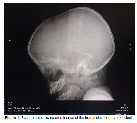
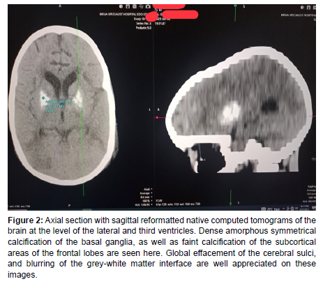
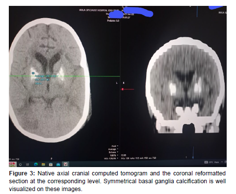
Native scans of the child’s brain were acquired in axial planes and reconstructed using a 32-slice Canon Aquilion Helical Computed Tomographic scanner (TSX-037A model, manufactured in Japan). It showed prominence of the frontal and occipital regions, with relative reduction in the biparietal distance giving an appearance of scaphocephaly. The brain symmetry was preserved with no demonstrable haemorrhage or dural collection. However, there were dense amorphous bilaterally symmetrical calcification in the basal ganglia with Hounsfield unit values up to 240. There was no perilesional edema. However, there was widespread blurring of the grey-white matter interface with global effacement of the sulci. Faint subcortical white matter calcification was also noted in the frontal lobes bilaterally. Mild dilatation of the lateral ventricles was noted. Wholistically considering the cranial CT findings, vis-a-viz the clinical history of delayed developmental milestones, impaired cognitive and motor functions, as well as the acute presentation with seizures and fluctuating degree of consciousness, we made a radiologic impression of Fahr’s disease/syndrome with acute encephalitis. We had difficulty in excluding cavernous sinus thrombosis due to the non-administration of intravenous contrast agent occasioned by the impairment of the patient’s renal function.
Our patient died barely 24 hours after the Computed tomographic scanning was done. CT images acquired are shown below (Figures 1-3).
Figure 2: Axial section with sagittal reformatted native computed tomograms of the brain at the level of the lateral and third ventricles. Dense amorphous symmetrical calcification of the basal ganglia, as well as faint calcification of the subcortical areas of the frontal lobes are seen here. Global effacement of the cerebral sulci, and blurring of the grey-white matter interface are well appreciated on these images.
Discussion
Fahr’s disease is a rare neurodegenerative disorder which can affect both genders with a male predilection. More cases have also been reported amongst older patients between the third and sixth decade of life, with only a few cases of pediatric Fahr’s disease reported worldwide [9-11]. Reports of cases from Africa are rare. Also, not many cases of Fahr’s disease have been reported in Nigeria. Even more sparse are reports of Fahr’s disease in pediatric patients in Nigeria [12]. However, we believe that the overall prevalence (though not yet reported in Africa) of this disease in Africa may actually be higher than the general belief. Low socioeconomic status of our patients which may hinder their assess to tertiary health care facilities, traditional belief system especially in Sub-Saharan Africa, and non availability of advanced imaging modalities like CT scanners, Magnetic resonance imaging (MRI) etc, are factors considered responsible for possible underdiagnosis.
Okike et al. [12] described and reported the first case of pediatric Fahr’s disease in Asaba, Southern Nigeria in the year 2020, three years before the report of this index case in Irrua, Nigeria, which appears to be the second reported case, as no other case has been reported in a Nigerian child after diligent literature search. The age of the child with this index case is quite similar to that reported by Okike etal, which was a 9-year old female child. However, the genders were different.
Manyam et al. [5] in their retrospective study of 38 reported cases in the year 2001, noted a predilection of the disease in males in a ratio of 2:1. However, no definite reason was adduced for this trend. They also observed significantly greater amount of calcification in symptomatic patients compared to asymptomatic patients. Our reported case was symptomatic with seizures and fluctuating level of consciousness at presentation. His condition progressively deteriorated until his death. The finding of extensive and dense basal ganglia calcification, may have been contributory to the severity of the disease. The diagnosis of Fahr’s disease could not have been easily made if not for the role of cross sectional imaging using Computed tomography (CT).
Krzysztof et al. [3] had previously noted that children with Fahr’s disease may present with severe encephalopathy, dwarfism, microcephaly and optic nerve atrophy. Our case presented with craniosynostosis and scaphocephaly, but the optic nerves were normal (diameter of 2.6mm). Features of encephalitis seen on the cranial CT of our patient could have been due to overwhelming septicaemia confirmed by a positive blood culture.
Conclusion
Although Fahr’s disease is a rare neurodegenerative disease entity in both adults and children, the presence of seizures, neurological and motor deficits, as well as childhood history of delayed developmental milestones, should raise the index of suspicion, and prompt brain imaging using CT/MRI.
In particular, Cranial CT scan is indispensable in identifying basal ganglia and cerebral calcifications, other intracerebral associations, as well as excluding differentials like intracerebral hemorrhages, intracranial tumours, hypoxia, hypo/hyper parathyroidism etc.
References
- https://www.ninds.nih.gov/health-information/disorders/fahrs-syndrome
- Fahr T (1930) Calcification of the cerebral vessels. Zentralbl Allg Pathol 50:129-133.
- Jaworski K, Styczyńska M, Mandecka M, Walecki J, Kosior DA (2017) Fahr syndrome-an important piece of a puzzle in the differential diagnosisof Mary Disease. Pol J Radiol 82: 490-493.
- Nilika S, Haren KV, Wu Y (2012) Fahr Disease: Pediatric presentation of a Rare Neurodegenerative Disorder (IN 10-1.008). Neurology 78.
- Manyam BV, Watters AS, Narla KR (2001) Bilateral Striopallidodentate Calcinosis: Clinical characteristics of Patients seen in a registry. Mov Disord 16: 258-264.
- Fenelon G, Gray F, Paillard F, Thibierge M, Mahieux F, et al. (1993) A Prospective study of patients with CT detected pallidal calcifications. J Neurol Neurosurg Psychiatry 56: 622-625.
- Ellie E, Julien J, Ferrer X (1989) Familial Idiopathic Striopallidodentate Calcifications. Neurology 39: 381-385.
- Saleem S, Aslam HM, Maheen A, Shahzad A, Maria S, et al. (2013) Fahr Syndrome: Literature review of current evidence. Orphanet J Rare Dis 8: 156.
- Nicolas G, Charbonnier C, Campion D, Veltram JA (2018) Estimation of minimal disease prevalence from population genomic data : application familial brain calcification. Am J Med Genet B Neuropsychtr Genet 177: 68-74.
- Kotan D, Aygul R (2009) Familial Fahr Disease in a Turkish Family. South Med J 102: 85-86.
- Thillaigovindan R, Arumugam E, Rai R, Prabhu R, Kesavan R, et al. (2021) Idiopathic Basal Ganglia Calcification: Farh’s syndreome, a rare disorder. Cureus 11: e5895.
- Okike OC, Ajaegbu CO, Origbo L, Muoneke VU (2021) Idiopathic basal ganglia calcification (Fahr’s disease) in a 9-year old Nigerian child. Niger J Pediatr 48: 46-49.
- Badilla CR (2023) Fahr Syndrome: Case study. Radiopedia.org
- Perugula ML, Lippmann S (2016) Fahr’s disease or Fahr’s Syndrome?. Innov Clin Neurosci 13: 45-46.
- Ho M, El-Feky M, Galliard F (2023) Fahr syndrome. Reference article, Radiopedia.org.
- Tai XY, Batla A (2015) Fahr’s disease: Current perspectives. Orphan Drugs: Research and Reviews 5: 43-49.
- Geschwind DH, Loginov M, Stem JM (1999) Identification of a locus on Chromosome 14q for idiopathic basal ganglia calcification (Fahr disease). Am J Hum Genet 65: 764-772.
Indexed at, Google Scholar, Crossref
Indexed at, Google Scholar, Crossref
Indexed at, Google Scholar, Crossref
Indexed at, Google Scholar, Crossref
Indexed at, Google Scholar, Crossref
Indexed at, Google Scholar, Crossref
Indexed at, Google Scholar, Crossref
Indexed at, Google Scholar, Crossref
Citation: Eluehike SU, Izevbekhai OS, Irabor PFI, Oriaifo B, Oshodin E (2023) Fahr’s Disease in a Nigerian Pediatric Patient: A Rare Neurodegenerative Disorder. OMICS J Radiol 12: 465. DOI: 10.4172/2167-7964.1000465
Copyright: © 2023 Eluehike SU, et al. This is an open-access article distributed under the terms of the Creative Commons Attribution License, which permits unrestricted use, distribution, and reproduction in any medium, provided the original author and source are credited.
Share This Article
Open Access Journals
Article Tools
Article Usage
- Total views: 1289
- [From(publication date): 0-2023 - Mar 29, 2025]
- Breakdown by view type
- HTML page views: 1049
- PDF downloads: 240


