Conference Proceeding Open Access
Evaluation of Multi-Slice Computed Tomography in Assessment of Cochlear Implant Electrode Position: A Pictorial Essay
| Fatemeh Nasri*, Hashem Sharifian, Mehrzad Mehdizadeh and Mohammad Pir Hayati | |
| Department of Radiology, Iranshahr University of Medical Science, Iran | |
| *Corresponding Author : | Fatemeh Nasri Department of Radiology, Iranshahr University of Medical Science, Iran Tel: +989122904262 E-mail: Fatemeh.nasri. 62@gmail.com |
| Received date: January 20, 2016; Accepted date: March 10, 2016; Published date: March 14, 2016 | |
| Citation: Nasri F, Sharifian H, Mehdizadeh M, Hayati MP (2016) Evaluation of Multi-Slice Computed Tomography in Assessment of Cochlear Implant Electrode Position: A Pictorial Essay. OMICS J Radiol 5:217. doi:10.4172/2167-7964.1000217 | |
| Copyright: © 2016 Nasri F, et al. This is an open-access article distributed under the terms of the Creative Commons Attribution License, which permits unrestricted use, distribution, and reproduction in any medium, provided the original author and source are credited. | |
Visit for more related articles at Journal of Radiology
Abstract
Cochlear implant has become the standard therapy to rehabilitate patients with severe to profound bilateral sensorineural hearing loss. The position of the electrode in the scala and the depth of its insertion have been shown to be predicting factors of hearing outcomes. This article serves to review the multi-slice computed tomography imaging characteristics and appearance of cochlear implant, their exact position and the depth of insertion in children who underwent cochlear implant surgery. Moreover, in order to evaluate the impact of the electrode position on clinical results following cochlear implant surgery, in this study we also compared the findings on the electrode location with the results of audiometry of children after one year of follow up. Finding the best location for the electrode results in better audiometric outcomes.
| Keywords |
| Multi slice computed tomography; Cochlear implant electrode array position; Audiometric outcome |
| Introduction |
| Electrical stimulation of the auditory nerve for auditory rehabilitation by cochlear implants with a multichannel electrode array has become the standard therapy to rehabilitate patients with severe to profound bilateral sensorineural hearing loss [1]. Surgical techniques in these patients require non traumatic insertion of the electrode array to preserve hearing and to avoid damage to the scala media, the organ of Corti, and other delicate inner ear structures. Over the past few years, many studies have focused on exact positioning of the electrode array in the cochlea to improve surgical techniques and audiometric outcomes [2-4]. |
| Many prior studies have determined significant predicting factors of hearing outcomes in patients with cochlear implants [5-7]. These include duration of deafness [8], level of pre-implant speech recognition [9], pre-lingual / post-lingual status [10], and electrode programming configuration [11,12]. One of the most important factors that can influence the clinical outcome of CI is shown to be the position of the electrode in the scala and the depth of its insertion [13]. Therefore, being expert in postoperative radiological assessment of the electrode position deserves the special attention of clinicians. |
| This article serves to review the multi-slice CT imaging characteristics and appearance of the cochlear implant and the exact position and depth of the insertion of the electrode array, their clinical presentations and complications, and their audiometric outcome. The optimal positioning of the cochlear implant |
| Theoretically, the loudness of the sound perceived by the patient may depend on the number of auditory nerve fibers activated by the electrode array and the proximity of electrodes to the nerve fibers (perimodiolar location) [9]. Whenever the electrode is inserted closer to the hair cells along the basilar membrane, the auditory stimulus, perceived by the auditory nerve will be stro nger. Insertion of the electrode into the scala tympani is the preferred route because the scalar lamina and basilar membrane provide the scala media a modest degree of protection. On the other hand, the scala tympani is of a larger size for the placement of electrodes; insertion of the electrode into the Scala vestibule may cause traumatic disruption of the Reissner’s membrane and damage the underlying hair cells, resulting in meningitis and loss of residual hearing ability [4]. |
| Modifications in surgical techniques have been advanced to increase the accuracy of scala tympani insertion, but there has not been a definite method for determining the intra-cochlear location after surgery [5]. |
| Post-operative imaging |
| Several imaging techniques such as multi-slice CT, cone-beam CT, high resolution CT (HRCT), digital volume tomography (DVT) and magnetic resonance imaging (MRI) are available for postoperative evaluation of cochlear implants [1]. The main purposes of this evaluation are to document electrode placement and its position and, quality control of cochlear implant surgery, and to evaluate the temporal bone in case of complications or additional central morbidities (e.g., acoustic neuroma, cerebral tumor, cerebral-vascular diseases, etc.). Each of the above-mentioned techniques has advantages and disadvantages. The selection of the technique is dependent on many factors including the accuracy of the images provided by the technique based on the goal of the examination, the rate of radiation exposure dose, its availability in the daily clinic routines and the costeffectiveness of each specific technique. For instance, postoperative application of MRI is a specific challenge because it depends on the implant type and can result in different types of motion and metal artifacts. That is to say, MRI is possible up to 1.5 tesla (with or without a magnet depending on the recommendations of the manufacturer) [14]. In general, the indication of postoperative MRI could be the occurrence of complications or the presence of other central comorbidities [1]. DVT does not provide sufficient delineation of soft tissue but it makes smaller metal artifacts. |
| In comparison to multi-slice CT, using Cone-beam Ct scan reduce the radiation dose to 14%.The major problem with Cone-beam CT is that acquisition of high resolution images requires a long exposure time of 40 seconds, which results in more motion artifacts. This can result in the need for “retakes,” causing a higher radiation dose exposure in patients. For reduction of motion artifacts, Cone-beam CT units that study patients in the supine position are preferred to those that study sitting and standing positions. The patient should also be trained not to swallow and to breathe through the nose [15]. So performing Cone- beam CT has dramatic limitations in children, who are the main group of patients. |
| Multi-slice CT is frequently used for postoperative assessment of the CI. It is an easily available non-invasive method that decreases motion artifacts and improves the patient’s comfort by reducing the examination time, which is an important aspect especially in children [16]. Moreover, it provides the ability to determine the exact position and depth of the insertion of the electrode. In a study of 17 patients who underwent cochlear implant surgery, a multi-detector 64-slice CT scan was used to determine the location of the electrode array in the cochlea. However, the study did not assess the depth of the insertion of the electrode, which is a very important factor for hearing low frequency signals [2]. In another study, the visibility of cochlear walls in postoperative images of multi-slice CT has also been considered an important factor in determining of electrode positioning [17]. In this study, we assessed both the exact position and the depth of the insertion of the electrode array with the help of multi-slice computed tomography. Therefore, it has been used in this study as a postprocedural evaluation technique. |
| Details about the imaging technique |
| All acquisitions were made with a 16-slice multi-detector computed tomography scanner (General Electronic, Bright Speed) in axial plane, parallel to the infra-orbito meatal base line and in some cases parallel to the orbitomeatal base line with 0 to 15 degrees angle to caudal with the purpose of not including the lens of orbit (voltage, 140 kv; amperage, 250 mA; pitch 0.56, matrix 512 × 512, slice thickness 0.6 mm, collimation 0.6 mm, rotation time 1s, and without an interslice gap). The field of view was 100 mm, and the images were reconstructed at the bony algorithm. |
| The suitable window width and window level were 4500 and 1050 CT number respectively. |
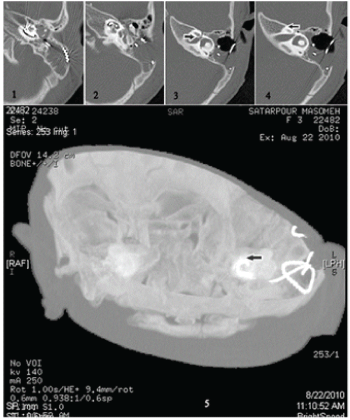
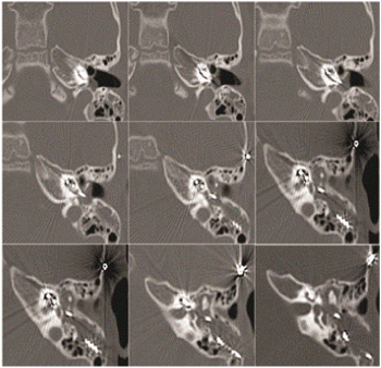
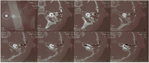
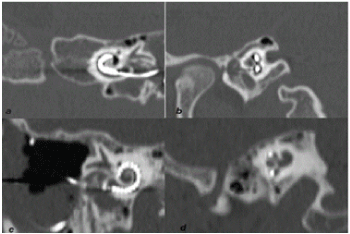
| Images were reconstructed with software AW4 / 4 in the oblique coronal plane, parallel to the cochlea (modified stenverse view) and oblique sagittal plane (perpendicular to the modiolus) with the help of a volume rendering technique. Three-dimensional images were reconstructed by maximal intensity projection. The field of view in the reconstructed images was 70 mm. Multi-planar reconstructions and three-dimensional images were then evaluated to determine the location of the electrode array and the depth of the electrode insertion. Oblique coronal and oblique sagittal reformations were helpful in detecting the location of the intra-cochlear electrode position, relative to the scala media and lateral wall of the scala tympani. True axial graphs were helpful in detecting the depth of insertion. Threedimensional reconstructs were not helpful in detecting the depth of insertion, and they underestimated the number of turns of implant array inside the cochlea (Figure 1). |
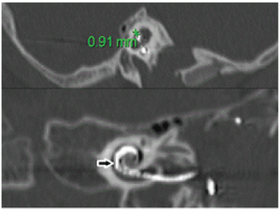
| Electrodes rotated at least two turns inside the cochlea were considered deeply inserted (Figures 2 and 3). |
| Clinical rehabilitation outcome |
| To evaluate the impact of the electrode position on rehabilitation results following cochlear implant surgery, in this study we also compared the findings on the electrode location with the results of audiometry of children after one year of follow up. Finding the best location for the electrode results in better audiometric outcomes. Patients were examined pre-operatively and one year after CI by pure tone audiometry (PTA) and speech reception threshold (SRT). As described in the figure legends, cases in which the electrodes were deeply inserted and located in the medial compartment had better clinical results after CI surgery. Results demonstrated that electrodes that were inserted deeply inside the cochlea and rotated at least two turns inside the cochlea produced better audiometry results (Figures 4 and 5). |
| Summary |
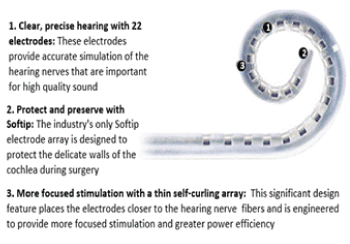
| MSCT is a useful tool for the exact positioning of an implant array inside the cochlea. High resolution and artifact-free images allow for determining the electrode position inside the perimodiolar location or lateral compartment of the scala tympani. The electrode position inside the medial compartment accompanies better audiometric results in children (Figures 6 and 7). |
| The common goal of all imaging procedures in cochlear implant patients is the improvement of surgical planning and results, the control of surgical quality, and the reduction of complications. Therefore, these findings may be a useful guideline for surgeons to obtain a better outcome of cochlear implant surgery. |
References
- Antje Aschendorff (2011) Imaging in cochlear implant patients. GMS Curr Top Otorhinolaryngol Head Neck Surg 10: 07.
- van Wermeskerken GK, Prokop M, van Olphen AF, Albers FW (2007) Intracochlear assessment of electrode position after cochlear implant surgery by means of multislice computer tomography. European Archives of Oto-rhino-laryngology 264: 1405-1407.
- Aschendorff A, Kubalek R, Turowski B, Zanella F, Hochmuth A, et al. (2005) Quality control after cochlear implant surgery by means of rotational tomography. Otology &Neurotology 26: 34-37.
- Richter B, Aschendorff A, Lohnstein P, Husstedt H, Nagursky H, et al. (2001) The Nucleus Contour electrode array: a radiological and histological study. The Laryngoscope 111: 508-514.
- Friedland DR, Venick HS, Niparko JK (2003) Choice of ear for cochlear implantation: the effect of history and residual hearing on predicted postoperative performance. OtolNeurotol 24: 582-589.
- Summerfield AQ, Marshall DH (1995) Preoperative predictors of outcomes from cochlear implantation in adults: performance and quality of life. Ann OtolRhinolLaryngol 166: 105-108.
- Shipp DB, Nedzelski JM (1995) Prognostic indicators of speech recognition performance in adult cochlear implant users. Ann OtolRhinolLaryngol 166: 194-196.
- Blamey P, Arndt P, Bergeron F, Bredberg G, Brimacombe J, et al. (1996) Factors affecting auditory performance of post linguistically deaf adults using cochlear implants. AudiolNeurotol 1: 293-306.
- Rubinstein JT, Parkinson WS, Tyler RS, Gantz BJ (1999) Residual speech recognition and cochlear implant performance: effects of implantation criteria. Am J Otol 20: 445-452.
- Tong YC, Busby PA, Clark GM (1988) Perceptual studies on cochlear implant patients with early onset of profound hearing impairment prior to normal development of auditory, speech, and language skills. J AcoustSoc Am 84: 951-962.
- Pfingst BE, Franck KH, Xu L, Bauer EM, Zwolan TA (2001) Effects of electrode configuration and place of stimulation on speech perception with cochlear prostheses. J Assoc Res Otolaryngol 2: 87-103.
- Mens LH, Berenstein CK (2005) Speech perception with mono- and quadrupolar electrode configurations: a crossover study. OtolNeurotol 26: 957-964.
- Shepherd RK, Hatsushika S, Clark GM (1993) Electrical stimulation of the auditory nerve: the effect of electrode position on neural excitation. Hear Res 66: 108-122.
- Crane BT, Gottschalk B, Kraut M, Aygun N, Niparko JK (2010) Magnetic resonance imaging at 1.5 T after cochlear implantation. Otology &Neurotology 31: 1215-1220.
- Casselman JW, Gieraerts K, Volders D, Delanote J, Mermuys K, et al. (2013) Cone beam CT: Non dental _applications. Journal Belge de Radiologie_BelgischTijdchriftvoorRadiologie 96: 1-22.
- Valvassori GA (2005) Imaging of the temporal bone. In: Mafee MF, Valvassori GE, Becker M (2ndedn) Imaging of the head and neck. Georg ThiemeVerlag, Stuttgart, pp: 30.
- Theunisse HJ, Joemai RM, Maal TJ, Geleijns J, Mylanus EA, et al. (2015) Cone-Beam CT Versus Multi-slice CT Systems for Postoperative Imaging of Cochlear Implantation-A Phantom Study on Image Quality and Radiation Exposure Using Human Temporal Bones. Otology &Neurotology 36: 592-599.
Tables and Figures at a glance
| Table 1 | Table 2 | Table 3 | Table 4 | Table 5 |
Figures at a glance
 |
 |
 |
 |
| Figure 1 | Figure 2 | Figure 3 | Figure 4 |
 |
 |
 |
| Figure 5 | Figure 6 | Figure 7 |
Relevant Topics
- Abdominal Radiology
- AI in Radiology
- Breast Imaging
- Cardiovascular Radiology
- Chest Radiology
- Clinical Radiology
- CT Imaging
- Diagnostic Radiology
- Emergency Radiology
- Fluoroscopy Radiology
- General Radiology
- Genitourinary Radiology
- Interventional Radiology Techniques
- Mammography
- Minimal Invasive surgery
- Musculoskeletal Radiology
- Neuroradiology
- Neuroradiology Advances
- Oral and Maxillofacial Radiology
- Radiography
- Radiology Imaging
- Surgical Radiology
- Tele Radiology
- Therapeutic Radiology
Recommended Journals
Article Tools
Article Usage
- Total views: 11029
- [From(publication date):
April-2016 - Jul 05, 2025] - Breakdown by view type
- HTML page views : 10165
- PDF downloads : 864
