Research Article Open Access
Evaluating the Effectiveness of Organic Coatings Based on Natural Polymers for the Treatment of Wounds in the Experiment
Kaulambayeva MZ*, Toleubekova AS and Akhmetsadykov NNAntigen Co Ltd, Almaty, Kazakhstan
- Corresponding Author:
- Marzhan Kaulambayeva
Head of laboratory
Antigen Co Ltd, 4 Azerbayev street
Almaty, --- 040905, Kazakhstan
Tel: +77074325763
E-mail: marzan61z@mail.ru
Received date: October 30, 2015; Accepted date: November 23, 2015; Published date: November 30, 2015
Citation: Kaulambayeva MZ, Toleubekova AS, Akhmetsadykov NN (2015) Evaluating the Effectiveness of Organic Coatings Based on Natural Polymers for the Treatment of Wounds in the Experiment. J Biotechnol Biomater 5:211. doi:10.4172/2155-952X.1000211
Copyright: © 2015 Kaulambayeva MZ, et al. This is an open-access article distributed under the terms of the Creative Commons Attribution License, which permits unrestricted use, distribution, and reproduction in any medium, provided the original author and source are credited.
Visit for more related articles at Journal of Biotechnology & Biomaterials
Abstract
This article presents the results of research on the effectiveness of a medical preparation «Biotangish®FT» in the treatment of wounds. The experiment was conducted by inflicting wounds on the skin of animals and subsequent treatment with a preparation «Biotangish®FT», a comparative analogue "Tachocomb" and an ointment "Levomekol." The results showed that the «Biotangish®FT» has a strong wound-healing and anti-inflammatory effect.
Keywords
Collagen; Hyaluronic acid; Fibrinogen; Thrombin; Wound healing; Wound covering
Introduction
Today, the treatment of burns and surgical wounds is one of the urgent problems of modern combustiology and surgery. One of the solutions to this problem is the use of wound coverings based on natural or synthetic polymers. Application of wound coverings can significantly improve the efficiency of the treatment of wounds and burns. Analysis of the literature sources showed that more than 300 wound coverings are at different stages of development [1-6]. However, so far there is no universal product that would be appropriate for the use in all phases of wound healing. Of the currently existing wide range of polymer coatings for wounds and burns, the absorbable coatings are the best answer to all medical and biological requirements. They can be beneficial at early stages of burn and wound healing as well as at the later stages. Currently, the development of absorbable adherent polymer coatings with high absorbability and various time of absorption is the most actual direction in the field of development of effective coatings for wounds and burns.
Under the subproject "Organic coatings based on natural polymers for the treatment of burns and surgical wounds", the wound covering technology "Biotangish®FT" was developed based on collagen, hyaluronic acid and components of fibrin glue.
The purpose of this work is to determine the efficiency of application of "Biotangish®FT" coating in the treatment of wounds. The work is executed under the grant "Technology commercialization project" of the World Bank and Ministry of Education and Science of the Republic of Kazakhstan.
Materials and Methods
24 healthy adult guinea pigs (male) weighing 350-500 g were used in the experiment. Animals were housed in ventilated complexes. Three animals were kept in each rack. The pressure and the air was automatically regulated. The diet of animals has been a standard (vegetables, pelleted feed). Air temperature was 20-23 °C. The fur on the experimental areas of the back skin was carefully cut out a day before the study began. The animals were divided into 4 groups: - three test and one control group, containing 6 animals in each group. At the time of wounding, the animals were anesthetized with 2% rometara solution.
Then a back skin flap with subcutaneous tissue till fascia was removed with scissors. Full layer skin wounds had a rounded shape with the diameter of 2.0 cm. To reduce the number of experimental animals in the groups, two wounds was applied to each animal.
The wounds of animals in the first control group were untreated. The wounds of the animals in second group were treated with the ointment "Levomikol" (1 g ointment contains active substances: chloramphenicol - 7.5 mg, methyluracil - 40mg). The "Biotangish®FT" coating was applied to animals from the third group, whereas the Tachocomb drug (active substances: fibrinogen - 5,5 mg, thrombin - 2 IU) was used on the wounds of the fourth group. The studied medications were applied once.
The wounds of animals in the first control group were untreated. The wounds of the animals in second group were treated with the ointment "Levomikol" (1 g ointment contains active substances: chloramphenicol - 7.5 mg, methyluracil - 40mg). The "Biotangish®FT" coating was applied to animals from the third group, whereas the Tachocomb drug (active substances: fibrinogen - 5,5 mg, thrombin - 2 IU) was used on the wounds of the fourth group. The studied medications were applied once.
The dynamics of the skin wounds healing process was monitored on the 4th, 7th, 11th, 15th and 23rd days following the time of wounding, which included these parameters: measurement of the diameter of a skin wound, the general assessment of animals' condition, the severity of the inflammatory response, the time of occurence of granulation tissue, epithelialization, wound healing. Obtained results were compared by groups. For histological studies skin regenerates were analyzed after 4, 7 and 11 days from the beginning of the experiment. The pieces were excised from the edges and from the central part of the skin defect on 4, 7 and 11 days of the experiment. The pieces were fixed in 10% aqueous solution of neutral formalin and sealed with paraffin. Microtome cuts with a thickness of 5 microns were stained with hematoxylin-eosin.
Results
Wound area was calculated by the formula: S = 2 πτ2 , where S - the area of the wound surface, π- 3,14 (constant), τ- radius of the circular wound.
The obtained results are given in Table 1.
| Animalgroups | Duration of observation(days) | |||||
| 1 | 4 | 7 | 11 | 15 | 23 | |
| 1.Withou ttreatment | 6,28cm2 | 4,52cm2 | 3,53cm2 | 1,57cm2 | 0,39±0,15cm2 | 0 |
| 2.The ointment "Levomikol" | 6,28cm2 | 5,10±0,20cm2 | 4,02±0,15cm2 | 1,88±0,21cm2 | 0,39±0,20cm2 | |
| 3. "Biotangish®FT"coating |
6,28cm2 | 3,53±0,50cm2 | 2,64 ±0,43cm2 | 1,0±0,14cm2 | 0 | |
| 4.«Tachacom»sponge | 6,28cm2 | 2,82±0,42cm2 | 2,26 ±0,30cm2 | 0,75±0,21cm2 | 0 | |
Table 1: According to data, the animals from third group with "Biotangish®FT" coating and animals from fourth group with "Tachacomb" sponge showed 100% healing of experimental wounds by 15th day, which provided a shorter period of healing in comparison with the ointment "Levomikol" (by 20th day). The full wound healing in animals from control group was observed on 23rd day from the beginning of the experiment.
Based on the above data, we can see that "Biotangish®FT" coating has a strong wound- healing as well as anti-inflammatory effect, has an advantage over "Levomikol", and is not in any way inferior to similar drug "Tachocomb".
Histomorphological study of wound surfaces
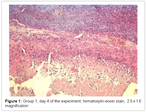
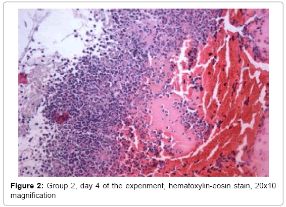
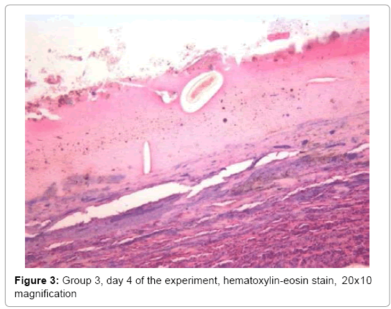
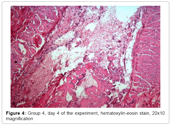
Histomorphological study of wounds on day 4 showed that the condition of the wound defect walls differed little between groups and was characterized by the presence of fibrin purulent leukocyte covering, necrotic layer and underlying connective tissue fibers with inflammatory lymph leukocyte infiltration and foci of hemorrhage (Figure 1-4).
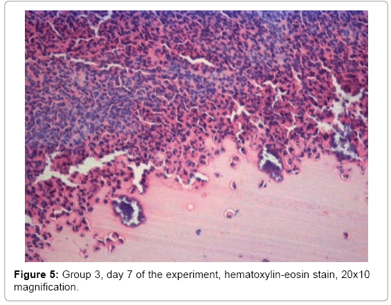
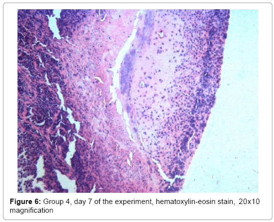
The marginal epithelialization of the wound defect was more explicit in the third and fourth groups on day 7 of the experiment in comparison with second and control groups. Superficial imposition of fibrin and leukocyte-necrotic layer with hemorrhages were thinner in the same groups, whereas developing granulation tissue was characterized by increased proliferation processes of newly formed capillaries and fibroblastic cell (Figure 5-6).
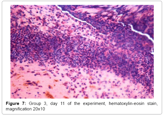
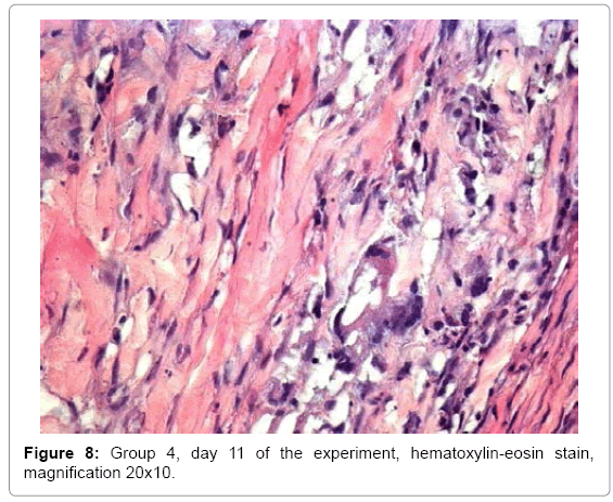
The tendency of advancing process of epithelialization of the wound surface was constant in third and fourth groups by the day 11 of the experiment. The acceleration of reparative processes was provided by a reduction of inflammatory response and an increase in the number of macrophages with high phagocytic activity. The well known important role of macrophages is not only in the implementation of the phagocytosis of tissue detritus, but also in the ability to allocate inducers of angiogenesis, as well as increasing the number of fibroblasts (Figure 7-8).
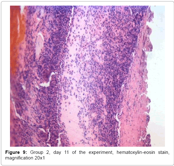
Morphological changes in the treatment of wounds with "Levomikol" were typical for sluggish healing of purulent wounds. During the application of Levomikol drug, the wound covered with a layer of fibrin organization events and impregnated leukocytes on 11th day of the experiment. Next, necrotic tissue, densely infiltrated by leukocytes and lymphoid cells.
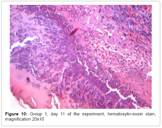
Granulation tissue without clear limitation of necrosis (Figure 9). The wound in the untreated control group was covered with a thin layer of fibrin with a large number of neutrophils, granulation tissue with a large number of thin collagen fibers on the 11th day of the experiment. The number of vessels in a large number of fibroblasts arranged horizontally, allows some of them to turn into fibroblasts (Figure 10). Macrophage response is not significant.
The use of "Biotangish®FT" showed a pronounced wound-healing, anti-inflammatory action and had a definite advantage over Levomikol, not yielding to similar "Tachocomb" drug.
Conclusion
1. A distinctive feature of wound healing is an acceleration of reparative process with an application of organic coverings: a prerequisites for this process are the reduction in the number of neutrophils and increase in the number of mononuclear cells, especially mature macrophages with high phagocytic activity. The granulation that produced in the wound during the application of organic coverings are rich in blood vessels and cellular elements (macrophages, fibroblasts), which significantly stimulates the regeneration of wound healing.
2. The histological image during the usage of "Tachocomb" drug revealed a decrease in the amount of bleeding, a moderate proliferation of the endothelium, the neutrophilic infiltration of granulation tissue, not a large number of polynuclears, delayed transformation of fibroblasts into fibrocytes, not enough fast elimination of necrotic masses.
3. The histological image of "Levomikol" usage in the treatment of wounds was typical for sluggish healing of purulent wounds.
4. "Biotangish®FT" covering has rather pronounced woundhealing as well as anti- inflammatory effects and has an advantage over "Levomikol", not yielding to similar "Tachocomb" drug.
Acknowledgements
The work was executed under the grant "Technology commercialization project" of the World Bank and Ministry of Education and Science of the Republic of Kazakhstan (GSNS #80)
References
- Dobysh SV, VasilyevAV, ShurupovaOV (2001) Modern bandage materials for the treatment of wounds in the second phase of wound healing. Materials of the International Conference.
- Andreev DY, Paramonov BA, Mukhtarova AM (2009)Themodern wound coverings Part I.VestnKhirIm I IGrek 168: 109-112.
- Kolsanov AV (2003) Comprehensive treatment of wound skin defects and soft tissues of different etiologies with the use of cell cultures and organic coverings. Experimental and clinical research.
- Albanova VI, Kogergina LD (1995) Effectiveness of usage of porous collagen containing coatings in dermatology. Modern approaches for developing an effective bandage materials, suture materials and polymeric implants.
- Eulay PI (1995) Biological systems for healing burns. In: Modern approaches for developing an effective bandage materials.
- Adamyan AA (2001) Modern biological active bandage materials in systematic treatment of wounds.
Relevant Topics
- Agricultural biotechnology
- Animal biotechnology
- Applied Biotechnology
- Biocatalysis
- Biofabrication
- Biomaterial implants
- Biomaterial-Based Drug Delivery Systems
- Bioprinting of Tissue Constructs
- Biotechnology applications
- Cardiovascular biomaterials
- CRISPR-Cas9 in Biotechnology
- Nano biotechnology
- Smart Biomaterials
- White/industrial biotechnology
Recommended Journals
Article Tools
Article Usage
- Total views: 12073
- [From(publication date):
December-2015 - Mar 31, 2025] - Breakdown by view type
- HTML page views : 11164
- PDF downloads : 909

