Case Report Open Access
Esophageal Perforation Secondary to Radiofrequency Catheter Ablation for Atrial Fibrillation
| James X. Liu1* and Maria C. Shiau2 | |
| 1New York University School of Medicine, Radiology - OBH C&D Bldg., NYU Langone Medical Center, New York, USA | |
| 2Department of Radiology, New York University Langone Medical Center, New York, USA | |
| Corresponding Author : | James X. Liu New York University School of Medicine, Radiology - OBH C&D Bldg. D-120 NYU Langone Medical Center 550 1st Avenue, New York NY 10016, USA Tel: 443-528-4143 Fax: 212-263-7454 E-mail: James.Liu@nyumc.org |
| Received April 12, 2012; Accepted May 14, 2012; Published May 20, 2012 | |
| Citation: Liu JX, Shiau MC (2012) Esophageal Perforation Secondary to Radiofrequency Catheter Ablation for Atrial Fibrillation. OMICS J Radiology. 1:106. doi: 10.4172/2167-7964.1000106 | |
| Copyright: © 2012 Liu JX, et al. This is an open-access article distributed under the terms of the Creative Commons Attribution License, which permits unrestricted use, distribution, and reproduction in any medium, provided the original author and source are credited. | |
Visit for more related articles at Journal of Radiology
Abstract
Radiofrequency catheter ablation of pulmonary veins is generally a safe procedure used in patients with atrial fibrillation. We report a case of a 67-year old woman who suffered esophageal perforation following radiofrequency catheter ablation. Salient symptoms, such as dysphagia, severe chest pain, palpitations, and malaise, as well as the radiological findings of pneumopericardium and esophageal perforation were consistent with the presence of an atrioesophageal fistula. This extremely rare complication warrants immediate surgical intervention. Atrio-esophageal fistula is best diagnosed by thoracic CT with contrast, and may be prevented during the procedure by careful esophageal temperature monitoring and reduction of radiofrequency energy.
| Keywords |
| Radiofrequency catheter ablation; Esophageal perforation; Pneumopericardium; Atrio-esophageal fistula; Atrial fibrillation |
| Introduction |
| Radiofrequency Catheter Ablation (RFCA) of the pulmonary veins is widely recognized as a safe and effective procedure for patients suffering from recurrent atrial fibrillation, atrial flutter, and some cases of supra-ventricular tachycardia [1-4]. Although the incidence of esophageal injury secondary to RF ablation is low, this is a severe complication that must be carefully considered, as the consequences are often fatal [1,3]. We report a case of esophageal perforation that resulted in an atrio-esophageal fistula secondary to a routine RF ablation procedure for drug-refractory atrial fibrillation. We outline the key clinical consequences of this complication, present radiological findings that assist with proper diagnosis, and describe and review the most effective methods for detection and prevention. |
| Case Report |
| A 67 year-old Caucasian female with past medical history of Paroxysmal Atrial Fibrillation (PAF), atrial flutter, hypertension, and dyslipidemia, was transferred from a local community hospital for management of atrial tachyarrhythmia. Two weeks prior to admission, she was discharged from the hospital following uneventful radiofrequency catheter ablation procedure for her PAF. One day prior to admission, she presented to the community hospital with palpitations and severe, intense chest pain that awoke her in the middle of the night. At this time, her heart rate was in the 130 s. Her chest pain was constant, pressure-like in quality, and lasted for 24 hours. The patient also complained of paroxysmal cough and odynophagia/ dysphagia when eating whole foods. The patient reports feeling well one week after the RFCA procedure before developing chest pain and odynophagia. She did not have a history of fever, emesis, or signs of gastrointestinal bleeding. |
| Upon admission, temperature was 99.1°F, heart rate was 160, blood pressure was 106/69, and O2 saturation was 96% on room air. Heart auscultation revealed irregular rate and rhythm, with normal S1/S2 and no abnormal heart sounds. Jugular venous distension was present, with prominent, rapid pulsations. |
| Lab results revealed normal troponin levels. ECG demonstrated atrial tachycardia with variable block and no ischemic changes. There was ST segment elevation in leads I, II, AVL, V5, and V6. Transthoracic echocardiogram (TTE) showed a moderate size pericardial effusion with compression of the right atrial free wall. Of note, patient’s INR was 5 (normal INR 0.9–1.2) upon admission. The patient was found to have leukocytosis (24,000, 86% neutrophils) in the setting of recent steroid administration from the community hospital. |
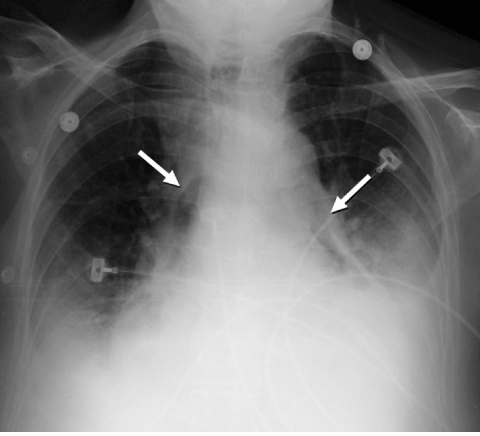
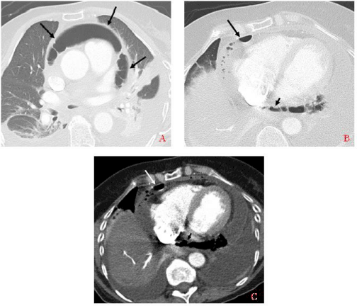
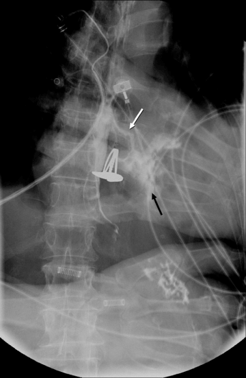
| Chest X-ray showed evidence suggestive of pneumopericardium (Figure 1). This finding, as well as the patient’s poor clinical status, prompted the decision for thoracic CT, which confirmed the diagnosis of pneumopericardium (Figure 2). Esophagram was performed, revealing esophageal perforation at the level of the left atrium (Figure 3). The patient’s radiological findings and clinical history/status were consistent with the diagnosis of atrio-esophageal fistula. The patient was immediately admitted to the operating room for drainage of pericardial fluid and pleural effusions, where she underwent pericardial window, sternotomy with mediastinal washout, and esophagogastroduodenoscopy with esophageal stent placement. |
| The patient’s deteriorating hospital course was complicated with development of septic shock secondary to mediastinitis despite immediate surgical intervention and aggressive broad antibiotic therapy. Subsequently, she developed low-grade fever, oliguric renal failure, severe hypotension, elevated liver enzymes secondary to ischemic liver disease, continued leukocytosis, persistent pleural effusions, and frequent intermittent seizure-like activity. Eight days following admission, the patient showed signs of altered mental status secondary to toxic metabolic encephalopathy. Non-contrast head CT and MRI revealed massive bilateral strokes in middle cerebral artery territories, consistent with the diagnosis of persistent vegetative state. The patient’s negligible chance for meaningful neurological recovery was conveyed to the family, who decided to withdraw medical care except hydration and tube feeding. 25 days after admission, the patient was discharged and transferred to hospice care. |
| Discussion |
| Radiofrequency catheter ablation for treatment of atrial fibrillation is recognized as a safe procedure with a low incidence and prevalence of death, with Cappato et al. [1] reporting a death rate of <0.1%. Using a probe set to a predetermined temperature and duration, a series of transmural lesions are created in the left atrium. Although the radiofrequency temperature and energy are easily controlled, lesion depth can be variable, which may lead to undesirable consequences: non-transmural lesions will result in an unsuccessful operation, whereas lesions that are too deep may cause damage to adjacent anatomical structures [2]. Thus, unique precautions must be considered to avoid potentially fatal complications. These complications include puncture of intracardiac septa, cardiac tamponade, pulmonary vein stenosis, vascular accidents, thromboembolic events, nerve damage, and esophageal injury due to the proximity of the posterior wall of the left atrium and the esophagus [3]. |
| Esophageal injury is a rare consequence of RF CA. Pappone et al. [4] reports a prevalence of 0.05% out of 4360 patients who have undergone circumferential pulmonary vein ablation, although an incidence rate of up to 1% has been reported. Despite the low incidence rate and prevalence of esophageal injury, development of atrio-esophageal fistula is the second most frequent cause of death that may be caused by RF catheter ablation, with cardiac tamponade found to be the most common fatal complication [1]. It is usually reported 3-35 days following the procedure [3,5,6], and is associated with mortality rates in excess of 70% [1]. |
| Clinically, esophageal damage and atrio-esophageal fistula presents with several non-specific symptoms, such as general malaise, fever, chest pain, convulsions or seizures, loss of consciousness, dysphagia/ odynophagia, septic shock, abdominal pain, anorexia, and hematemesis [3]. These patients often subsequently develop endocarditis/ pericarditis and embolism leading to possible myocardial and/or cerebral infarctions. |
| Our patient displayed many of the signs and symptoms associated with esophageal damage following RF ablation of the pulmonary veins. Possible risk factors that predispose patients to these events may include body habitus and general health status [2]. In a healthy individual, there exists a large area of contact between the posterior left atrium and esophagus, with an intervening fat pad of 0.9 ± 0.2 mm [7]. However, our patient was malnourished and cachectic, and likely had a thinner than usual left atrium, with a very thin and variable layer of tissue between the left atrium and esophagus, predisposing to RF heat damage. There are no identified specific predictors of esophageal damage following RF CA, although previous studies report that overlap of ablation lines in the posterior wall of the left atrium could create enough RF energy accumulation to cause esophageal trauma [4,8]. Furthermore, Scanavaccca et al. [9] propose that due to specific anatomic relationships, RF energy directed a few millimeters outside of the inferior pulmonary vein ostia could precipitate esophageal perforation. |
| An early warning sign for esophageal perforation is dysphagia, as seen in our patient’s intolerance of solid food. This symptom could be a sign of inflammatory esophagitis, eventually leading to atrioesophageal fistula, even in the presence of a normal MRI following the ablation procedure [9]. For these patients, both transesophageal echocardiography and gastro-esophageal endoscopy are contraindicated due to risk of further damage and/or massive air embolism resulting from CO2 inflation [3,4,9]. Our patient did not experience hematemesis, suggesting the presence of a one-way valve covering the fistula. |
| Prevention and early diagnosis of atrio-esophageal fistula is absolutely critical. Reducing RF power, in addition to simultaneously inserting an esophageal lead, could help the clinician better identify the position of the esophagus to avoid injury [9]. Other prevention methods include CT angiography with oral contrast prior to ablation to better identify the position of the esophagus and left atrium, careful esophageal temperature monitoring, adjusting the position of the transverse posterior left atrial line to the roof where the atrium is thicker [4], and use of a water-irrigated intraesophageal balloon for the purposes of esophageal cooling [3]. Takahashi et al. [3] report an effective esophageal temperature monitoring method: a deflectable temperature probe is maneuvered as close as possible to the ablation electrode via an endogastric tube. The RF energy is set to 35 Watts for 40 seconds at the posterior left atrial wall; RF energy application is terminated when esophageal temperature reaches 42°C, and reinitiated only when esophageal temperature returns to under 38°C. Other studies recommend RF generator settings of no more than 50 Watts and 55°C to be used during ablation to avoid excessive left atrial posterior wall injury [4]. |
| The most useful method for identification of injury is thoracic CT enhanced with oral or intravenous contrast; an air pocket or fistula can be observed in the mediastinum [3,4]. Transthoracic echocardiography is generally not useful due to nonspecific findings, although it can occasionally be effective if it reveals specific signs such as gas bubbles in the left atrial appendage or vegetation-like appearance of the posterior wall of the left atrium [3]. If discovered, immediate surgical intervention is mandatory to avoid fatal consequences. |
| Rapid diagnosis, preferably by non-invasive techniques such as MRI, transthoracic echocardiography, and thoracic CT with watersoluble oral contrast, followed by immediate surgical intervention is critical in preventing patient death. By considering the patient’s unique anatomic relationships prior to the procedure, using lower energy applications, and employing careful esophageal temperature monitoring, we hope that future cases of atrio-esophageal fistula will be avoided. |
References |
|
Figures at a glance
 |
 |
 |
| Figure 1 | Figure 2 | Figure 3 |
Relevant Topics
- Abdominal Radiology
- AI in Radiology
- Breast Imaging
- Cardiovascular Radiology
- Chest Radiology
- Clinical Radiology
- CT Imaging
- Diagnostic Radiology
- Emergency Radiology
- Fluoroscopy Radiology
- General Radiology
- Genitourinary Radiology
- Interventional Radiology Techniques
- Mammography
- Minimal Invasive surgery
- Musculoskeletal Radiology
- Neuroradiology
- Neuroradiology Advances
- Oral and Maxillofacial Radiology
- Radiography
- Radiology Imaging
- Surgical Radiology
- Tele Radiology
- Therapeutic Radiology
Recommended Journals
Article Tools
Article Usage
- Total views: 6884
- [From(publication date):
May-2012 - Jul 19, 2025] - Breakdown by view type
- HTML page views : 2258
- PDF downloads : 4626
