Epispadiac Duplication of the Urethra in an Adult Male
Received: 08-Mar-2022 / Manuscript No. roa-22-57383 / Editor assigned: 10-Mar-2022 / PreQC No. roa-22-57383 (PQ) / Reviewed: 24-Mar-2022 / QC No. roa-22-57383 / Revised: 29-Mar-2022 / Manuscript No. roa-22-57383 (R) / Published Date: 05-Mar-2022 DOI: 10.4172/2167-7964.1000371
Abstract
Urethral duplication and Epispadias are one of the rare anomalies of the genitourinary system, usually diagnosed in childhood, but some adult presentations have been reported. There are several anatomical variants of urethral duplications. Therefore, a detailed evaluation of the urinary tract is necessary to characterize the type of duplication and to better guide the therapeutic strategy. We report a case of a 32 year old man with an Epispadias variant of urethral duplication
Introduction
Urethral duplicity is a very rare congenital anomaly of the genitourinary system; affect more frequently the male fetus and is nearly always diagnosed in childhood or adolescence. It can be complete or incomplete, isolated or associated with Anorectal malformations, Epispadias, hypospadias, bladder duplicity, Bladder exstrophy and other urinary excretory system anomalies [1, 2]. An Epispadias is a rare type of malformation of the penis, considered to be the least severe defect of the exstrophy–Epispadias complex in which the urethra terminates on the upper aspect of the penis [2].
The presence of multiple anatomical variants of urethral duplication makes a detailed study of the anatomy of the urinary tract necessary to guide surgical management. There are a multitude of radiological or endoscopic examinations that can be used to study the anatomy of the urethra. These include Micturating Cystourethrogram (MCUG), Intravenous urography, Sonourethrography, Nuclear scintigraphy and Cystourethroscopy [3].
Case Report
We report a case of a 32 year old man with a history of recurring urinary tract infections, presented with dysuria evolving over 3 years.
General physical examination was unremarkable, but the examination of the external genitalia showed a normal foreskin, glans with two meatus, one opening at normal position and the second on the dorsal surface of the penis.
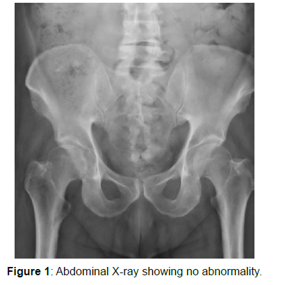
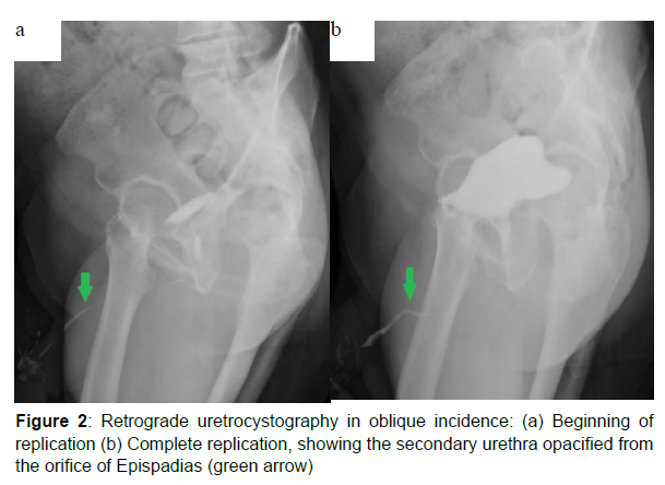
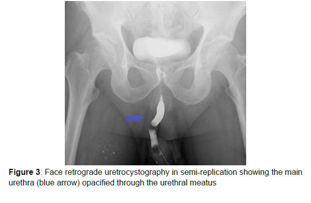
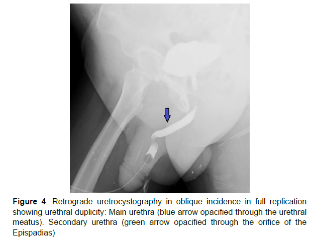
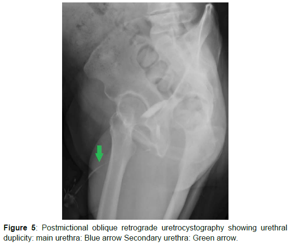
Retrograde urethrocystography was performed (Figure 1-5) with double catheterization, the first Foley’s catheter was placed and the normal meatus and the second catheter in the Epispadias meatus. The results shows duplicated urethra with an accessory arising from the normal urethra, describes an anterior trajectory, and terminates at the dorsal surface of the penis (Type IIA2 according to Effman’s classification). The orthotopic urethra is normal with no stricture. The cystogram reveal a bladder of good capacity with regular wall with no vesicoureteral reflux.
The patient underwent surgery for excision of the accessory Epispadias urethra (Figure 1-5).
Discussion
Urethral Duplicity (UD) and Epispadias are very rare congenital abnormalities of the genitourinary system with an unclear pathophysiology. These anomalies can be observed in both associated and isolated forms [2]. The accessory urethra can be placed on ventral or dorsal position, when the accessory urethra is in the dorsal position, the external meatus can be Epispadias [3].
The UD occurs frequently in a sagittal plane, in this case the duplicated urethra is in dorsal or ventral position in relation to the orthotopic urethra, and the ventral urethra is usually the more functional and contains the sphincteric mechanism. Duplications occurring in the coronal plane are rare and they are frequently associated with bladder duplication. The cases of urethral duplication in the sagittal plane could be separated into 3 forms: pre pubic congenital sinus, Epispadias urethral duplication (as in our case), hypospadiac urethral duplication. Epispadiac urethral duplications are considered to be a variant of the bladder-Epispadias exstrophy complex [4].
Clinical symptoms vary according to the type of UD. The patients can be asymptomatic, or may present with a double urinary stream, urinary incontinence, and urinary infection or outflow obstructive symptoms [4].
Several classifications of UD have been suggested, but the most adopted is Effman’s classification which divides UD into three types: In type I (usually asymptomatic) there is blind accessory urethra (incomplete urethral duplication), IA is the most common type, with distal duplicated urethras opening on the dorsal or ventral surface of the penis without communication with the urethra or bladder, in type IB there is a proximal accessory urethra that derives from the functional urethra but terminates blindly in the peri-urethral tissues. In type II, there is completely patent accessory urethra. It is divided into two subtypes: A (two meatuses) and B (one meatus) in type IIA1 there is two non-communicating urethras arising independently from the bladder, for the type IIA2, accessory urethra arises from the first and coursing independently into a second meatus (Y-type). In type III, there are accessory urethras arising from duplicated or septated bladders [3].
There are a variety of radiological or endoscopic procedures; the first-line radiologic study is a RUG (retrograde urethrography), and should be performed in lateral projections for visualization of the size, shape and position of the two urethras [3, 5].
The Intravenous pyelogram (IVP) represents an alternative modality however, compared with RUG, IVP is more invasive and requires intravenous injection of the contrast agent as well as more radiation and time to evaluate the kidneys and bladder. IVP may other associated abnormalities like unilateral renal agenesis, ureteral duplication and bladder duplication [3, 5].
MRI is an excellent modality for exploring duplicated urethras and periurethral soft tissues. It demonstrates accurately the size, shape and positions of both urethras as well as other associated genitourinary anomalies. In fact, MR is the best radiologic modality for detailed assessment of anomalies, fistulas and other associated genitourinary anomalies allowing a better orientation of the surgical approach [3, 5].
The indication for cystoscopy arises in certain ambiguous cases where it remains difficult to determine the functional urethra. Cystoscopy allows direct visualization of the veru Montanum to determine the functional urethra [6].
The treatment must be planned considering both the function of the urinary system and the aesthetic defect. Surgical treatment is indicated when symptoms of urethral duplication are severe, and consists of total resection of the accessory urethra [7].
Conclusion
Epispadiac duplication of the urethra is a very rare type of the urethral duplication, and commonly diagnosed in childhood. Retrograde urethrocystography is the first-line examination for the assessment of the normal and the accessory urethra.
References
- Rimtebaye K, Sillong FD, Tashkand AZA, Kaboro M, Niang L, et al. (2015) Complete Urethral Duplicity: A Rare Cause of Urinary Incontinence, New Type According to Effman’s Classification. Open J Urol 5: 182.
- Polukhov R, Mahammadov V, Baghirli M (2020) Epispadias associated with urethral duplication: Our practice. Int J Surg Case Rep 71: 199-201.
- Bhadury S, Parashari UC, Singh R, Kohli N (2009) MRI in congenital duplication of urethra. The Indian J Radiol Imaging 19 : 232-234.
- Onofre LS, Gomes AL, de Souza Leão JQ, Leão FG, Cruz TMA, et al. (2013) Urethral duplication–a wide spectrum of anomalies. J Pediatr Urol 9: 1064-1071.
- Shittu OB, Kamara TB (1999) Urethral duplication associated with Epispadias in a child. Niger J paediatr 26: 53-55.
- Frankel J, Sukov R (2017) Diagnosing urethral duplication including a novel radiological diagnostic algorithm. BJR case R 3: 20150506.
- Rimtebaye K, Sillong FD, Tashkand AZA, Kaboro M, Niang L, et al. (2015) Complete Urethral Duplicity: A Rare Cause of Urinary Incontinence, New Type According to Effman’s Classification. Open J Urol 5: 182.
Indexed at, Google Scholar, Crossref
Indexed at, Google Scholar, Crossref
Indexed at, Google Scholar, Crossref
Indexed at, Google Scholar, Crossref
Indexed at, Google Scholar, Crossref
Citation: Imrani K, Oubaddi T, Nassar I, Billah NM (2022) Epispadiac Duplication of the Urethra in an Adult Male. OMICS J Radiol 11: 371. DOI: 10.4172/2167-7964.1000371
Copyright: © 2022 Imrani K, et al. This is an open-access article distributed under the terms of the Creative Commons Attribution License, which permits unrestricted use, distribution, and reproduction in any medium, provided the original author and source are credited.
Share This Article
Open Access Journals
Article Tools
Article Usage
- Total views: 3255
- [From(publication date): 0-2022 - Mar 29, 2025]
- Breakdown by view type
- HTML page views: 2709
- PDF downloads: 546

