Make the best use of Scientific Research and information from our 700+ peer reviewed, Open Access Journals that operates with the help of 50,000+ Editorial Board Members and esteemed reviewers and 1000+ Scientific associations in Medical, Clinical, Pharmaceutical, Engineering, Technology and Management Fields.
Meet Inspiring Speakers and Experts at our 3000+ Global Conferenceseries Events with over 600+ Conferences, 1200+ Symposiums and 1200+ Workshops on Medical, Pharma, Engineering, Science, Technology and Business
Case Report Open Access
Epicardial Fat Mimicking Pericardial Effusion: A Patient with Gastrointestinal Bleeding
| Ozcan Basaran* | |
| Division of Cardiology, Mugla Sitki Kocman University Education and Research Hospital, Mugla, Turkey | |
| Corresponding Author : | Ozcan Basaran Division of Cardiology, Mugla Sitki Kocman University Education and Research Hospital Orhaniye Mahallesi Ismet Catak Caddesi, Mugla, Turkey Tel: +905065359013 Fax: +9025221307 62 E-mail: basaran_ozcan@yahoo.com |
| Received January 23, 2013; Accepted January 26, 2014; Published January 30, 2014 | |
| Citation: Basaran O (2014) Epicardial Fat Mimicking Pericardial Effusion: A Patient with Gastrointestinal Bleeding. OMICS J Radiol 3:159. doi:10.4172/2167-7964.1000159 | |
| Copyright: © 2014 Basaran O. This is an open-access article distributed under the terms of the Creative Commons Attribution License, which permits unrestricteduse, distribution, and reproduction in any medium, provided the original author and source are credited. | |
Visit for more related articles at Journal of Radiology
| Keywords |
| Gastrointestinal bleeding; Epicardial fat; Pericardial effusion |
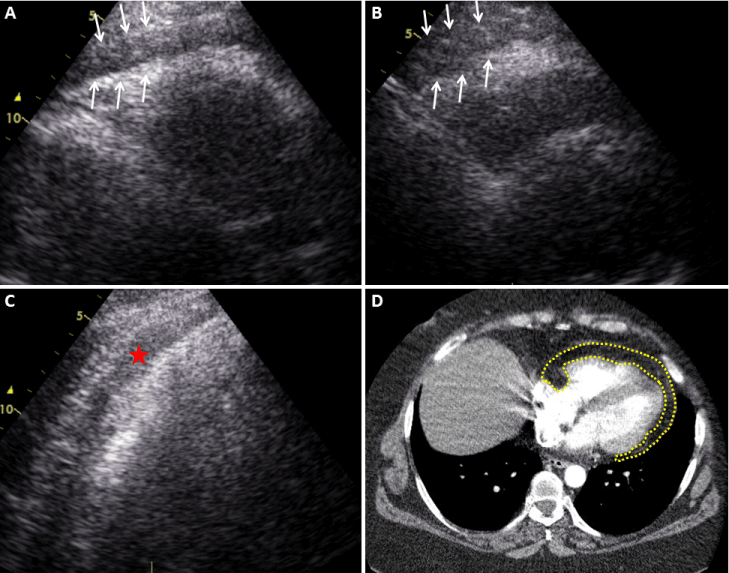
| A 49-year-old morbid obese woman admitted our hospital with upper gastrointestinal bleeding. She had been diagnosed with pericardial effusion at two different visits, and had been taking ibuprofen 800 mg twice daily for a month. She had haemetemesis with coffee ground vomiting. Her detailed questioning revealed that she had stomach pain and heartburn occasionally. On her physical examination she was pale and diaphoretic. A complete blood count revealed a white blood cell count of 8.2×10⁶/mL with 54.1% neutrophils, and a hemoglobin level of 8.2 g/dL and hematocrit of 25.8%. Proton pump inhibitor therapy was initiated and ibuprofen was stopped. Her upper gastrointestinal system endoscopy revealed erythematous gastritis. A transthoracic echocardiography (TTE) was performed and pericardial echo-free zone was thought to be epicardial fat rather than pericardial effusion (Figure 1A-1C). Chest contrast-enhanced computed tomography (CT) was performed for further evaluation because of poor image quality on TTE. Epicardial fat was clearly differentiated from pericardial effusion by CT findings (Figure 1D). |
| Epicardial fat may have a similar echolucent appearance to that of a pericardial effusion. While TTE is usually sufficient for detecting pericardial abnormalities, CT imaging also can be helpful in some cases. Taking account the difficulty of the clinical scenario (obese patient with insufficient image quality on TTE) like in our patient, one should bear in mind CT imaging for differential diagnosis. It is also important to question patients for gastrointestinal symptoms before initiating a nonsteroidal anti-inflammatory drug. This case presentation highlights the need for caution in diagnosing pericardial effusion by echocardiography. |
Figures at a glance
 |
| Figure 1 |
Post your comment
Relevant Topics
- Abdominal Radiology
- AI in Radiology
- Breast Imaging
- Cardiovascular Radiology
- Chest Radiology
- Clinical Radiology
- CT Imaging
- Diagnostic Radiology
- Emergency Radiology
- Fluoroscopy Radiology
- General Radiology
- Genitourinary Radiology
- Interventional Radiology Techniques
- Mammography
- Minimal Invasive surgery
- Musculoskeletal Radiology
- Neuroradiology
- Neuroradiology Advances
- Oral and Maxillofacial Radiology
- Radiography
- Radiology Imaging
- Surgical Radiology
- Tele Radiology
- Therapeutic Radiology
Recommended Journals
Article Tools
Article Usage
- Total views: 13479
- [From(publication date):
April-2014 - Apr 04, 2025] - Breakdown by view type
- HTML page views : 8970
- PDF downloads : 4509
