Enteric Duplication Cyst in Children: Imaging Finding
Received: 06-Jun-2023 / Manuscript No. roa-23-105397 / Editor assigned: 08-Jul-2023 / PreQC No. roa-23-105397 (PQ) / Reviewed: 22-Jul-2023 / QC No. roa-23-105397 / Revised: 24-Jul-2023 / Manuscript No. roa-23-105397 (R) / Published Date: 31-Jul-2023 DOI: 10.4172/2167-7964.1000466
Abstract
Rare congenital malformations known as enteric duplication cysts occur during the early stages of the digestive tract’s development. The distal ileum and prenatal diagnosis are more common. The most reliable diagnosis is made with the use of clinical examination, imaging, particularly ultrasonography, which also helps to prevent future complications.
Case History
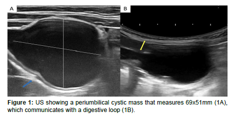
15-month-old boy operated 3 weeks ago for inguinal hernia and circumcision, who presented abdominal pain vomiting and rectal bleeding. An Ultrasound was performed showing a periumbilical cystic mass that measures 69x51mm (figure 1A) which communicates with a digestive loop (Figure 1B) (Figure 1).
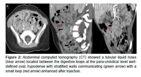
Abdominal computed tomography (CT) showed a tubular liquid mass (Figure 2 blue arrow), located between the digestive loops at the para-umbilical level, well-defined oval, hypodense, with stratified walls, communicating (Figure 2 green arrow) with a small loop (Figure 2 red arrow), enhanced after injection (Figure 2).
Commentary
Enteric duplication cysts (EDCs) are rare congenital anomalies found anywhere along the gastrointestinal tract (GT) from the mouth to the rectum; most commonly in the ileum (33%), followed by the oesophagus (20%), colon (13%), jejunum (10%), stomach (7%) and duodenum (5%). Structurally, there are two types of gastrointestinal duplication cyst: cystic is the most common (80%) typically do not communicate with the adjacent lumen, and tubular duplications (20%) communicating directly with the bowel lumen [1].
US is the imaging method of choice in the diagnosis of EDCs, only limited in the evaluation of oesophageal EDCs. Classical findings of uncomplicated EDC well described in the literature: double wall or muscular rim sign, which refers to the appearance of a cyst mimicking the gastrointestinal tract with an echogenic inner margin corresponding to mucosa surrounded by a hypoechoic rim of tissue representing the smooth muscle layer. this sign has been regarded as characteristic of an EDC, also US is a dynamic study and allows to visualize the peristalsis of the cyst wall [2].
Complicated EDCs rarely present the classic five layers or doublewall sign. In the event of haemorrhage or infection occurs: fluid levels, echogenic debris and an important inflammatory change in the surrounding mesentery fat can be seen.
CT is not usually done to evaluate EDC due to radiation. CT can show the location and extent of cysts, complications, associated abnormalities, and their anatomical relationship to surrounding structures. On CT, EDC appears as a cystic mass with a thin, slightly strengthened wall adjacent to the gastrointestinal wall. Severe weakness within the cyst may be seen due to bleeding or proteinaceous material. Thick reinforced walls, internal air bubbles, and cysts surrounding inflammation may indicate EDC complicated by infection [1,2].
EDCs are typically managed with surgical excision, particularly if found prenatally or during infancy [1].
References
- Cheng G, Soboleski D, Daneman A, Poenaru D, Hurlbut D (2005) Sonographic pitfalls in the diagnosis of enteric duplication cysts. Am J Roentgenol 184: 521-525.
- Nebot CS, Salvador RL, Palacios EC, Aliaga SP, Pradas VL (2018) Enteric duplication cysts in children: varied presentations, varied imaging findings. Insights Imaging 9: 1097-1106.
Indexed at, Google Scholar, Crossref
Citation: Rostoum S, Zhim M, Naggar A, Haddad SE, Allali N, et al. (2023) Enteric Duplication Cyst in Children: Imaging Finding. OMICS J Radiol 12: 466. DOI: 10.4172/2167-7964.1000466
Copyright: © 2023 Rostoum S, et al. This is an open-access article distributed under the terms of the Creative Commons Attribution License, which permits unrestricted use, distribution, and reproduction in any medium, provided the original author and source are credited.
Share This Article
Open Access Journals
Article Tools
Article Usage
- Total views: 1004
- [From(publication date): 0-2023 - Mar 29, 2025]
- Breakdown by view type
- HTML page views: 782
- PDF downloads: 222


