Case Report Open Access
Endodontic Treatment of Single Rooted Maxillary First Molar with C Shaped Canal Configuration
Kanupriya Dhingra1* and Rakshith S Shetty2
1Consultant Endodontist, Ceramco Dental Clinic, Mumbai, Maharashtra, India
2Kannur Dental College, Kannur, Kerala
- Corresponding Author:
- Kanupriya Dhingra
MDS (Conservative Dentistry & Endodontics)
Consultant Endodontist, Ceramco Dental Clinic
Mumbai, Maharashtra, India
Tel: +912232573682
E-mail: kp.dhingra@gmail.com
Received date: July 01, 2014; Accepted date: August 04, 2014; Published date: August 11, 2014
Citation: Dhingra K, Shetty RS (2014) Endodontic Treatment of Single Rooted Maxillary First Molar with C Shaped Canal Configuration. J Interdiscipl Med Dent Sci 2:139. doi: 10.4172/2376-032X.1000139
Copyright: © 2014 Dhingra K, et al. This is an open-access article distributed under the terms of the Creative Commons Attribution License, which permits unrestricted use, distribution, and reproduction in any medium, provided the original author and source are credited.
Visit for more related articles at JBR Journal of Interdisciplinary Medicine and Dental Science
Abstract
The C shaped canal configuration is an anatomic variant most commonly seen in mandibular second molars. The presence of this configuration in a single rooted maxillary permanent first molar is rare. This case report describes the endodontic management of a single rooted infected maxillary first molar having a c shaped root canal configuration.
Keywords
Maxillary molar; Single root; C shaped canal
Introduction
The basic objectives of endodontic therapy include a thorough cleaning and debridement of the fine intricacies of the root canal system followed by complete obturation to provide a an apical and coronal seal. Anatomic variations in the morphology of the root canal system often complicate endodontic treatment. Treatment failure may occur if the clinician fails to recognize these variations and treat them accordingly.
Generally, it is accepted that the maxillary first permanent molar has three roots- the palatal, mesiobuccal and distobuccal and four canals - one each in the palatal and distobuccal and two canals in the mesiobuccal root [1]. Rare cases of single rooted conical maxillary first molars [2,3] and maxillary first molars having a c shaped canal configuration [4,5] have been reported in literature, but the presence of a c shaped canal in a single rooted maxillary first molar has not been reported.
Cooke and Cox in 1979 were the first to report the presence of c shaped canals. The cross sectional morphology of these roots and root canals has a “C- shape”. In such cases, instead of separate canal orifices, there is a single ribbon shaped opening with a 180°arc [6].
Case Report
A 37 year old female patient reported to the clinic with a complaint of pain in the upper right back tooth region. The pain was spontaneous in onset, throbbing in nature, aggravated on chewing and radiated to the temple. The patient had taken an NSAID to relieve the pain, but the symptoms were relieved only temporarily. Clinical examination revealed a deep carious lesion with the tooth 16. The tooth was tender on vertical percussion. Nothing abnormal was detected on periodontal evaluation. Thermal and electrical pulp vitality tests were negative.
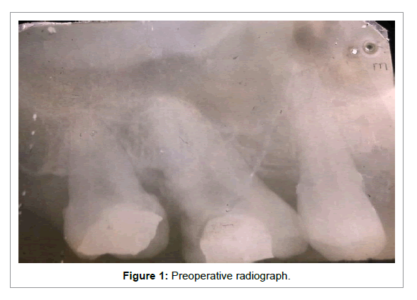
A preoperative periapical radiograph (Figure 1) revealed a radiolucency involving the pulp and periapical widening. The radiograph also indicated that the tooth had a single, fused root. Additional radiographs were taken at 30° mesial and distal angulations to confirm the presence of a single root. A final diagnosis of irreversible pulpitis with apical periodontitis was made.
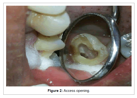
Local anesthesia was administered and the access cavity was prepared. The pulp chamber was very large and profuse bleeding was encountered. The coronal pulp was excavated using a small spoon excavator to reveal the pulp chamber floor. The chamber floor showed the presence of a single c shaped orifice in the centre of the root (Figure 2). The chamber was further explored with a DG 16 explorer to identify any additional orifices, however, no other orifices were located.
The single c shaped orifice formed a 180° arc extending from the palatal to the distobuccal aspect of the tooth. A fin of pulp tissue was present extending throughout the length of the root, connecting the fused palatal and distal roots.
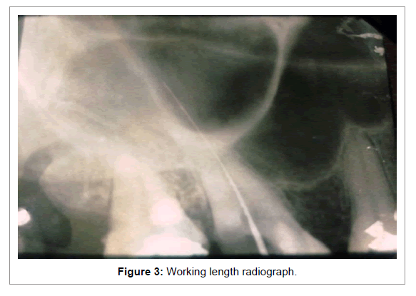
The root canal was initially negotiated with a no. 15 K file, the pulp tissue was removed. Two files were placed in the root canal, but they converged at a single point. The absence of additional canals was confirmed using multiple angulated radiographs. A single file passed unobstructed from one end of the orifice to the other- indicating the presence of a continuous c shaped canal (Figure 3). The working length was calculated using Ingle’s technique and confirmed with an apex locator.
The canal was biomechanically prepared using hand and rotary Protaper files. 3% Sodium hypochlorite solution and normal saline were used for irrigating the canals between files and the canal was finally rinsed with 17% EDTA solution. A calcium hydroxide based intra canal medicament was placed in the canal and the access cavity was sealed temporarily using Cavit. The patient was recalled after one week. She was prescribed analgesics and antibiotics during this period.
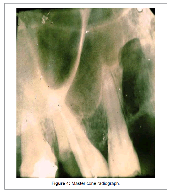
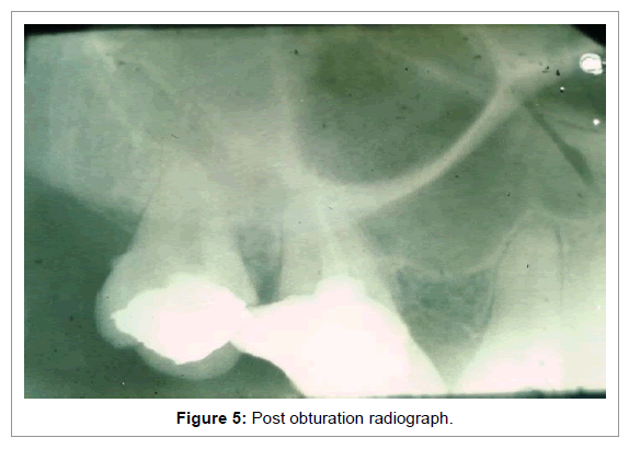
A week later the patient was asymptomatic and the root canal was dry. Master cone radiograph was taken (Figure 4) and Obturation was done using Protaper gutta percha cones and cold lateral compaction. AH Plus root canal sealer was used (Figure 5). The access cavity was then restored with composite resin and a full coverage restoration was planned.
Discussion
A Thorough knowledge and respect of the intricate root canal anatomy along with meticulous cleaning and debridement are the keystones of successful endodontic therapy. Anatomic variations within the root canal system are a challenge for the dental practitioner. Extra roots, missed canals, variable morphology, ramifications, isthmus etc result in an incomplete cleaning and disinfection of the pulp space [7]. This may lead to treatment failures.
In multi rooted teeth, the presence of additional canals is more commonly reported; however, sometimes fusion of the roots occurs resulting in fewer canals [2]. This anatomic variation has to be confirmed using various angulated radiographs. The pulp chamber in such cases should be thoroughly inspected for any additional canals.
In 2006, Gopalkrishna et al. had reported the presence of a single rooted maxillary first molar [2]. They confirmed the root canal morphology using spiral CT. Chabbra et al. [2] reported a similar case, and CBCT was used to confirm morphology [8]. The key feature of a of C-shaped canal is a fin or web which forms a connection between individual root canals. This configuration is generally associated with conical or square roots [6].
In the presented case, the maxillary first molar had a single conical root with a c -shaped canal configuration. The c extended from the palatal to the distobuccal aspect of the tooth. The presence of c shaped canals is most commonly associated with mandibular 2nd molars [6]. Cases involving the mandibular first molars [9], mandibular and maxillary 3rd molars [10], and maxillary first molars have been described.
In 1984, the first case of a c shaped canal in a maxillary molar was reported by Newton and McDonald [11]. De Moor [12] published a paper about the c shaped root canal configuration in maxillary molars. He concluded that the presence of c shaped canals in maxillary molars was very rare- the incidence being 0.091%. His article also mentioned that the c shaped canal configuration generally occurred in the distal portion of the pulp chamber.
A c shaped canal may be suspected if radicular fusion is evident on preoperative radiographs. Additional angulated radiographs are necessary. A final diagnosis can only be made after access cavity preparation. . The pulp chamber is large occluso- cervically with a low bifurcation. Two distinct orifices may be probed initially, which may be linked by a wide or fine isthmus- forming the typical c configuration.
Biomechanical preparation of a c configuration requires utmost care- so as not to over instrument and cause strip perforations. Also, the central fin needs to be thoroughly debrided. Copious irrigation is essential and ultrasonic activation of irrigants is recommended. Complete Obturation of the canal is difficult to achieve. Lateral compaction with sealer and heated spreaders or use of thermoplasticised gutta percha is indicated.
Conclusion
Variations in root canal anatomy are rare. These however should not be overlooked- regarding the prognosis of endodontic therapy. Cases of the maxillary first molar with varied no. of root canals and internal anatomy have been reported [2,3,8,12]. But, the presence of a c shaped canal in a single rooted maxillary molar has not been documented so far.
A thorough knowledge of the internal anatomy, with a keen eye for variations is necessary for the success of therapy. Early recognition of morphological variations is essential. This is aided by using additional angulated preoperative and intra-operative radiograph.
References
- Walton R Torabinejad M (1996) Principles and practice of endodontics. (2nd edn), W.B. Saunders Co., Philadelphia.
- Gopikrishna V, Bhargavi N, Kandaswamy D (2006) Endodontic management of a maxillary first molar with a single root and a single canal diagnosed with the aid of spiral CT: a case report. J Endod 32: 687-691.
- Cobankara FK, Terlemez A, Orucoglu H (2008) Maxillary first molar with an unusual morphology: report of a rare case. Oral Surg Oral Med Oral Pathol Oral RadiolEndod 106: e62-65.
- Dankner E, Friedman S, Stabholz A (1990) Bilateral C shape configuration in maxillary first molars. J Endod 16: 601-603.
- De Moor RJ (2002) C-shaped root canal configuration in maxillary first molars.IntEndod J 35: 200-208.
- Jafarzadeh H, Wu YN (2007) The C-shaped root canal configuration: a review. J Endod 33: 517-523.
- Cantatore G, Berutti E, Castelluchi A (2009) Missed anatomy: frequency and clinical impact. Endodontic Topics 15: 3-31.
- Chhabra N, Singbal KP, Chhabra TM (2013) Type I canal configuration in a single rooted maxillary first molar diagnosed with an aid of cone beam computed tomographic technique: A rare case report. J Conserv Dent 16: 385-387.
- Barnett F (1986) Mandibular molar with C-shaped canal.Endod Dent Traumatol 2: 79-81.
- Sidow SJ, West LA, Liewehr FR, Loushine RJ (2000) Root canal morphology of human maxillary and mandibular third molars. J Endod 26: 675-678.
- Newton CW, McDonald S (1984) A C-shaped canal configuration in a maxillary first molar. J Endod 10: 397-399.
- De Moor RJ (2002) C-shaped root canal configuration in maxillary first molars.IntEndod J 35: 200-208.
Relevant Topics
- Cementogenesis
- Coronal Fractures
- Dental Debonding
- Dental Fear
- Dental Implant
- Dental Malocclusion
- Dental Pulp Capping
- Dental Radiography
- Dental Science
- Dental Surgery
- Dental Trauma
- Dentistry
- Emergency Dental Care
- Forensic Dentistry
- Laser Dentistry
- Leukoplakia
- Occlusion
- Oral Cancer
- Oral Precancer
- Osseointegration
- Pulpotomy
- Tooth Replantation
Recommended Journals
Article Tools
Article Usage
- Total views: 14796
- [From(publication date):
October-2014 - Jul 15, 2025] - Breakdown by view type
- HTML page views : 10194
- PDF downloads : 4602

