Research Letter Open Access
Encapsulation of N-Nitroso-melatonin with Poly(lactide-co-glycolide)
Michael Kirsch1*, Hans-Gert Korth2, Joachim Fandrey3and Katja B Ferenz11Institut für Physiologische Chemie, Universitätsklinikum Essen, Hufelandstrasse 55, 45122 Essen, Germany
2Institut für Organische Chemie, Universität Essen, 45117 Essen, Germany
3Institut für Physiologie, Universität Duisburg-Essen, Hufelandstrasse 55, 45122 Essen, Germany
- *Corresponding Author:
- Kirsch M
Institut für Physiologische Chemie
Universitätsklinikum Essen, Hufelandstrasse 55
45122 Essen, Germany
Tel: 49- 201-723-4108/5943
E-mail: michael.kirsch@uni-due.de
Received date: March 13, 2017; Accepted date: May 09, 2017; Published date: May 16, 2017
Citation: Kirsch M, Korth HG, Fandrey J, Ferenz KB (2017) Encapsulation of N-Nitroso-melatonin with Poly(lactide-co-glycolide). J Biotechnol Biomater 7:258. doi:10.4172/2155-952X.1000258
Copyright: © 2017 Kirsch M, et al. This is an open-access article distributed under the terms of the Creative Commons Attribution License, which permits unrestricted use, distribution, and reproduction in any medium, provided the original author and source are credited
Visit for more related articles at Journal of Biotechnology & Biomaterials
Abstract
N-Nitrosomelatonin (NOMela) is well known for its capabilities to transnitrosate nucleophiles such as thiols and ascorbate thereby generating nitric oxide (NO)-releasing compounds. Like molsidomine, NOMela is one of the few NO-releasing substrates not inducing nitrate tolerance and may be therefore highly suitable as NO-therapeutical. As the physical and chemical properties of NOMela do not allow its direct application (oral or intravascular) in animals/ humans, the encapsulation with biodegradable poly(lactide-co-glycolide) (PLGA) polymers was performed and NOreleasing kinetics were studied. NOMela could be successfully encapsulated in PLGA (NOMela-PLGA) with an efficiency of 85% thereby prolonging its half-life time in aqueous solution (e.g. in the cytoplasm of endothelial and smooth muscle cells) about 3-fold. In the presence of “activated hydroxy compounds“ like vitamin C and thus under physiological conditions, NOMela-PLGA yielded two therapeutically relevant hormones, melatonin and nitric oxide, via reactions only known (until now) for unencapsulated, freely diffusing NOMela. Importantly, in the absence of any activated hydroxy compound the unwanted hydrolysis reaction of NOMela dominated, generating the non-functional nitrite (and not nitric oxide). These findings suggested that PLGA-encapsulated NOMela will be highly attractive as a novel NO-releasing drug lacking common side-effects of classical NO-releasing molecules such as glyceroltrinitrate.
Keywords
Nitric oxide; N-nitrosomelatonin; Vitamin E; laserscanning microscope; Vitamin C; Centrifugation; Melatonin
Introduction
Nitric oxide (NO) fulfills multiple physiological functions such as in neurotransmission, in the immune response by activated macrophages, and in vasodilatation through smooth muscle relaxation [1]. In the blood NO is a short-lived intermediate because it is irreversibly oxidized to nitrate by oxyhemoglobin [2] and reversibly but strongly bound by deoxyhemoglobin (deoxyhemoglobin binds NO 105 times stronger than molecular oxygen) [3]. Because of these highly effective mechanisms, it has been suggested that in vivo NO is transported by carrier molecules in order to increase its efficacy [4,5].
In 2001, Blanchard-Fillion et al. observed that N-nitrosomelatonin (NOMela) can act as a NO releasing compound [6]. There is now compelling evidence that a shift of the nitroso function from NOMela to “activated hydroxy compounds” (enols or phenols such as vitamin C [7,8], vitamin E [9], catechols [10] or serotonin [11]) generates melatonin and NO via intermediary alkyl or aryl nitrites, respectively.
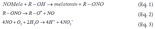
At physiological pH such reactions can even out compete the hydrolysis reaction of NOMela.

In fact, in a complex biological environment of a cell culture system, NOMela simultaneously generates melatonin and biologically active NO [12]. Importantly, all mentioned reactions (Eqs. 1-4) proceed spontaneously. Because of this fact, release of NO from NOMela cannot be hampered by so-called “nitrate tolerance”, a common side-effect of other NO-releasing drugs [1].
Unfortunately, the physical and chemical characteristics of NOMela are unfavorable for its application in animals and humans. An oral administration is obviously senseless because conditions, as present in the stomach, are well known to accelerate the degradation of N-nitroso-indole compounds in general [13]. The alternative intravenous application with a NOMela-enriched salt solution seems to be unpracticable because the solubility in water is too low. NOMela is slightly soluble in ethanol, but the use of this vehicle in animals would alter both metabolism and systemic parameters. In order to confect a pharmacological acceptable approach with a prolonged life-time of NOMela, its encapsulation seems to be a promising alternative. In fact, the encapsulation of sparingly water-soluble drugs into biodegradable poly(lactide-co-glycolide) (PLGA) polymers is an established procedure [14-16].
In the present study, we report on the encapsulation of NOMela with PLGA polymers (NOMela-PLGA capsules) and discuss the releasing kinetics of both NO and melatonin from this system with the idea of placing a useful application form of those two hormones at the disposal of clinicians.
Materials and Methods
Reagents
Poly (DL-lactide-co-glycolide) (50:50) produced by LACTEL (B6013-2) was purchased from NRC Nordmann Rassmann GnbH (Hamburg, Germany). Vitamin C, Trolox, Nile-red and hemoglobin came from Sigma-Aldrich (Deisenhofen, Germany).
Synthesis of N-nitrosomelatonin
Melatonin (276 mg) was dissolved in acetone (3 mL) and H2O (9 mL) was added by vortexing. NaNO2 (208 mg) was dissolved in H2O (3 mL) and was then added to the melatonin solution by vortexing for about 5 min. Afterwards HCl (1.5 mL, 1 M) was added under rigorous stirring and this mixture was cooled down to 4°C. The generated product N-nitrosomelatonin was extracted by shaking with dichloromethane (20 mL, 4°C) and the separated, water insoluble organic phase was dried by adding Na2SO4 (750 mg). After removing Na2SO4 this mixture was cooled down to –20°C and taken to evaporation under reduced pressure (18 Torr) to yield about 250 mg N-nitrosotryptophan (Ɛ346=7070 M–1cm–1 [17]).
Capsule preparation
The preparation of the PLGA-NOMela microcapsules basically followed the emulsion-evaporation procedure reviewed by Pisani et al. for capsules with sizes in the low μm range [18]. An organic solution of PLGA (100 mg) in the absence or in the presence of NOMela (26.1 mg) in dichloromethane (4 ml) at 4°C was emulsified into an aqueous solution (20 ml) of 35 mM sodium cholate by using an Ultraturrax T25 (IKA Werke, Staufen, Germany) operating with a S25KV-25G-IL dispersing tool at a velocity of 3200 rpm. For laser-scanning microscopy, 100 μL of a stock solution of Nile-red (0.057 mg/ml in dichloromethane) was added to the organic solution prior to emulsification [19]. The dichloromethane was then removed by bubbling with synthetic air for 20 min at 4°C. Such a procedure yielded polydisperse dispersions containing capsules with mean sizes of about 2 μm which were used for all experiments.
Laser-scanning microscopy (LSM)
A laser-scanning microscope equipped with a helium/neon laser was used to study the formation of microcapsules. Imaging of nanocapsules (d<0.5 μm) was not possible due to the resolution limit of the optical system. Nile red-stained NOMela-PLGA capsules were diluted 50- 200-fold with 0.9% NaCl solution and placed on an object slide or within a modified Pentz chamber. The objective lens was a 63x NA 1.40 plan-apochromat. Red fluorescence of Nile red excited at 543 nm was collected through a 585 nm long-pass filter. Image processing and evaluation were performed using the software of the LSM 510 imaging system. The diameter of the capsules was analyzed by evaluating the LSM pictures with the CEEXO 2006 CellExplorer software (BioSciTec GmbH, Frankfurt a.M., Germany).
Deoxyhemoglobin solutions
Commercially available methemoglobin (4 mM) from bovine was reduced with vitamin C (20 mM) under aerobic conditions in phosphate buffered solution (100 mM, pH 7.0) [20]. After 2 h of reaction, oxyhemoglobin was formed and the solution was purified over a Sephadex G-50 column with the same buffer as the eluant. Deoxyhemoglobin solutions were prepared by carefully bubbling oxyhemoglobin solutions for 30 min with gaseous nitrogen in order to avoid foaming of the solution.
Determination of nitrite
The nitrite yields from decomposition of NOMela were quantified by the Griess assay [21].
Determination of Nitric Oxide with EPR spectrometry
NO formation was determined using deoxyhemoglobin and EPR spectrometry. EPR spectra were recorded at 77 K on a Bruker ESP- 300E X-band spectrometer (Bruker, Rheinstetten, Germany) equipped with a TM110 wide-bore cavity. Solutions were prepared from 1 ml of the phosphate buffered solution (pH 7.0) containing either NOMela (2 mM) or NOMela-PLGA capsules (100 mg/mL) and deoxyhemoglobin (1.5 mM) in the absence and in the presence of vitamin C (20 mM). The reaction solutions were quickly transferred to an aqueous solution quartz cell (Willmad, Buena, NY). Recording conditions were as follows: microwave frequency, 9.635 GHz; modulation, 0.2 mT; signal gain, 1 × 10⁵; sweep range, 20 mT; microwave power, 20 mW; sweep time, 6 min. Spectrum simulation was performed using the WinSim program [22].
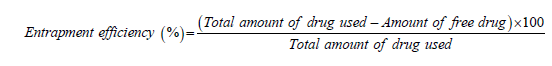
Determination of the entrapment efficiency
All experiments were performed in phosphate buffer (50 mM, pH 7.4, 37°C). Time-dependent adsorption of either melatonin or NOMela (220 μM each) by empty PLGA capsules (capsule material=100 mg/mL) was analyzed. An aliquot of 1 mL sample was taken from the supernatant after centrifugation (5000 rpm) and the concentration of either free melatonin or NOMela was determined by fluorescence (λex=276 nm/λem ≥ 354 nm) or UV-vis absorption (Ɛ346=7070 M-1 cm-1 [23]), respectively. The % entrapment efficiency was determined indirectly by using the following formula:
Results
The modified oil-in-water (o/w) emulsion/solvent evaporation technique encapsulated NOMela with an entrapment efficiency of 85 ± 8%. Capsules with a mean size between 1 and 3 μm were obtained at the employed velocity of 3200 rpm (Figure 1).

Figure 1: Formation of NOMela-PLGA capsules.
In order to demonstrate that the encapsulated NOMela is accessable for solvated, “activated” hydroxy compounds, the release of melatonin was studied in the absence and in the presence of Trolox or vitamin C, respectively, as additives (Figure 2). In general, the presence of the selected additives facilitated the release of melatonin from NOMela- PLGA capsules. Within a reaction period of 5 h, the amount of melatonin released from NOMela had been increased two- to three-fold. Although both hydroxy compounds react with solvated nitrosoindole derivatives with nearly the same rate constant [8,9,17,24], Trolox was a bit more effective than vitamin C in releasing melatonin.

Figure 2: Release of melatonin from NOMela-PLGA capsules.
In order to demonstrate that the decay of encapsulated NOMela proceeds via reactions outlined in equations 1-4, the formation of nitrite was monitored (Figures 3A and 3B). Since vitamin C interferes with the formation of the nitrite reporter dye formation reaction, these measurements were only performed with Trolox. In the absence of Trolox (Figure 3A), nitrite was simultaneously generated with melatonin over a reaction period of 5 h, but the yield of nitrite was always about 21% higher than the yield of melatonin. For instance, after 5 h of reaction 1490 ± 71 μM nitrite and 1229 ± 19 μM melatonin were detected. The presence of Trolox increased the yield of both products but did not change the enhanced yield of nitrite by about 21% (Figure 3A). For instance, after a reaction period of 5 h 2780 ± 62 μM nitrite and 2300 ± 53 μM melatonin were detected, respectively. Although melatonin and nitrite were simultaneously formed in the absence as well as in the presence of Trolox, they were obviously not generated in the expected 1:1 stoichiometric ratio. In contrast, melatonin and nitrite were released in a strict 1:1 stoichiometric ratio from uncapsulated NOMela in the absence as well as in the presence of Trolox (Figure 3B). The data of Figures 3A and 3B also demonstrate that encapsulation significantly increased the half-life time of NOMela from the known value of about 140 min (Figure 3B, [17]) to >5 h (Figure 3A).

Figure 3: Release of melatonin and nitrite from solvated or encapsulated NOMela.

Figure 4: Effect of PLGA capsules on the concentrations of melatonin and nitrite.
A reasonable explanation for the apparent non-stoichiometric production of melatonin and nitrite in the aqueous phase is a slightly preferred accumulation of melatonin but not of nitrite in the PLGA capsule material. In order to clarify this possibility, experiments with PLGA capsules and melatonin were performed (Figures 4A and 4B). In the first set of experiments melatonin was encapsulated in PLGA with an entrapment efficiency of 25 ± 4% and the kinetic of melatonin release in buffer was monitored (Figure 4A). Melatonin was released in an exponential manner but leveled off to about 500 μM after an incubation period of 3 h. This time dependence reflected the onset of absorptiondesorption equilibrium with a steady-state concentration of melatonin in the capsules somewhat (about 25%) higher than in the bulk aqueous phase. This was further confirmed by the effect which unloaded PLGA capsules exerted on the concentration of both melatonin and nitrite (220 μM each) in the surrounding aqueous phase (Figure 4B). In the presence of unloaded PLGA capsules (capsule material 100 mg/ml) the melatonin concentration decreased exponentially by about 27% from 220 μM to ca. 160 μM to level off after 3 h of reaction, whereas the nitrite concentration remained essentially unaffected over the whole reaction period (Figure 4B). Noticeably, unloaded PLGA capsules decreased the concentration of 220 μM NOMela by about 90% within 15 min (data not shown). Both decreases (27% for melatonin and 90% for NOMela) corresponded very well with the observed entrapment efficiencies (25% for melatonin and 85% for NOMela) in PLGA.
So far the proof is missing that according to equations 1 and 2, NO is released from NOMela-loaded PLGA capsules. Unfortunately, it was not possible to monitor NO production electrochemically because the NO-sensitive electrodes were impaired by adsorption of PLGA. In order to exclude any artefacts driven by PLGA and to unequivocally prove the intermediacy of nitric oxide, deoxyhemoglobin was used as NO scavenger and the corresponding nitrosylhemoglobin was detected by EPR spectrometry. Experiments were performed in the absence and in the presence of vitamin C (in order to maintain iron ions in the ferrous state) as an additive (Figure 5). In the absence of vitamin C formation of nitrosylhemoglobin from decaying NOMela- PLGA capsules could not be detected (data not shown). This failure is in agreement with observations from the literature that hydrolytic decomposition of NOMela does not produce ANY (eq. 4) [7-9]. However, in the presence of vitamin C, the characteristic three-line spectrum of nitrosylhemoglobin (g=2.011; a (14N)=1.67 mT) was recorded from encapsulated NOMela (lines a and b) as well as from an aqueous solution of neat NOMela (line c). The observation that NO is in fact released in the presence of an “activated hydroxy compound” (Eq. 2) is in agreement with data from the literature [7-9]. The prolonged half-life time of PLGA-encapsulated NOMela is also reflected by the EPR data (compare line a with line c; from the relative ESR intensities an about ten-fold faster production of NO from uncapsulated NOMela is estimated).

Figure 5: Vitamin C mediated release of NO from solvated or encapsulated NOMela.
Discussion and Conclusion
Here we demonstrated for the first time that NOMela can be encapsulated in PLGA capsules and that such a procedure prolonges the life-time of NOMela in aqueous solution. PLGA-encapsulated NOMela yielded in the presence of either, was generated by hydrolysis of NOMela (Eq. 4). The apparent excess production (by about 21%) of nitrite over melatonin can satisfactorily be explained by a slight accumulation of the formed melatonin in the capsules material (i.e., [melatonin]caps/[melatonin]aq>1), whereas nitrite does not accumulate in the capsules, i.e., its concentration is the same in both phases ([nitrite]caps/[nitrite]aq=1). Noticeably, PLGA is highly effective in transporting hormones [14,25]. Since NOMela-PLGA capsules can be separated from the synthesis solution and thus can be disperesed in any other aqueous solution, the low solubility of NOMela in an aqueous environment is, of course only in principle, no longer a hindrance for its in vivo application.
Generally, there are two possibilities by which activated hydroxy compounds would liberate melatonin and nitric oxide from NOMela- PLGA. On the one side, the activated hydroxy compound may to some degree enter the PLGA capsule in order to react in its interior with NOMela, and the reaction products are then released to the surrounding aqueous phase. This mode would in part explain why the more lipophilic Trolox released melatonin somewhat faster than did the hydrophilic vitamin C (Figure 2). Since both activated hydroxy compounds are known to react with nitrosoindoles with similar rate constant [8,9], an equal distribution of vitamin C and Trolox between PLGA capsules and the water phase would result in an identical rate of melatonin production. If this is not the case, the relative steadystate concentration of Trolox in the capsules must be expected to be higher than that of vitamin C. On the other hand, PLGA-capsules may donate NOMela to the bulk aqueous phase where a rapid reaction with the solvated hydroxy compound takes place. The fact that addition of unloaded PLGA capsules to an aqueous solution of NOMela decreased the NOMela concentration by about 90% indicates that PLGAencapsulated NOMela is in a favourable equilibrium with free, solvated NOMela ([NOMela]caps/[NOMela]aq ≈ 9/1). Of course, both processes must be expected to operate simultaneously, however, the strong enhancement of the half-life time of NOMela in the presence of PLGA capsules indicates that the former mode is of minor importance, i.e., NO release from PLGA-NOMela by reaction with activated hydroxy compounds is primarily controlled by the reduced release of NOMela into the bulk aqueous phase. Such a behavior is desirable for a good transporter system (Figure 6).
In any case, melatonin is expected to support the physiological action of NO because melatonin counteracts the vasospastic effect induced by lysophosphatidylcholine as well as enhances the endothelium-dependent vasodilatation in mesenteric artery and aorta observed in spontaneously hypertensive rats [17].
Initially, PLGA polymers have been developed for use as degradable surgical sutures [26]. Characteristics such as biocompatibility and established regulatory approval for clinical use have attracted interest in PLGA for controlled-release applications [27]. A typical implementation was the injection of hormone-loaded PLGA microcapsules into the musculature [14]. Particle size was shown to be an important effector. At optimized conditions, i.e., particle size >10 μm, non-circulating PLGA microcapsules remained active for several months [14,27,28]. Such a strategy might be favorable for the encapsulation of melatonin but not for the chemically labile NOMela.

Figure 6: PLGA as a transporter for NOMela.
Therefore, due to the relative short half-life time of NOMela, circulating entities (capsules) are obviously a matter of choice. Since circulating particles with a mean size of >5 μm are lethal in animals [29-31], the preferable particle size must be lower than that limiting value. Additionally, the circulatory half-life time of intravenously administered particles is generally limited by phagocytic ingestion. Evidence has been presented that maximal uptake of microparticles into macrophages occurred with particles between 1-3 μm [32-34]. Antibodies and complement proteins which can associate to foreign particles render microcapsules more attractive to phagocytes, thereby largely enhancing their clearance from the circulation system [35,36]. Since the mean size of our model NOMela-PLGA capsules presently lies between 1 and 3 μm (Figure 1), it must be expected that they would rapidly be cleared (within a few minutes [37]) from the blood by the reticuloendothelial system. According to Eq.s 1-4 NOMela is a NOdonor generating compound but it is not a direct NO releasing entity. Because of this an expected circulation time of a few minutes would be too short to effectively generate NO-releasing entities. In order to avoid such undesirable effects, the properties of the capsules have to be improved for later in vivo use. So to solve the two problems (i) fast recognition by the RES and (ii) to large size, at first, removal of the capsules by macrophages of the reticuloendothelial system can be minimized by covering their surface with hydrophilic polymers such as covalently bound polyethyleneglycols [37-39]. Although principally, non-covalently bound polymers like Tetronic-908 have been shown to operate similarly in experimental systems [40] because of the replacement of the synthetic macromolecules by plasma proteins, such polymers are not suitable for in vivo applications due to their high toxicity. At second, particles with a lower size (<1 μm, so-called nanoparticles) are removed with a significantly lower rate from the circulation, because they are favorably internalized by endothelial and smooth muscle cells [41]. This is why we are currently working on the preparation of NOMela-PLGA nanocapsules. Covering the surface of those nanoparticles with polyethyleneglycol will additionally down regulate the opzonization process [42]. As we have already successfully developed long circulating polyethyleneglycol-coated perfluorodecalinfilled PLGA-capsules as artificial oxygen carriers [43], we are sure that the preparation of NOMela-PLGA nanocapsules with a modified surface will result in a clinically relevant application form of NOMela.
References
- Moncada S, Palmer RMJ, Higgs EA (1991) Nitric oxide: Physiology, pathophysiology and pharmacology. Pharmacol Rev 43: 109-142.
- Moller JKS, Skibsted LH (2004) Mechanism of nitrosylmyoglobin autoxidation: Temperature and oxygen effects on the two consecutive reactions. Chem Eur J 10: 2291-2300.
- Grubina R, Huang Z, Shiva S, Joshi MS, Azarov I, et al. (2007) Concerted nitric oxide formation and release from the simultaneous reactions of nitrite with deoxy- and oxy-hemoglobin. J Biol Chem 282: 12916-12927.
- Ignarro LJ (1990) Biosynthesis and metabolism of endothelium-derived nitric oxide. Annu Rev Pharmacol Toxicol 30: 535-560.
- Czapski G, Goldstein S (1995) The role of the reactions of NO with superoxide and oxygen in biological systems: A kinetic approach. Free Radic Biol Med 19: 785-794.
- Blanchard-Fillion B, Servy C, Ducrocq C (2001) 1-Nitrosomelatonin is a spontaneous NO-releasing compound. Free Radic Res 35: 857-866.
- Kirsch M, Fuchs A, de Groot H (2003) Regiospecific nitrosation of N-(terminal) blocked tryptophan derivatives by N2O3 at physiological pH. J Biol Chem 278: 11931-11936.
- Kytzia A, Korth HG, Sustmann R, de Groot H, Kirsch M (2006) On the mechanism of the ascorbic acid-induced release of nitric oxide from nitrosated tryptophan derivatives. Scavenging of NO by ascorbyl radicals. Chem Eur J 12: 8786-8797.
- Müller K, Korth HG, de Groot H, Kirsch M (2007) Reaction of vitamin E compounds with N-nitrosated tryptophan derivatives and its analytical use. Chemistry 13: 7532-7542.
- Kytzia A, Korth HG, de Groot H, Kirsch M (2006) Catecholamine-induced release of nitric oxide from N-nitroso-tryptophan derivatives: A non-enzymatic way of catecholamine-oxidation. Org Biomol Chem 4: 257-267.
- Kytzia A, Korth HG, de Groot H, Kirsch M (2007) N-Nitroso-Melatonin releases nitric oxide in the presence of serotonin and its derivatives. J Pineal Res 43: 343-350.
- Berchner-Pfannschmidt U1, Tug S, Trinidad B, Becker M, Oehme F, et al. (2008) The impact of N-nitrosomelatonin as nitric oxide donor in cell culture experiments. J Pineal Res 45: 489-496.
- Williams DLH (2004) Nitrosation reactions and the chemistry of nitric oxide. Elsevier, Amsterdam.
- Beck LR, Pope VZ, Flowers CE, Cowsar, DR, Tick, TR et al. (1983) Poly(DL-lactide-co-glycolids/norethisterone microcapsules: An injectable biodegradable contraceptive. Biol Reprod 28: 186-195.
- Garvin K, Feschuk C (2005) Polylactide-polyglycolide antibiotic implants. Clin Orthop Relat Res 105-110.
- Singh M, Kazzaz J, Ugozzoli M, Malyala, P, Chesko J, et al. (2006) Polylactide-co-glycolide microparticles with surface adsorbed antigens as vaccine delivery systems. Curr Drug Deliv 3: 115-120.
- Kirsch M, de Groot H (2009) N-Nitrosomelatonin: Synthesis, chemical properties, potential prodrug. J Pineal Res 46: 121-127.
- Li M, Rouad O, Poncelet D (2008) Microencapsulation by solvent evaporation: State of the art for process engineering approaches. Int J Pharm 363: 26-39.
- Pisani E, Tsapis N, Paris J, Nicolas, V, Cattel, L et al. (2006) Polymeric nano/microcapsules of liquid perfluorocarbons for ultrasonic imaging: Physical characterization. Langmuir 22: 4397-4402.
- Tomoda A, Yoneyama Y, Takeshita M (1976) Stimulative effect of 2,3-diphosphoglycerate on methemoglobin reduction by ascorbic acid. Experientia 32: 932-933.
- Ioannidis I, de Groot H (1993) Cytotoxicity of nitric oxide in Fu5 rat hepatoma cells: Evidence for co-operative action with hydrogen peroxide. Biochem J 296: 341-345.
- Duling DR (1994) Simulation of multiple isotropic spin-trap EPR spectra. J Magn Reson B 104: 105-110.
- Turjanski AG, Leonik F, Estrin DA, Rosenstein RE, Doctorovich F (2000) Scavenging of NO by melatonin. J Am Chem Soc 122: 10468-10469.
- Kirsch M, Korth HG (2007) Generation, basic chemistry and detection of N-nitrosotryptophan derivatives. Org Biomol Chem 5: 3889-3894.
- Wischke C, Schwendeman SP (2008) Principles of encapsulating hydrophobic drugs in PLA/PLGA microparticles. Int J Pharm 364: 298-327.
- Frazza EJ, Schmitt EE (1971) A new absorbable suture. J Biomed Mater Res 5: 43-58.
- Gupta RK, Singh M, O'Hagan DT (1998) Poly(lactide-co-glycolide) microparticles for the development of single-dose controlled-release vaccines. Adv Drug Deliv Rev 32: 225-246.
- Ruan G, Feng SS (2003) Preparation and characterization of poly(lactic acid)-poly(ethylene glycol)-poly(lactic acid) (PLA-PEG-PLA) microspheres for controlled release of paclitaxel. Biomaterials 24: 5037-5044.
- Riess JG (2001) Oxygen carriers raison d'etre, chemistry and some physiology. Chem Rev 101: 2797-2920.
- Tadross TF (2005) Applied Surfactants. WILEY-VCH Verlag GmbH & Company, Weinheim.
- Slack JD, Kanke M, Simmons GH, de Luca PP (1981) Acute hemodynamic effects and blood pool kinetics of polystyrene microspheres following intravenous administration. J Pharm Sci 70: 660-664.
- Tabata Y, Ikada Y (1988) Macrophage phagocytosis of biodegradable microspheres composed of L-lactic acid/glycolic acid homo- and co-polymers. J Biomed Mater Res 22: 837-858.
- Singh M, Kazzaz J, Ugozzoli M, Chesko J, O'Hagan DT (2004) Charged polylactide co-glycolide microparticles as antigen delivery systems. Expert Opin Biol Ther 4: 483-491.
- Ikada Y, Tabata Y (1986) Phagocytosis of bioactive microspheres. J Bioactive Compatible Polym 1: 32-46.
- Moghimi SM (1995) Mechanisms regulating body distribution of nanospheres conditioned with pluronic and tetronic block-copolymers. Adv Drug Delivery Rev 16: 183-193.
- Todd CW, Balusubramanian M, Newman MJ (1998) Development of adjuvant-active non-ionic block copolymers. Adv Drug Deliv Rev 32: 199-223.
- Panagi Z, Beletsi A, Evangelatos G, Livaniou E, Ithakissios DS, et al. (2001) Effect of dose on the biodistribution and pharmacokinetics of PLGA and PLGA-mPEG nanoparticles. Int J Pharm 221: 143-152.
- Moghimi SM, Porter CJH, Muir IS, Illum L, Davis SS (1991) Non-phagocytic uptake of intravenously injected microspheres in rat spleen: influence of particle size and hydrophilic coating. Biochem Biophys Res Commun 177: 861-866.
- Avgoustakis K, Beltsi A, Panagi Z, Klepetsanis P, Livaniou E, et al. (2003) Effect of copolymer composition on the physicochemical characteristics, in vitro stability and biodistribution of PLGA-mPEG nanoparticles. Int J Pharm 259: 115-127.
- Kirsch M, Bramey T, Waack IN, Petrat F, Mayer C, et al. (2012) The necessity for the coating of perfluorodecalin-filled poly(lactide-co-glycolide) microcapsules in the presence of physiological cholate concentrations: Tetronic-908 as an exemplary polymeric surfactant. J Microencapsulation 29: 30-38.
- Nguyen KT, Shukla KP, Moctezuma M, Braden ARC, Zhou J et al. (2009) Studies of the cellular uptake of hydrogel nanospheres and microspheres by phagocytes, vascular endothelial cells and smooth muscle cells. J Biomed Mater Res 88A: 1022-1030.
- Soppimath KS, Aminabhavi TM, Kulkarni AR, Rudzinski WE (2001) Biodegradable polymeric nanoparticles as drug delivery devices. J Control Release 70: 1-20.
- Ferenz KB, Waack IN, Mayer C, de Groot H, Kirsch M (2013) Long-circulating poly(ethylene glycol)-coated poly(lactid-co-glycolid) microcapsules as potential carriers for intravenously administered drugs. J Microencapsulation 30: 632-642.
Relevant Topics
- Agricultural biotechnology
- Animal biotechnology
- Applied Biotechnology
- Biocatalysis
- Biofabrication
- Biomaterial implants
- Biomaterial-Based Drug Delivery Systems
- Bioprinting of Tissue Constructs
- Biotechnology applications
- Cardiovascular biomaterials
- CRISPR-Cas9 in Biotechnology
- Nano biotechnology
- Smart Biomaterials
- White/industrial biotechnology
Recommended Journals
Article Tools
Article Usage
- Total views: 3056
- [From(publication date):
June-2017 - Jul 11, 2025] - Breakdown by view type
- HTML page views : 2185
- PDF downloads : 871