Case Report Open Access
Empyema Caused by Unusual Pathogen Capnocytophaga
Emhemmid Karem, Hazim Bukamur*, Yousof Elgaried, and Mahmoud Shorman
Internal Medicine Department, Joan C. Edwards School of Medicine, Marshall University, Huntington, WV, USA
- *Corresponding Author:
- Hazim Bukamur
Internal Medicine Department
Joan C. Edwards School of Medicine
Marshall University, Huntington
WV, USA
Tel: 8608419018
E-mail:Bukamur@marshall.edu
Received date: July 19, 2016; Accepted date: August 23, 2016; Published date: September 03, 2016
Citation: Emhemmid K, Hazim B, Yousof E, Mahmoud S (2016) Empyema Caused by Unusual Pathogen Capnocytophaga . J Emerg Infect Dis 1:112. doi:10.4172/2472-4998.1000112
Copyright: © 2016 Karem E, et al. This is an open-access article distributed under the terms of the Creative Commons Attribution License, which permits unrestricted use, distribution, and reproduction in any medium, provided the original author and source are credited.
Visit for more related articles at Journal of Infectious Disease and Pathology
Abstract
Empyema is a collection of pus in the pleural space. Empyema caused by Capnocytophaga species is extremely rare, and after extensive review of the literature, we found only four reported cases of Capnocytophaga empyema. In our case report, we present a case of 71-year-old male with Capnocytophaga empyema initially presented with signs and symptoms of acute congestive heart failure (CHF), with no history of splenectomy or animal bite. The pleural fluid Cultures grew Capnocytophaga, no other organisms were identified and blood cultures were negative. People at increased risk of developing Capnocytophaga infections include post splenectomy patients, Diabetics, male sex, age over 50, and alcohol abuse. The risk factors for our case were age, male sex, congestive heart failure, and history of alcohol abuse. The patient was treated with chest tube drainage and appropriate antibiotics with complete resolution of his empyema.
Keywords
Empyema; Capnocytophaga; Zoonotic
Introduction
Empyema is a collection of pus in the pleural space. It is typically a serious complication of pneumonia with high morbidity and mortality rates.The most common organisms causing empyema are gram positive organisms such as Staphylococcus and Streptococcus. Management of empyema includes drainage by chest tube or surgery and use of appropriate antibiotics [1]. Capnocytophagaempyema is extremely rare [2] and to our knowledge there is only few cases that have been reported. Capnocytophaga is a group of long, thin, fusiform, slowly growing, facultative anaerobic, Gram-negative rods with gliding motility whose growth is optimal in a CO2-enriched atmosphere; hence the name Capnocytophaga (consumption of CO2). Capnocytophagaspecies are part of the normal oral flora in dogs, cats, and humans and were once associated with periodontal disease but are now considered commensals in dental plaque of humans. Capnocytophagainfections can have varied clinical presentations, such as periodontal disease, respiratory tract infections, ophthalmic lesions, traumatic pericarditis, mediastinal abscess, brain abscess, meningitis, and peritonitis [3].
Case
A 71-years-old, white male with significant past medical history of Congestive heart failure, Diabetes, Non-Hodgkin’s Lymphoma in remission, and Chronic Kidney disease stage 3. He presented to our facility with worsening shortness of breath, orthopnea, and lower extremity swelling with weeping stasis dermatitis. By reviewing his social history, He did not report animal bite or scratch but regularly cared for four dogs at his house who used to lick his legs. On physical examination, his vital signs were only remarkable for hypotension. Chest examination revealed decrease air entry bilaterally with scattered rhonchi all over the chest. Left leg showed skin breakdown with bilateral weeping stasis dermatitis. The reminder of physical examination was unremarkable.
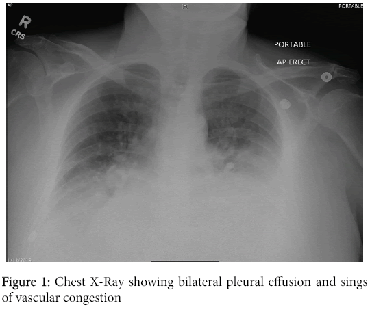
Complete blood count and metabolic panel was only significant for anemia (HGB 9.8), and high Cr. 1.9, as well as high B-type natriuretic peptide 1536. Chest x-ray on the day of admission was noted to be positive for bilateral pleural effusions with possible bilateral atelectasis (Figure 1).
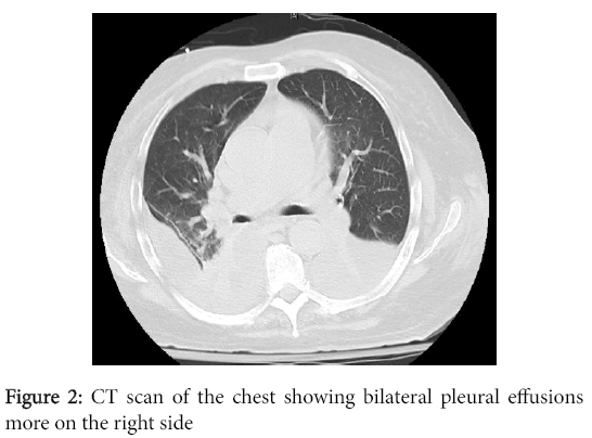
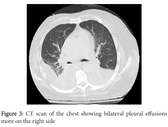
Initially the patient was admitted to the medical floor as a case of acute exacerbation of CHF. He was distressed and seen by Cardiology who optimized his meds. Overnight the patient had deterioration in his level of consciousness with decrease in his O2 saturation to the low 70s. Arterial blood gas showed severe respiratory acidosis. Serial chest X-rays showed worsening pleural effusion. He was transferred to the ICU, emergently intubated, and was treated for septic shock with Pipercillin-Tazobactam, Levofloxacin, vancomycin, and Vasopressors. CT scan of the chest was done and showed bilateral pleural effusions (Figures 2 and 3).
Pleural fluid was drained from the left lung with a pig tail catheter which showed exudative Para-pneumonic effusion followed by Right sided chest tube. After four days, the pleural fluid Cultures came back positive for Capnocytophaga spp. , no other organisms have been identified. Blood cultures were negative. Patient clinically improved, extubated, and the antibiotics were de-escalated to ampicillinsulbactam. No growth was detected on the repeated pleural fluid culture few days later. Chest tubes were removed, and he was transferred to the floor. The serial chest x rays showed significant improvement without serious pulmonary complications. Patient was discharged on amoxicillin-clavulanic acid for a total duration of four weeks.
Discussion
The most common organisms causing empyema are gram positive bacteria such as Staphylococci and Streptococci. Empyema caused by Capnocytophaga species is extremely rare. Up to our knowledge with extensive literature review, there have been only four previous case reports of Capnocytophaga empyema in the literature. The first one was secondary to laparoscopic Nissen fundoplication, which was sensitive to clindamycin, penicillin G, and fosfomycin. The second was a case of spontaneous empyema in a patient who suffered from hepatitis C-induced liver cirrhosis, and the patient responded to imipenem-cilastatin. The third one was a case Capnocytophagasputigena empyema, was found to be susceptible to ampicillin and ceftriaxone [3]. Last one was empyema due to Capnocytophagamixed with Actinomycesisraeli in Japan.
Capnocytophaga is the name of a group of Gram-negative bacteria that live in the mouth of some animals and humans. It is long, thin gram-negative bacilli that are slow growing, with gliding ability on agar media. They are facultative anaerobic and require enrichment with CO2 (5-10%) for optimum growth. Clinical isolates of Capnocytophaga species are classified into two broad groups: Those species found in the human oral cavity including C. gingivalis, C. ochracea, and those two species that life in the oral cavities of dogs: C. canimorsus and C. cynodegmi [4].
Capnocytophagacanimorsus is the species most commonly involved in zoonotic infections. It can be transmitted from pets to people via bites or contact of saliva with broken skin. Immunocompromised individuals are highly susceptible for infection. Persons at increased risk of developing C. canimorsus infections include patients who have undergone a splenectomy, Diabetics, being male, over the age of 50, and those who abuse alcohol [5]. The risk factors for our case were age, Non- Hodgkin’s lymphoma, CHF and history of alcohol abuse.
Almost all the Capnocytophagaempyema cases that have been reported occurred in the absence of bacteremia, as is in our case. This augment the possibility of the lung as the primary source of infection. Other possibility is that the patient might have had transient bacteremia. In this patient we still suspect that the port of entry was his left leg and patient had unrecognized transient bacteremia. Many laboratories still have difficulty identifying this species as it requires special media. Capnocytophaga sp. need special environment to grow in the laboratory, including media rich with iron and carbon dioxide, that is why they may find difficulty to detect this kind of bacteria unless specific testing is requested, typically based on the patient’s risk factors and a history of animal exposure. To diagnose those species, Physicians should have high index of suspicion and consider C. canimorsus in patients with bacterial sepsis and a recent history of a dog bite or animal exposure as well as the characteristic features of C. canimorsus which include positive test results for oxidase, catalase, arginine dihydrolase, and lack reactions for urease, nitrates, and indole [5].
Beta-lactamase producing strains of Capnocytophaga spp., are increased in recent years so it wise to treat with beta-lactamase inhibitors such as amoxicillin-clavulanic acid [6]. The majority of Capnocytophaga spp.are susceptible to cefoxitin, linezolid and clindamycin, imipenem, cefotaxime, ceftazidime, and ceftriaxone. Ampicillin, penicillin G, ticarcillin, aztreonam, and temocillin showed moderate activity whereas aminoglycosides, polymyxin B, vancomycin, and trimethoprim were resistant to Capnocytophaga spp. [7].
Conclusion
Capnocytophaga empyema is extremely rare with Very few cases have been reported, and it can result in high mortality rate if not recognized early and treated appropriately as it may cause septic shock. The pathogen is susceptible to different antibiotic agents, including beta lactams, clindamycin, fluoroquinolones, and treatment of patients is dependent on the severity of the disease empirically pending culture results. Finally, with dealing with older patient’s population, and better diagnostic techniques, including polymerase chain reaction, and the different epidemiology and exposures to different pet animals, clinicians should entertain the possibility of different zoonotic infections [8].
References
- Rosenstengel A (2012) Pleural infection-current diagnosis and management. JThorac Dis 4: 186-193.
- Bonatti H, Rossboth DW, Nachbaur D, Fille M, Aspock C, et al. (2003) A series of infections due to Capnocytophaga spp. in immunosuppressed and immunocompetent patients. ClinMicrobiol Infect 9: 380-387.
- Thirumala R, Rappo U, Babady NE, Kamboj M, Chawla M (2012)Capnocytophaga lung abscess in a patient with metastatic neuroendocrine tumor. JClinMicrobiol 50: 204-207.
- Li A, Tambyah P, Chan D, Leong KK (2013) Capnocytophagasputigena empyema. J ClinMicrobiol 51: 2772-2774.
- Mally M, Cornelis GR (2008) Genetic tools for studying Capnocytophagacanimorsus.Appl Environ Microbiol 74: 6369-6377.
- Janda JM, Graves MH, Lindquist D, Probert WS (2006) Diagnosing Capnocytophagacanimorsus infections. Emerg Infect Dis 12: 340-342.
- JolivetGougeon A (2007) Capnocytophaga Species. Antimicrobe 29: 367-373.
- Rummens JL,GordtsB, Van Landuyt HW (1986) In vitro susceptibility of Capnocytophaga species to 29 antimicrobial agents. Antimicrob Agents Chemother 30: 739-742.
Relevant Topics
Recommended Journals
Article Tools
Article Usage
- Total views: 12138
- [From(publication date):
September-2016 - Apr 05, 2025] - Breakdown by view type
- HTML page views : 11243
- PDF downloads : 895


