Research Article Open Access
Efficacy Testing of Orally Administered Antibiotics against an Inhalational Bacillus anthracis Infection in BALB/C Mice
| Graham J Hatch, Simon R Bate, Anthony Crook, Nicola Jones, Simon G Funnell and Julia Vipond* | ||
| Public Health England, Porton Down, Salisbury, SP4 0JG, UK | ||
| Corresponding Author : | Julia Vipond Public Health England, Porton Down, Salisbury, UK Tel: +4401980 612584 Fax: +44 (0)1980 619894 E-mail:julia.vipond@phe.gov.uk |
|
| Received August 15, 2014; Accepted October 16, 2014; Published October 28, 2014 | ||
| Citation: Hatch GJ, Bate SR, Crook A, Jones N, Funnell SG, et al. (2014) Efficacy Testing of Orally Administered Antibiotics against an Inhalational Bacillus anthracis Infection in BALB/C Mice. J Infect Dis Ther 2:175. doi: 10.4172/2332-0877.1000175 | ||
| Copyright: © 2014 Hatch GJ, et al. This is an open-access article distributed under the terms of the Creative Commons Attribution License, which permits unrestricted use, distribution, and reproduction in any medium, provided the original author and source are credited. | ||
Related article at Pubmed Pubmed  Scholar Google Scholar Google |
||
Visit for more related articles at Journal of Infectious Diseases & Therapy
Abstract
Anthrax is a potentially fatal disease in man, and events of the last decade have highlighted the possibility for Bacillus anthracis to be used as a biological weapon through deliberate release. Whilst outbreaks of anthrax occur in several countries, there are only a few areas in which outbreaks of inhalational anthrax occur, so it is not possible to test therapies using conventional clinical trials. To overcome these problems, the FDA published the ‘Animal Rule’ in 2002; a rule designed to permit licensure of drugs and biologics intended to reduce or prevent serious conditions caused by exposure to biological and other agents. Due to limited proven post-exposure prophylaxis after inhalational exposure for use in humans, it is essential to have a robust animal model of disease. In a series of three studies conducted in BALB/c mice, the efficacy of postexposure, orally administered antibiotics in preventing disease following inhalational exposure to B. anthracis was evaluated. Mice treated with either azithromycin or clarithromycin displayed a near identical disease progression to the control, untreated mice; in contrast, those treated with levofloxacin, amoxicillin and co-amoxiclav were generally protected with survival of 97%, 95% and 79%, respectively. The details of the assessments and future direction of these studies are presented.
| Keywords |
| Bacillus anthracis; Anthrax; Co-amoxiclav; Orally administered antibiotics |
| Introduction |
| Bacillus anthracis, a Gram-positive, spore-forming bacillus, is the causative agent of anthrax, a disease that manifests via the gastrointestinal, cutaneous or inhalational route of exposure. Whilst human infection is rare, it generally occurs with low lethality via the cutaneous or gastrointestinal route following people handling infected animals or by ingesting infected meat. The disease, however, is potentially lethal via the inhalational route due to internalization of the spores [1], and the threat posed to public health and safety have caused it to be included on the US Centers for Disease Control and Prevention (CDC) list of select agents as a Category A biological threat agent [2]; indeed the human inhalational mortality rate ranges from 45%-80% [3,4]. |
| The current recommended antibiotic (ciprofloxacin) of choice against B. anthracis is based upon empirical treatments for sepsis, as there are no clinical studies of the treatment of anthrax in humans [4]; although recently the CDC has updated their guidelines on treatment of post-exposure inhalational anthrax, to include doxycycline and levofloxacin as well as ciprofloxacin [5]. Ciprofloxacin has been investigated in many animal models of inhalational infection, ranging from mice, guinea pigs and rabbits [6-10] through to marmoset and rhesus macaques [11-16] and it has proved effective. Whilst antibiotics are still considered to be the most effective therapeutic approach in the treatment of many pathogens, it is important to look at different antibiotics or indeed different combinations of antibiotics to combat potential antibiotic resistance. In addition, treatment with ciprofloxacin alone during the Amerithrax incident was seen to fail to protect 5 out of 11 individuals against inhalational anthrax. Some have speculated that this apparent antibiotic failure was due to continued action of toxin in the absence of viable organisms [17] and suggest a combination of antibiotics (e.g. bactericidal agents with protein synthesis inhibitors) would be more effective. This suggestion is supported by the fact that 53% of the human cases of inhalational anthrax that survived were treated with a combination of antibiotics [5]. |
| Inhalational anthrax has an incredibly rapid disease course, with death occurring within a few days of exposure if no treatment is administered, thereby making this a lethal form of the disease [18]. To evaluate the efficacy of different antibiotics following aerosol exposure to virulent B. anthracis (Ames strain), a model utilising BALB/c mice was established. This manuscript focuses on the survival and clinical signs of infection in BALB/c mice following orally administered antibiotics post-aerosol challenge with B. anthracis spores. |
| Material and Methods |
|
|
| Bacillus anthracis, AMES (NR-2324) was obtained from the Biodefense and Emerging Infections Research Resources Repository (BEI Resources, Manassas, VA). B. anthracis spores were produced by fed batch culture in a 2 L bioreactor for approximately 26 h at 37ºC ± 2ºC, 400 rpm ± 10 rpm. The spores were concentrated and washed in sterile distilled water by centrifugation, with final suspension in sterile distilled water to an approximate titre of 8E+09 CFU/ml. Confirmation of spores was by phase-bright microscopy; the final suspension was 100% phase-bright. |
| Experimental animals |
| All animal studies were carried out in accordance with the UK Animals (Scientific Procedures) Act, 1986 and the Codes of Practice for the Housing and Care of Animals used in Scientific Procedures, 1989. Female BALB/c mice aged over 8 weeks (sexually mature), free of inter-current infection, were obtained from a commercial supplier (Charles River, UK) with an approximate body weight of 20 grams. Mice were housed in groups of five on a 12 hour day-night light cycle, with food and water available ad libitum. |
| Aerosol challenge |
| Over three sequential experiments, groups of up to 20 mice (Table 1) were challenged for 10 minutes with a dynamic aerosol of B. anthracis spores using the AeroMP-Henderson apparatus; the challenge aerosol was generated using a three-jet Collison nebuliser (BGI, Waltham, USA) containing 20 ml B. anthracis spores in sterile water. The resulting aerosol was mixed with conditioned air (65 ± 5% relative humidity, 21 ± 1ºC) in the spray tube [19] and delivered to the nose of each animal via an exposure tube (sow) in which the un-anaesthetised mice were held in restraint tubes. Samples of the aerosol were taken using an All Glass Impinger (AGI30, Ace Glass, USA), [20] operating at 6 L/min containing 20 ml sterile water and an Aerodynamic Particle Sizer (TSI Instruments Ltd, Bucks, UK); these processes were controlled and monitored using the AeroMP management platform (Biaera Technologies LLC, MD, USA). |
| Back titrations of the aerosol samples, taken at the time of challenge, were performed by serial dilution and plating out onto trypticase soy agar (TSA) incubated at 37ºC for 16-24 h. These titrations were used to calculate the inhaled dose using a derived respiratory minute volume of 19.9 ml estimated from the average weight of the animals [21]. |
| Antibiotic Dosing |
| Antibiotics were obtained from a registered pharmacy. Levofloxacin tablets were dissolved with the aid of sonication in sterile water to a concentration of 8 mg/ml. Liquid preparations of amoxicilin, co-amoxiclav, clarithromycin and azithromycin were prepared in sterile water according to manufacturer’s instructions. Immediately prior to administration, amoxicillin, co-amoxiclav and clarithromycin were further diluted in sterile water to achieve concentrations of 3 mg/ml; azithromycin was diluted to 1.2 mg/ml for the first dose and 0.6 mg/ml thereafter (Table 2). |
| From one day post-exposure, once or twice daily oral gavage of 0.2 ml of either the test antibiotic or carrier control commenced. Treatment continued for thirty days after which the animals were observed for a further fourteen days. Each treatment dose was verified from peak and trough serum samples taken from a separate group of three animals, 1 h after treatment and 1 h prior to the following treatment dose. Sera were filter sterilised prior to analyses. These animals were exposed to B. anthracis prior to antibiotic administration as in the main studies. Analyses were performed at Huntington Life Sciences, UK. Briefly, protein precipitation was used to extract test antibiotic from the mouse serum. Mouse serum (20 µl) was spiked with 10 µl of ciprofloxacin (internal standard), then precipitated with 300 µl of acetonitrile. Samples were centrifuged for 5 minutes at +4ºC (ca 12000 g). The supernatant was transferred and evaporated to dryness, after which the sample was reconstituted in 300 µl of mobile phase, and ca. 10 µl was then injected onto the LC MS/MS system. |
| Monitoring/Sampling |
| Challenged mice were returned to their home cages within an ACDP (U.K. Advisory Committee on Dangerous Pathogens) animal containment level 3 facility. Animals were closely observed at least twice daily over the 45-day period for the development of clinical signs. A clinical score was attributed based upon ruffled fur, eyes closed, arched back and immobility. The humane endpoint of immobility was strictly observed so that no animal became distressed. ‘Time to death’ figures include those animals killed according to the humane endpoint. Cause of death was confirmed by the presence of bacteraemia by streaking 10 µl blood onto TSA and incubating at 37ºC for 24 h. Any animals that reached the end of the study (45 days post-challenge) were designated as survivors. Survivors were humanely euthanized and lung tissue subsequently removed and frozen at less than minus 60ºC. |
| Bacterial Enumeration |
| Lung tissue was thawed, weighed and homogenised in sterile water using a Precellys24 tissue homogenizer (Bertin Technologies, Villeurbanne). Tissue samples were serially diluted in sterile water and then plated out onto TSA. Plates were incubated at 37ºC for 16-24 h before enumeration using a well-developed and characterised standard procedure. |
| Minimum Inhibitory Concentration (MIC) |
| The MICs of the test antibiotics were assessed using 12 B. anthracis isolates recovered from murine survivors’ lung tissue. Antibiotics were serially diluted two-fold in Mueller-Hinton broth so that four wells of a 96-well test plate contained 100 µl in the following concentration range; amoxicillin (4-0.004 µg/ml), co-amoxiclav (4-0.004 µg/ml), levofloxacin (2-0.002 µg/ml), clarithromycin (4-0.004 µg/ml), azithromycin (16-0.015 µg/ml) and ciprofloxacin (2-0.002 µg/ml). Lung tissue isolates and B. anthracis controls (B. anthracis, Ames working cell bank) were grown for 20-24 h at 37ºC on Columbia blood agar. A sample from each was suspended in 10 ml Mueller-Hinton broth to an optical density measured at 600 nm of 0.08 to 0.12. The bacterial suspensions were further diluted 150-fold; 100 µl was added to each well of the test plates and then incubated for 18-24 h at 35 ± 2ºC. Following incubation, the optical density of each well was measured at 600 nm and the MIC curve and values calculated. |
| Statistical Analysis |
| Analysis of survival rates between mice in treatment groups was performed using the log-rank test. |
| Results and Discussion |
|
|
| Groups of 20 mice were challenged with a mean inhaled dose of B. anthracis of 1.2E+06 CFU equating to 20 LD50 (range 11-31 LD50); the mass median aerodynamic diameter of the challenge aerosols was <2 µm. |
| Clinical observations and mortality |
| Administration of antibiotics by oral gavage once or twice daily for 30 days was well tolerated. In addition to mortality data, in order to generate a numerical assessment of the observable well-being of groups of animals, a cumulative overall scoring system was devised which was based on clinical observations indicative of the disease. Clinical observations were scored in the following manner; healthy, score=0; ruffled fur, score=2; eyes closed, score=3; arched back, score=3; immobility, score=9 and death in cage or humane euthanasia, score=10. |
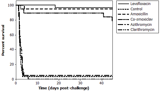
| A graphical summary of the survival of animals from all of these studies is displayed in Figure 1. Analysis of the combined Kaplan Meier curves with the Log-Rank statistic shows that, compared to the negative control, levofloxacin, amoxicillin and co-amoxiclav all conferred significant protection (p<0.001) from inhalational anthrax when administered for 30 days post-aerosol challenge. Azithromycin and clarithromycin failed to protect (p=0.198 and 0.815, respectively) from morbidity or mortality in this murine model. |
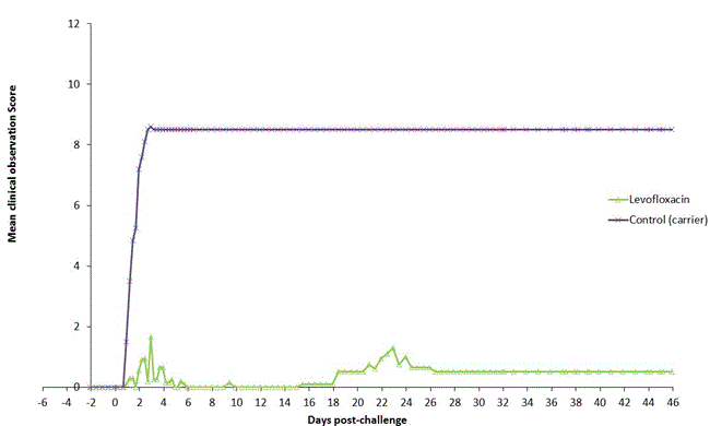
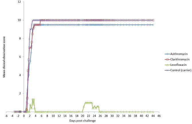
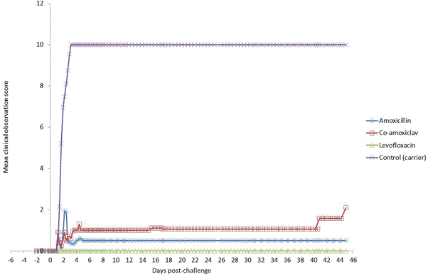
| In all experiments, any mouse showing signs of severe disease (immobility) was immediately humanely euthanized. In animals that did not succumb to disease, clinical signs were largely absent with the exception of two levofloxacin-treated animals (Figures 2 and 3). Control, sham-treated mice generally displayed a rapid onset of clinical signs from 1 day-post-challenge with the appearance of ruffled fur followed by immobility/death/euthanasia with a median time of 1.92 days post-challenge (Figures 2-4). Mice treated with either azithromycin or clarithromycin displayed a near identical disease progression to that of the control mice with rapid onset and escalation of clinical signs leading to immobility/death/euthanasia in a median time of 1.92 and 1.67 days, respectively (Figure 3 and Table 3). Levofloxacin, amoxicillin and co-amoxiclav generally protected mice from disease conferring survival 97%, 95% and 79%, respectively (Table 2). A summary of the protective efficacy of the test antibiotics is presented in Table 2. |
| The presence of a low but detectable level of anthrax persistence was noted in all groups of survivors, irrespective of antibiotic treatment. The probability that the presence of these few colonies was due to cross-contamination from inside the containment isolator during manual enumeration can be discounted due to the fact that the colonies tended only to grow from plates inoculated with most concentrated lung homogenate samples. This is because cross-contamination from within the isolator would have presented randomly across all plates. |
| An increase in antibiotic resistance in these persistent colonies was not detected. The reason for the presence of these persistent anthrax cells in apparently healthy survivors cannot, therefore, be attributed to an increase in antibiotic resistance. It is possible that these colonies represent spores which did not germinate in the lung or they could represent viable partially-germinated anthrax spores which resided in an intracellular or otherwise sheltered location which was protected from the bactericidal concentration of test antibiotics. This interpretation of our data is consistent with the opinion of others [5] and is further supported by the recent observations of Jenkins and Xu [22] that anthrax spores may reside intracellulary, specifically within epithelial cells of the respiratory tract, in BALB/c mice after intranasal inoculation with B. anthracis spores. |
| Serum concentrations of antibiotics |
| The antibiotic concentrations present in the mouse sera at peak and trough times are displayed in Table 3. Levels of azithromycin and clarithromycin were below that expected, even taking into consideration the potential for the development of resistance in Gram positive bacteria to macrolides (including clarithromycin); however, this may have been a result of an additional freeze/thaw cycle of the mouse serum that was required to prove sterility prior to analyses. Confirmation that no antibiotic resistance was incurred during the animal study is confirmed by the results presented in Table 4. The MIC of the B. anthracis isolates recovered from the lungs of surviving mice were the same or very similar to the MIC of the control cell bank; thus indicating that the persistence of these viable anthrax cells was not attributable to the development of increased antibiotic resistance. |
| Conclusions |
| In this set of studies, BALB/c mice were demonstrated to be reproducibly susceptible to aerosol challenge with virulent B. anthracis (Ames) spores. In addition, this BALB/c model was found to be appropriate for the assessment of different antibiotic treatment regimes. Orally delivered levofloxacin, amoxicillin and to a slightly lesser extent, co-amoxiclav, were found to significantly reduce mortality when compared against azithromycin and clarithromycin at the doses evaluated. The presence of a low, but detectable, level of anthrax in some survivors from treatment groups was unexpected and could not be attributed to an increase in antibiotic resistance. |
| Based on the presented data (and corresponding bacterial burden; not presented), levofloxacin, amoxicillin and co-amoxiclav will be evaluated further in a cynomolgus macaque model. The FDA 2002 draft guidance recommends the use of an appropriate macaque model [2] and subsequent guidance and publications support the use of the African green, rhesus or cynomolgus macaque [23,12-16]. For this reason we have characterised the cynomolgus macaque model and will be evaluating the post-exposure efficacy of these three orally administered antibiotics in terms of animal survival, bacterial burden and time course of progression of diseases (including disease manifestations e.g. pathologic features and immune status) ultimately aiming to increase the therapeutic options to support further post-exposure prophylaxis indications for inhalational anthrax. In addition we will investigate if these antibiotics have the ability to prevent systemic replication and dormancy, as this aspect has not been clearly defined to date. |
| Acknowledgement |
| These studies have been funded in whole with Federal funds from the National Institute of Allergy and Infectious Diseases contract number N01-AI-30062. The authors would like to thank all PHE employees from the Biological Investigations Group and bacteriology group who helped support this work. They would also like to acknowledge the work conducted by the Bio-analytical group at HLS. |
| Ethical approval: All animal studies were carried out in accordance with the Scientific Procedures Act of 1986 and the Codes of Practice for the Housing and Care of Animals used in Scientific Procedures 1989. Work was authorised by a Home Office Project Licence. |
References
- Mock M, Fouet A (2001) Anthrax.Annu Rev Microbiol 55: 647-671.
- Food and Drug Administration, HHS (2002) New drug and biological drug products; evidence needed to demonstrate effectiveness of new drugs when human efficacy studies are not ethical or feasible. Final rule.Fed Regist 67: 37988-37998.
- Holty JE, Bravata DM, Liu H, Olshen RA, McDonald KM, et al. (2006) Systematic review: a century of inhalational anthrax cases from 1900 to 2005.Ann Intern Med 144: 270-280.
- Inglesby TV, Henderson DA, Bartlett JG, Ascher MS, Eitzen E, et al. (1999) Anthrax as a biological weapon: medical and public health management. Working Group on Civilian Biodefense.JAMA 281: 1735-1745.
- Hendricks KA, Wright ME, Shadomy SV, Bradley JS, Morrow MG, et al. (2014) Centers for disease control and prevention expert panel meetings on prevention and treatment of anthrax in adults.Emerg Infect Dis 20.
- Steward J, Lever MS, Simpson AJ, Sefton AM, Brooks TJ (2004) Post-exposure prophylaxis of systemic anthrax in mice and treatment with fluoroquinolones.J AntimicrobChemother 54: 95-99.
- Altboum Z, Gozes Y, Barnea A, Pass A, White M, et al. (2002) Postexposure prophylaxis against anthrax: evaluation of various treatment regimens in intranasally infected guinea pigs. Infect Immun 70: 6231-6241
- Heine HS, Bassett J, Miller L, Hartings JM, Ivins BE, et al. (2007) Determination of antibiotic efficacy against Bacillus anthracisin a mouse aerosol challenge model. Antimicrob Agents Ch 51: 1373-1379
- Peterson JW, Moen ST, Healy D, Pawlik JE, Taormina J, et al. (2010) Protection Afforded by Fluoroquinolones in Animal Models of Respiratory Infections with Bacillus anthracis, Yersinia pestis, and Francisellatularensis.Open Microbiol J 4: 34-46.
- Vietri NJ, Purcell BK, Tobery SA, Rasmussen SL, Leffel EK, et al. (2009) A short course of antibiotic treatment is effective in preventing death from experimental inhalational anthrax after discontinuing antibiotics. J Infect Dis 99: 336-341
- Nelson M, Stagg AJ, Stevens DJ, Brown MA, Pearce PC, et al. (2011) Post-exposure therapy of inhalational anthrax in the common marmoset.Int J Antimicrob Agents 38: 60-64.
- Friedlander AM, Welkos SL, Pitt ML, Ezzell JW, Worsham PL, et al. (1993) Postexposure prophylaxis against experimental inhalation anthrax.J Infect Dis 167: 1239-1243.
- Fritz DL, Jaax NK, Lawrence WB, Davis KJ, Pitt ML, et al. (1995) Pathology of experimental inhalation anthrax in the rhesus monkey.Lab Invest 73: 691-702.
- Kao LM, Bush K, Barnewall R, Estep J, Thalacker FW, et al. (2006) Pharmacokinetic considerations and efficacy of levofloxacin in an inhalational anthrax (postexposure) rhesus monkey model.Antimicrob Agents Chemother 50: 3535-3542.
- Vasconcelos D, Barnewall R, Babin M, Hunt R, Estep J, et al. (2003) Pathology of inhalation anthrax in cynomolgus monkeys (Macacafascicularis).Lab Invest 83: 1201-1209.
- Twenhafel NA, Leffel E, Pitt ML (2007) Pathology of inhalational anthrax infection in the african green monkey.Vet Pathol 44: 716-721.
- Karginov VA, Robinson TM, Riemenschneider J, Golding B, Kennedy M, et al. (2004) Treatment of anthrax infection with combination of ciprofloxacin and antibodies to protective antigen of Bacillus anthracis.FEMS Immunol Med Microbiol 40: 71-74.
- Twenhafel NA (2010) Pathology of inhalational anthrax animal models.Vet Pathol 47: 819-830.
- Druett HA (1969) A mobile form of the Henderson apparatus.J Hyg (Lond) 67: 437-448.
- May KR, Harper GJ (1957) The efficiency of various liquid impinger samplers in bacterial aerosols.Br J Ind Med 14: 287-297.
- Guyton AC (1947) Measurement of the respiratory volumes of laboratory animals.Am J Physiol 150: 70-77.
- Jenkins SA, Xu Y (2013) Characterization of Bacillus anthracispersistence in vivo.PLoS One 8: e66177.
- Food and Drug Administration; HHS (2009) Guidance for Industry: Animal Models – Essential Elements to Address Efficacy Under the Animal Rule.
Tables and Figures at a glance
| Table 1 | Table 2 | Table 3 | Table 4 |
Figures at a glance
 |
 |
 |
 |
|||
| Figure 1 | Figure 2 | Figure 3 | Figure 4 |
Relevant Topics
- Advanced Therapies
- Chicken Pox
- Ciprofloxacin
- Colon Infection
- Conjunctivitis
- Herpes Virus
- HIV and AIDS Research
- Human Papilloma Virus
- Infection
- Infection in Blood
- Infections Prevention
- Infectious Diseases in Children
- Influenza
- Liver Diseases
- Respiratory Tract Infections
- T Cell Lymphomatic Virus
- Treatment for Infectious Diseases
- Viral Encephalitis
- Yeast Infection
Recommended Journals
Article Tools
Article Usage
- Total views: 15901
- [From(publication date):
December-2014 - Nov 21, 2024] - Breakdown by view type
- HTML page views : 11481
- PDF downloads : 4420
