Research Article Open Access
Effects of Practicing Difficult Movements of the Unilateral Arm on the Excitability of Spinal Motor Neurons in the Contralateral Arm
Naoki Kado1*, Masanori Ito1, Satoshi Fujiwara1, Yuki Takahashi1, Makoto Nomura2 and Toshiaki Suzuki31Department of Physical Therapy, Kobe College of Rehabilitation and Welfare, Japan
2Department of Rehabilitation, Tanabe Central Hospital, Japan
3Graduate School of Health Sciences, Graduate School of Kansai University of Health Sciences, Japan
- *Corresponding Author:
- Dr. Naoki Kado
Department of Physical Therapy
Kobe College of Rehabilitation and Welfare 1-2-2 Kominatodori
Chuo-ku, Kobe 650-0026,Japan
Tel: +81 78-361-2888
Fax: +81 78-361-2880
E-mail: kado@sumire-academy.ac.jp
Received date: December 20, 2016; Accepted date: January 28, 2017; Published date: February 04, 2017
Citation: Kado N, Ito M, Fujiwara S, Takahashi Y, Nomura M, et al. (2017) Effects of Practicing Difficult Movements of the Unilateral Arm on the Excitability of Spinal Motor Neurons in the Contralateral Arm. J Nov Physiother 7:330. doi: 10.4172/2165-7025.1000330
Copyright: © 2017 Kado N, et al. This is an open-access article distributed under the terms of the Creative Commons Attribution License, which permits unrestricted use, distribution, and reproduction in any medium, provided the original author and source are credited.
Visit for more related articles at Journal of Novel Physiotherapies
Abstract
Background: The excitability of the spinal motor neurons of the contralateral upper limb increases during voluntary movement of the upper limbs. However, reports on the changes in the facilitation effects of the movements of the unilateral upper limb on the spinal motor neurons in the contralateral upper limb that are associated with motor learning are few. Methods: Sixteen right-handed healthy adults were randomly assigned to either the control group or the practice group. The F-waves were derived from the right abductor pollicis brevis muscle during the tasks before and after practice. The tasks included repetitive movements of the left upper limb between two small targets placed 20 cm apart on a desk. The subjects were instructed to accurately touch the targets with the tip of a pen. The practice of the practice group included repetitive movements using the same targets. The practice of the control group included repetitive movements without targets. The practice was performed for five sessions with each session consisting of 30 movements. The F-waves were analyzed for the amplitude ratio of F/M and latency. In addition, the number of times the tip of the pen touched the outside of the target was counted. Results: The amplitude ratio of F/M during post-practice significantly decreased compared with that during prepractice in the practice group. Latency showed no significant differences. The number of failures during post-practice decreased significantly compared with that during pre-practice in the practice group. Conclusion: This study suggests that the facilitation effects of the voluntary movements of the unilateral upper limb that were performed at a high difficulty level on the spinal motor neurons in the contralateral upper limb decreased with motor learning.
Keywords
F-wave; Spinal motor neuron; Motor learning
Introduction
One of the objectives of physical therapy is the recovery of reduced function and the relearning of previously learned movement patterns. In the central nervous system, various plastic changes occur with movement learning. For example, reorganization is observed in the primary motor cortex by practicing complex movements [1,2]. In addition, spinal reflexes are reduced by exercise training that requires accurate movements [3]. In this way, if exercises that require high skills are practiced, the spinal cord receives strong control from the cortex; therefore, the gain of spinal reflexes is estimated to decrease in the spinal cord.
On the other hand, when performing high-difficulty movements, movements of parts that are not directly involved in the movements may be induced involuntarily. Such a phenomenon is rarely observed in the automatization phase of motor learning. The mechanism of the facilitation effect of muscle contraction of the remote part in healthy subjects has been analyzed using the H-reflex, F-waves, and motorevoked potentials (MEPs) evoked by transcranial magnetic stimulation (TMS) [4-9]. Previous studies revealed that voluntary muscle contraction enhances the excitability of the spinal motor neurons and motor areas in the cerebral cortex that are not directly associated with the contracting muscle. In addition, we examined the influence of voluntary movements with various difficulties on the muscles that are not directly involved in the movement and reported that the excitability of the spinal motor neurons of the contralateral upper limb increases during high-difficulty, voluntary upper limb movements [10]. However, reports on the changes in the facilitation effects of the unilateral upper limb movements on the spinal motor neurons in the contralateral upper limb associated with motor learning are few. Thus, the aim of the present study was to examine the changes in the excitabiljity of the spinal motor neurons in the contralateral upper limb caused by practicing high-difficulty movements of the unilateral upper limb using the F-waves in evoked electromyography (EMG). The Fwaves originate from the retrograde excitations of the α motor neurons through the stimulation of peripheral motor nerve axons and are used as an indicator of excitability of spinal motor neuron pools [11]. We hypothesized that practicing difficult movements reduces the promoting effect on the spinal motor neurons in the contralateral upper limb.
Subjects and Methods
Participants
Sixteen right-handed healthy adults (12 men and 4 women; mean age, 26.1 ± 6.0 years) with no orthopedic or neurological abnormalities participated in this study. They were randomly assigned equally to either a control group (6 men and 2 women; mean age, 26.4 ± 7.2 years) or a practice group (6 men and 2 women; mean age, 26.0 ± 4.9 years). The Edinburgh handedness inventory [12] was used to determine their dominant hands.
In addition to explanations of the objectives of this study, the subjects were informed that the test data would be strictly confidential and that they could withdraw from the study at any time during the course of the study. The subjects’ signatures on the study consent forms were obtained once they had agreed to participate. This study was conducted with the approval of the ethics committee of Kobe College of Rehabilitation.
Procedure
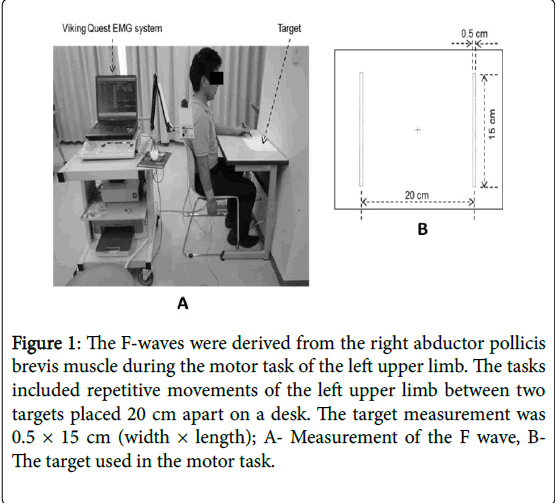
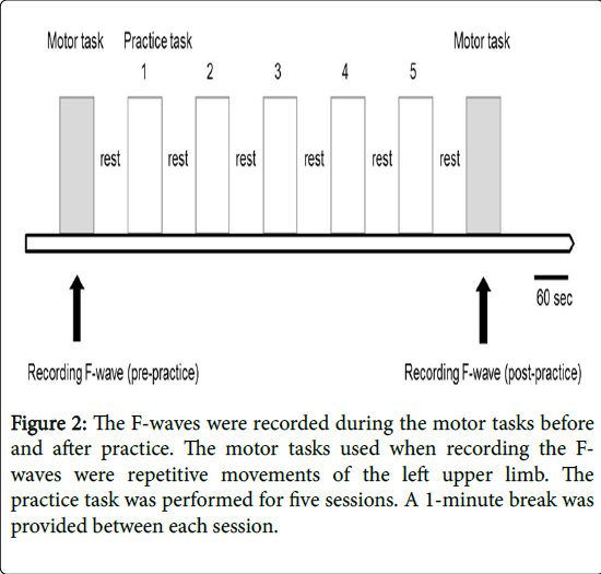
The F-waves were derived from the right abductor pollicis brevis muscle during the motor tasks of the left upper limb before and after the practice task using the Viking Quest EMG system (Nicolet Biomedical, WI, USA; Figures 1 and 2). The subjects were seated on a chair during the test, with the bilateral hip and knee joints flexed at 90° and the bilateral ankle, right shoulder, right elbow, and right hand joints at 0°. The subjects were instructed not to move body parts other than the left arm throughout the study.
Figure 1: The F-waves were derived from the right abductor pollicis brevis muscle during the motor task of the left upper limb. The tasks included repetitive movements of the left upper limb between two targets placed 20 cm apart on a desk. The target measurement was 0.5 × 15 cm (width × length); A- Measurement of the F wave, BThe target used in the motor task.
The motor tasks used when recording the F-waves were repetitive movements of the left upper limb to the left and right. The subjects were asked to hold a pen with their left hand and to accurately place its tip in contact within each of the two targets placed laterally 20 cm apart on the desk. This movement was performed 30 times in a reciprocating lateral movement to reach either the left or the right target in time with an auditory sound of a metronome at a frequency of 1 Hz. The index of task difficulty was defined by the distance to the target and the width of the target [13]. The target width used in the present study was 0.5 × 15 cm (width × length), which showed facilitation effects on the spinal nerve function in the contralateral upper limb in the previous study [10]. In addition, the number of times the tip of the pen touched the outside of the target was counted.
The practice task included repetitive movements at a frequency of 1 Hz. The practice group performed repetitive movements using the same targets when recording the F-wave, and the control group performed repetitive movements without the targets. The practice task was performed for five sessions with each session consisting of 30 movements. A 1-minute break was provided between each session.
The stimulation conditions for F-wave elicitation were 30 consecutive stimulations of the median nerve at the right wrist, with an intensity 120% of the stimulation intensity required to evoke the maximum M-wave, frequency of 0.5 Hz, and duration of 0.2 ms. The bipolar surface stimulating electrodes used in general motor nerve conduction velocity tests were used as the stimulating electrodes. In addition, electrical stimulation for F-wave elicitation was given when the upper limb was moving toward the left target. For the recording conditions, the exploring electrode was placed over the belly of the right abductor pollicis brevis muscle; the reference electrode, on the proximal phalanx of the thumb; and the ground electrode, on the forearm. The electrode sites were first rubbed with a cotton pad moistened with alcohol to remove oil, and the horny layer of the skin was removed with a skin preparation gel. Silver-silver chloride disk electrodes 10 mm in diameter were fixed using conductive pastes on the skin surfaces. Bioelectrical signals obtained at the electrodes were conducted to the input via lead wires, amplified with a bioamplifier, converted from analog signals to digital signals with an AD converter, and displayed on a personal computer as waveforms. All the signals were digitized at a sampling frequency of 24 kHz and recorded on the hard disk. The data were band-pass filtered between 20 Hz and 3 kHz; the amplitude sensitivity was 200 μV/div; and the sweep speed was 5 ms/div.
Data analysis
The F-waves were analyzed for the amplitude ratio of F/M and latency. The amplitude ratio of F/M was calculated as the ratio of the average peak-to-peak F-wave amplitude and the maximum M-wave amplitude. This parameter represents the percentage of motoneurons activated by the antidromic stimulation [14]. Furthermore, the amplitude of the averaged F-responses at rest in healthy subjects is about 1% of the M-wave amplitude [15]. Latency was the mean time from the stimulus pulse to the F-wave onset, which measures the conduction in the motor axons [14].
Statistical analysis
The normality of the F-wave data and the number of failures were confirmed using the Shapiro-Wilk tests. As the normality was not confirmed, the Mann-Whitney test was used to compare the F-wave parameters (amplitude F/M ratio and latency) and the number of failures between the control and practice groups. The Wilcoxon signedrank sum test was used to compare the F-wave parameters and the number of pre- and post-practice failures. P values of < 0.05 were considered statistically significant. Statistical analyses were performed using the SPSS version 19 (SPSS Inc., Chicago, IL, USA).
Results
The F-wave parameters and the number of failures are shown in Table 1. The amplitude ratio of F/M during post-practice in the practice group significantly decreased from the pre-practice value in the practice group (p = 0.025). In addition, the post-practice values in the practice group were significantly lower than those in the control group (p = 0.012).
| Control group | Practice group | |||
| Pre | Post | Pre | Post | |
| Amplitude ratio of F/M (%) | 1.51 ± 0.47 | 1.72 ± 0.43 | 1.43 ± 0.51 | 1.12 ± 0.26 * |
| Latency (ms) | 25.6 ± 1.7 | 25.6 ± 1.7 | 26.4 ± 1.4 | 26.3 ± 1.5 |
| Number of failures (times) | 8.4 ± 5.6 | 7.1 ± 5.5 | 8.8 ± 3.5 | 3.8 ± 4.2 * |
| Data are presented as mean ± SDs. *: p < 0.05 | ||||
Table 1: F-wave parameters and number of failures in the control and practice groups.
Latency showed no significant difference between pre- and postpractice in both groups.
The number of failures during post-practice in the practice group decreased significantly from the pre-practice number in the practice group (p = 0.012).
Discussion
In the practice group, the amplitude ratio of F/M during postpractice decreased significantly compared with that during prepractice. Moreover, the number of failures during post-practice decreased significantly from the pre-practice number in the practice group. Thus, we suggest that the facilitation effects on the spinal motor neurons in the contralateral upper limb in the practice tasks of highdifficulty unilateral upper limb movements can be reduced by practicing the movements.
The influence of the muscle spindle activity involved in movements has been reported to be the mechanism of the facilitation effects due to the contraction of the remote muscles, which are not directly related to the test muscles, such as synergists and antagonists [16,17]. On the other hand, Hess et al. [18] reported the involvement of the facilitation effects of the intracortical mechanism because the MEPs by the contralateral TMS in the amputated patient with a phantom limb increased by imagining the muscle contraction of the amputated limb. Thus, facilitation effects of the input of proprioceptive sense and the upper central nervous system associated with voluntary movements of the upper limb were considered as the factors of excitability of the spinal motor neurons in the contralateral upper limb that increase during movements of the unilateral upper limb.
In this study, the difficulty level of the motor task was set high using the target width of 0.5 cm. The previous study confirmed that the task where the targets of 0.5 × 15 cm (width × length) were used appeared to be more difficult than the task where the targets of 5 × 15 cm were used, considering that the success rate of the latter task was 100% and that of the former task was 83% [10]. Shibasaki et al. [19] reported that not only the contralateral sensorimotor cortex but also the ipsilateral sensorimotor area was activated in the execution of complex sequential finger movements. Winstein et al. [20] examined the relationship between task difficulty and brain activity and reported that activities in areas related to complex movement planning requiring visual motion processing, including the ipsilateral dorsal premotor area, increase as the task difficulty increases. In addition, the excitability of the spinal motor neurons in the contralateral upper limb increased during the difficult task where targets of 0.5 × 15 cm were used when compared with that during the easy task where targets of 5 × 15 cm were used [10]. Thus, the excitability of the spinal motor neurons in the contralateral upper limb is increased by enhancing the facilitation effect of the ipsilateral motor-related areas during high-difficulty unilateral upper limb movements.
Motor learning is considered dependent on the plasticity in the motor and sensory areas of the brain. Therefore, the facilitation effects with movements of the unilateral upper limb on the spinal motor neurons in the contralateral upper limb can be reduced with motor learning. Suzuki et al. [21] examined the changes in learning the task of rotating two balls with the hands using MEP induced by TMS and reported that the excitability of the primary motor cortex ipsilateral to the movements was reduced with an enhanced performance. Winstein et al. [20] examined the relationship between the efficiency of motor tasks and brain activity and reported that the localization of movement-related areas occurs when exercise efficiency increases. In addition, Nelson et al. [22] recorded the somatosensory-evoked potentials (SEP) during the motor tasks, such as adjusting the angle of the joint to the correct position, and reported that the input of sensory information to the cerebrum in the central nervous system is reduced when motor tasks are acquired by motor learning; this was because the short latency SEP amplitude decreases with acquisition of the tasks. The present study considered that the facilitation effects of the sensory input and the upper central nervous system associated with voluntary movements of the upper limb on the spinal motor neurons in the contralateral upper limb decrease with the acquisition of tasks through practice.
Conclusion
The present study suggests that the facilitation effects of voluntary movements of the unilateral upper limb that were performed at a high difficulty level on the spinal motor neurons in the contralateral upper limb decrease with motor learning. When performing physiotherapy, understanding the influence of voluntary movements of the unilateral upper limb on the spinal motor neurons in the contralateral upper limb is important. This facilitation effect may be a factor impeding accurate movements. For example, association reactions observed in hemiplegic patients with cerebrovascular disorders increase the muscle tone of the limbs not related to the movement. Selective movement is restricted when muscle tone is increased. Therefore, practicing efficient implementation of difficult movements is necessary. The limitation of this study is that there is a lack of consideration on sex difference and lateral difference. Detailed analyses on these issues will be necessary in the future.
Acknowledgment
None declared.
References
- Karni A, Meyer G, Jezzard P, Adams MM, Turner R, et al. (1995) Functional MRI evidence for adult motor cortex plasticity during motor skill learning. Nature 377: 155-158.
- Pascual-Leone A, Nguyet D, Cohen LG, Brasil-Neto JP, Cammarota A, et al. (1995) Modulation of muscle responses evoked by transcranial magnetic stimulation during the acquisition of new fine motor skills. J Neurophysiol 74: 1037-1045.
- Nielsen J, Crone C, Hultborn H (1993) H-reflexes are smaller in dancers from The Royal Danish Ballet than in well-trained athletes. Eur J ApplPhysiolOccupPhysiol 66: 116-121.
- Bussel B, Morin C, Pierrot-Deseilligny E (1978) Mechanism of monosynaptic reflex reinforcement during Jendrassik maneuver in man. J NeurolNeurosurg Psychiatry 41: 40-44.
- Kawamura T, Watanabe S (1975) Timing as a prominent factor of the Jendrassikmanoeuvre on the H reflex. J NeurolNeurosurg Psychiatry 38: 508-516.
- Boroojerdi B, Battaglia F, Muellbacher W, Cohen LG (2000) Voluntary teeth clenching facilitates human motor system excitability. ClinNeurophysiol 111: 988-993.
- Sugawara K, Kasai T (2001) Facilitation of motor evoked potential and H-reflexes of flexor carpi radialis muscle induced by voluntary teeth clenching. Hum MovSci 21: 203-212.
- Muellbacher W, Facchini S, Boroojerdi B, Hallett M (2000) Changes in motor cortex excitability during ipsilateral hand muscle activation in humans. ClinNeurophysiol 111: 344-349.
- Stinear CM, Walker KS, Byblow WD (2001) Symmetric facilitation between motor cortices during contraction of ipsilateral hand muscles. Exp Brain Res 139: 101-105.
- Kado N, Ito M, Suzuki T, Ando H (2012) Excitability of spinal motor neurons in the contralateral arm during voluntary arm movements of various difficulty levels. J PhysTherSci 24: 949-952.
- Kimura J (2001) Electrodiagnosis in diseases of nerves and muscles: principles and practice, FA Davis Company, Philadelphia.
- Oldfield RC (1971) The assessment and analysis of handedness: the Edinburgh inventory. Neuropsychologia 9: 97-113.
- Fitts PM (1954) The information capacity of the human motor system in controlling the amplitude of movement. J ExpPsychol 47: 381-391.
- Mesrati F, Vecchierini MF (2004) F-waves: neurophysiology and clinical value.NeurophysiolClin 34: 217-243.
- Eisen A, Odusote K (1979) Amplitude of the F wave: a potential means of documenting spasticity. Neurology 29: 1306-1309.
- Hayashi A, Konopacki RA, Hunker CJ (1992) Remote facilitation of H-reflex during voluntary contraction of orofacial and limb muscles. In: Stelmach GE, Requin J (eds) Tutorials in motor behavior II. Elsevier Science Publishers BV, Amsterdam.
- Delwaide PJ, Toulouse P (1981) Facilitation of monosynaptic reflexes by voluntary contraction of muscle in remote parts of the body. Mechanisms involved in the JendrassikManoeuvre. Brain 104: 701-709.
- Hess CW, Mills KR, Murray NMF (1986) Magnetic stimulation of the human brain: facilitation of motor responses by voluntary contraction of ipsilateral and contralateral muscles with additional observations on an amputee. NeurosciLett 71: 235-240.
- Shibasaki H, Sadato N, Lyshkow H, Yonekura Y, Honda M, et al. (1993) Both primary motor cortex and supplementary motor area play an important role in complex finger movement. Brain 116: 1387-1398.
- Winstein CJ, Grafton ST, Pohl PS (1997) Motor task difficulty and brain activity: investigation of goal-directed reciprocal aiming using positron emission tomography. J Neurophysiol 77: 1581-1594.
- Suzuki T, Higashi T, Takagi M, Sugawara K (2013) Hemispheric asymmetry of ipsilateral motor cortex activation in motor skill learning. Neuroreport 24: 693-697.
- Nelson AJ, Brooke JD, McIlroy WE, Bishop DC, Norrie RG (2001) The gain of initial somatosensory evoked potentials alters with practice of an accurate motor task. Brain Res 890: 272-279.
Relevant Topics
- Electrical stimulation
- High Intensity Exercise
- Muscle Movements
- Musculoskeletal Physical Therapy
- Musculoskeletal Physiotherapy
- Neurophysiotherapy
- Neuroplasticity
- Neuropsychiatric drugs
- Physical Activity
- Physical Fitness
- Physical Medicine
- Physical Therapy
- Precision Rehabilitation
- Scapular Mobilization
- Sleep Disorders
- Sports and Physical Activity
- Sports Physical Therapy
Recommended Journals
Article Tools
Article Usage
- Total views: 3103
- [From(publication date):
February-2017 - Apr 02, 2025] - Breakdown by view type
- HTML page views : 2291
- PDF downloads : 812


