Case Report Open Access
Effects of a Prefabricated Functional Appliance in the Early Mixed Dentition Period
Iwata T*Graduate School of Dentistry, Kanagawa Dental University, Yokosuka-city, Kanagawa Prefecture, Japan
- *Corresponding Author:
- Iwata T
Doctor of Philosophy (Dentistry, Orthodontics), Graduate School of Dentistry
Kanagawa Dental University, Yokosuka-city, Kanagawa Prefecture, Japan
Tel: +81-46-825-8885
E-mail: t.iwata@kdu.ac.jp
Received: February 23, 2016; Accepted: March 16, 2016; Published: March 22, 2016
Citation: Iwata T (2016) Effects of a Prefabricated Functional Appliance in the Early Mixed Dentition Period. Pediatr Dent Care 1:104. doi:10.4172/pdc.1000104
Copyright: © 2016 Iwata T. This is an open-access article distributed under the terms of the Creative Commons Attribution License, which permits unrestricted use, distribution, and reproduction in any medium, provided the original author and source are credited.
Visit for more related articles at Neonatal and Pediatric Medicine
Abstract
Objective: We present a case in which a prefabricated functional appliance was used for two years, and evaluate its mechanism of action, indications for treatment, and a case requiring attention. Cases: The patient was a 9-year 9-month-old boy. This patient showed skeletal maxillary protrusion with excessive over jet and overbite and crowding in the maxilla-mandibular anterior tooth area. Clinical examination and results: This patient showed correction of over jet, overbite, molar relationship, cephalographic ∠ ANB, and maxillary anterior tooth axis. However, this patient presented slight correction of ∠ SNB due to mandibular clockwise rotation. Furthermore, marked labial tipping movement of the mandibular anterior tooth axis was noted. Conclusion: Regarding this appliance, greater effects can be achieved through understanding the disadvantages of the appliance and its application based on appropriate diagnosis of treatment cases.
Keywords
Orthodontic treatment; Prefabricated functional appliance; Early mixed dentition; Eruption guidance
Introduction
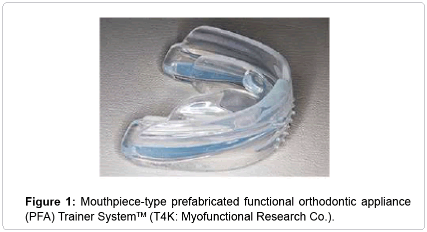
We reported the use of a mouthpiece-type prefabricated functional orthodontic appliance (PFA) (Figure 1) for about 6 months for the alignment of incisors and bite-opening and lingual inclination of maxillary incisors [1]. The PFA was effective for a prognathous case with anterior crowding and a deep overbite.
The effects of this appliance are considered to be not only functional correction [2,3], but also lateral expansion of the maxillo-mandibular dentition, correction of crowding of the anterior teeth, and facilitation of growth of the mandibular bone [2-9].
However, labial tipping of the mandibular anterior teeth and clockwise rotation of the mandible produced these changes at the same time, and therefore it was thought important to consider the possibility of a lack of stability of the anterior incisor and skeletal vertical changes for high angle cases with a steep mandibular plane.
The PFA is a functional appliance for improving malocclusion, correcting function for a patient by moving the teeth and the mandibular position at the same time.
In this case report, we evaluated patient who had used PFA in their deciduous and mixed dentition periods for two years to understand growth control of the maxillary-mandibular dentition, which was one of the effects of the functional appliance.
Case Report
Diagnosis and etiology
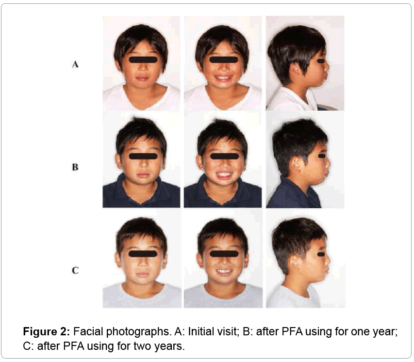
At the initial examination, a boy, aged 9 years 9 months, had a chief complaint of anterior crowding. His face was symmetric from the frontal view and his smile line was good, but his profile was convex because of upper lip protrusion and mandibular retrognathism. Lip incompetence was observed and the perioral muscles were strained on lip closure (Figure 2).
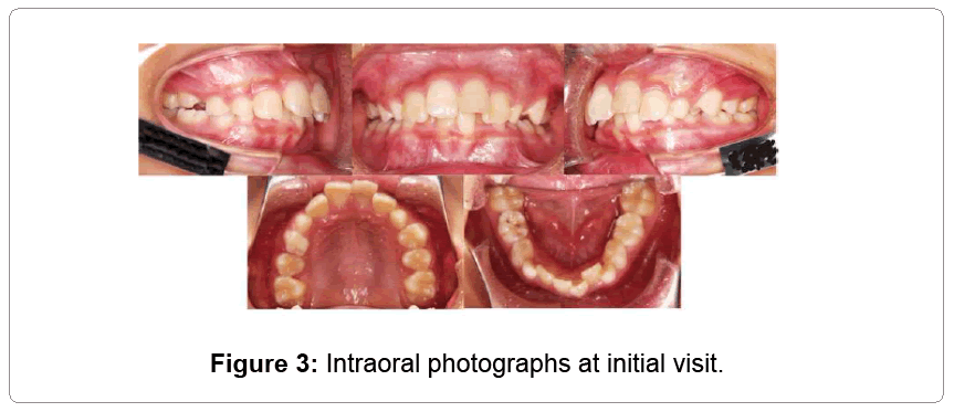
Intraoral findings included a 7.0 mm over jet and a 5.0 mm overbite. The molar relationship was Angle Class II. The maxillary and mandibular dental arches were narrow, and marked crowding was observed in the anterior regions. The arch length discrepancies (ALD) were -3.4 mm in the maxilla and -3.8 mm in the mandible (Figure 3).
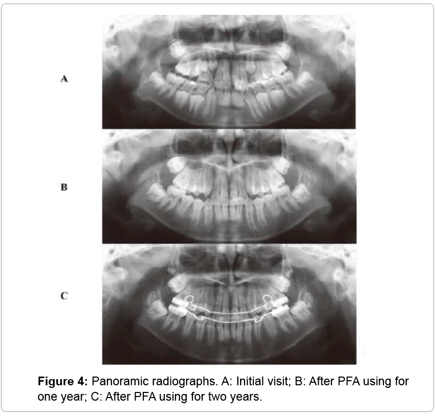
Panoramic radiography did not show tooth germs of the bilateral maxillary third molars (Figure 4). A lateral cephalometric radiograph showed an SNA angle of 84.2°, an SNB angle of 77.5°, and an ANB angle of 6.7°, producing a skeletal Class II relationship due to maxillary protrusion and mandibular retrognathism. In addition, the gonial angle was 123.9°, the ramus inclination was 86.9°, and the Frankfort-mandibular plane angle (FMA) was 24.9°, showing a standard angle. Dentally, the maxillary central incisor to the SN plane (SN) was 114.6°, the incisor mandibular plane angle (IMPA) was 95.6.0°, the Frankfort mandibular-incisor angle (FMIA) was 59.5°, and the interincisal angle was 119.1°. Labial inclination of the maxillary central incisors was observed (Table 1).
| Measurement | Initial visit | After 1 year | After 2 year |
|---|---|---|---|
| overjet | +7.0 | +3.0 | +3.0 |
| overbite | +5.0 | +3.0 | +2.0 |
| SNA | 84.2 | 83.8 | 83.8 |
| SNB | 77.5 | 77.8 | 78.3 |
| ANB | +6.7 | +6.0 | +5.5 |
| FMA | 24.9 | 27.7 | 26.2 |
| FMIA | 59.5 | 52.4 | 55.8 |
| IMPA | 95.6 | 99.9 | 98.0 |
| Y-axis | 64.2 | 65.8 | 64.5 |
| Gonial ang. | 123.9 | 123.0 | 121.2 |
| Ramusu (SN) | 86.9 | 89.1 | 91.0 |
| U-1 to SN | 114.6 | 106.4 | 105.0 |
| Occl.pl. | 12.6 | 15.2 | 10.1 |
| Interincisal | 119.1 | 121.5 | 124.9 |
Table 1: Cephalometoric analysis.
The patient was diagnosed with maxillary protrusion and dental crowding and deep-bite.
Treatment objectives
1) In the mandible, maximize anterior growth with PFA.
2) In the maxilla, minimize anterior growth with PFA.
3) In the mandible, maintain or reduce the anterior vertical dimension.
4) In the maxillary and mandibular dentition, maintain the intermolar and intercanine widths, expand the canine width with PFA, and slightly decrease the molar width during extraction space closure with an edgewise appliance.
5) Improve the facial esthetics by reducing lip protrusion and mentalis strain.
6) As for the patient, treatment effects will be re-examined at a Hellman dental age of IIIC.
Treatment progress
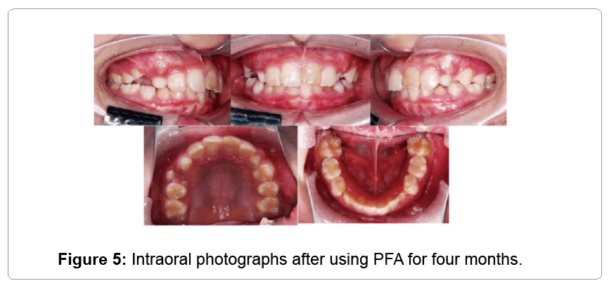
After three months using the PFA, correction of the anterior crowding of maxillary and mandibular dentition, and an effect of bite jumping were seen. The over jet was reduced to +4.5 mm and the overbite to +4.0 mm by the lingual inclination of the maxillary anterior teeth after use of the PFA for 4 months, and the crowding of the maxilla and mandible anterior teeth was almost corrected. The scissors bite was corrected at the upper first premolar and the lower first deciduous molar (Figure 5). The patient was diagnosed again after using the PFA for one year.
The frontal view and smile line were good, but the profile was convex, and upper lip protrusion was still observed. However, mandibular retrognathism and lip incompetence were corrected slightly (Figure 1).
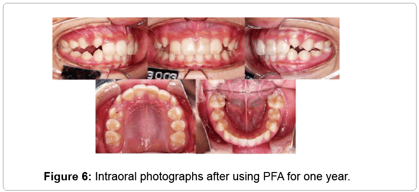
Intraoral findings showed that the over jet was reduced to +3.0 mm and the overbite to +3.0 mm by the lingual inclination of the maxillary anterior teeth after use of PFA for 1 year. The crowding of the maxilla and mandible anterior teeth was corrected.
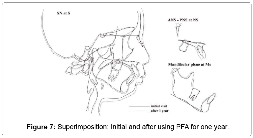
The molar relationship was Angle Class I. The maxillary and mandibular dental arches were expanded. All the deciduous teeth had been replaced by permanent teeth, and most permanent teeth had erupted at the appropriate position, but a lack of eruption space for the right and left maxillary canines was predicted (Figure 6). A panoramic radiograph showed good parallelism of the dental roots. Abnormal findings such as root resorption were not observed and showed tooth germs of the bilateral maxillary third molars (Figure 4). The lateral cephalometric radiograph showed improvement in SNA from 84.2° to 83.8°, SNB from 77.5° to 77.8°, and ANB from +6.7° to 6.0°. Growth of the mandible was slightly forward, but the ramus inclination was increased from 86.9° to 89.1°, FMA was increased from 24.9° to 27.7°, and clockwise mandibular rotation was observed (Figure 7 and Table 1). Dentally, the maxillary incisor to the SN plane (SN) was decreased from 114.6° to 106.4°, IMPA was increased from 95.6° to 99.9°, and FMIA was 52.4°, and the interincisal angle was 119.1°. The angle of tooth axis inclination was normal for the maxillary central incisors, but the mandibular central incisors had a labial inclination.
The second-year treatment plan
Increase of the lingual inclination of the maxillary central incisors should be prevented, and alignment space for the maxillary canine must be created. The relationship between the maxilla and mandible should be corrected by performing mandibular bone front growth stimulation.
To this end, a lingual arch with U-loop (LA) was worn on both the maxilla and mandible in addition to using PFA sequentially.
Treatment progress (after using PFA for 2 years)
Use of LA in the maxilla and in the mandible was initiated 14 and 17 months, respectively, after commencing PFA treatment. As the bone expanded anteriorly and/or laterally by LA, the PFA was changed for a hard type, and its use was continued.
The alignment of the permanent teeth except the second molar was completed approximately 22 months after the initiation of PFA use. PFA used was continued for the purpose of mandibular growth stimulation effect.
Treatment results
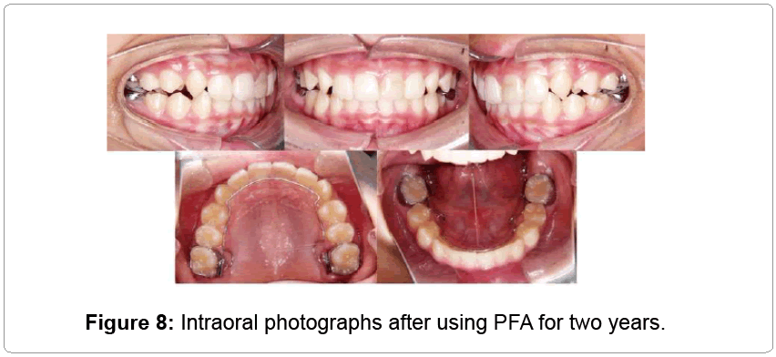
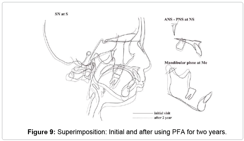
Upper lip protrusion and mandibular retrognathism were improved and perioral muscle tension disappeared (Figure 2). The over jet was reduced to 3.0 mm and the overbite to 2.0 mm. The molar relationship was Class I (Figure 8). The lateral cephalometric radiograph showed improvement in SNA to 83.8°, SNB to 78.3°, and ANB to 5.5°. Growth of the mandible was forward. In addition, the gonial angle was 121.2°, the ramus inclination was 91.0°, FMA was 26.2°, and mandibular counterclockwise rotation was not observed (Figure 9). Dentally, the maxillary incisor to the SN plane (SN) was 105.0°, IMPA was 98.0°, FMIA was 55.8°, and the interincisal angle was 124.9°. The angle of tooth axis inclination was normal for the maxillary central incisors, but the mandibular central incisors had a labial inclination.
Discussion
The PFA is a functional orthodontic appliance, and can be used to control tooth movement and maxillomandibular growth.
The appliance comprises integrated maxillomandibular mouthpieces. For the maxillomandibular antero-posterior occlusion in this patient, the PFA produced an edge-to-edge bite, which is a construction bite taken at the time of use of functional orthodontic appliances. The vertical bite was raised by approximately 3.5 mm in the most distal area, 4 mm in the molar area, 3 mm in the canine area, and 2 mm in the anterior tooth area.
Furthermore, to facilitate training for an appropriate tongue position, a tongue tag was fitted at the spot regarded as an appropriate position of the tongue apex. Because an appropriate inclination is established in the tongue guard to prevent tongue thrust, the antero-lateral expansion of the dentition can be conducted by occlusal force. In addition, a projection termed a ‘lip bumper’ was added in this PFA to suppress the activity of the lower lip and mental is muscle.
This patient demonstrated mouth breathing and a tongue thrusting habit at the initial examination, and his face photographs showed thickening and drying of the top and bottom lips. Strain of the lips neighborhood line was observed at the lips closedown. These views were corrected after two years of using the PFA.
Using this PFA, the patient could achieve a corrected occlusion from an early stage, because of the relationship between dentition and maxillo-mandibular correction. We believe that this encouraged the patient and motivated him to continue using the appliances. If the use time for this appliance were longer, we could expect a synergy in which the occlusion and function were corrected simultaneously.
1. Short-term effect (using PFA for one year)
McNamara [10] said that the labial wire did not touch the labial side of the maxillary anterior teeth when the bionator, a functional appliance, was applied for a prognathous case, and preparation of the occlusal surface on this appliance was not performed after wearing.
The mandible is advanced for maxillary protrusion cases because this PFA is made in a contraction bite position, like a functional appliance. Therefore, this PFA will apply an orthodontic force to the lingual side for the maxillary anterior teeth and to the labial side for the mandibular anterior teeth by moving the mandible backward.
In the present patient, the upper and lower anterior teeth were corrected after use of the PFA for four months. The maxillary incisor to the SN plane (SN) changed from 114.6° at initial examination to 106.4° after using the PFA for 1 year, and IMPA changed from 95.6° to 99.9°.
We thought that the movement of these anterior teeth was due to the use of this appliance from an early stage, because the tipping movement predominated.
However, the upper and lower anterior teeth came into contact by these fast reactions after one year the use of the PFA. We considered that mandibular growth stimulation was likely to be disturbed by these contacts, and therefore it was important to examine the timing for use of the LA in order to prevent excessive lingual tipping of the maxillary anterior teeth.
Furthermore, the effects of this PFA included lateral expansion of the dental arch [9]. In a simulation experiment that we conducted [1] using a typodont model, the mandibular dentition was expanded particularly well in the lateral plane.
The ALD of the mandibular arch was -3.8 mm at the time of the initial examination, and the mandibular dentition was well aligned after using the PFA for one year. We thought that space was acquired because the mandibular dentition was extended laterally in addition to the labial inclination of the mandibular anterior teeth. However, this lateral expansion caused clockwise rotation of the mandible. We considered that the risk of mandibular clockwise rotation was small because the FMA at the time of the initial examination was 24.9 degrees, i.e., a standard vertical relation of maxilla-mandible in this case. However, the FMA increased to 27.7 degrees one year later, and the mandible had turned 2.8 degrees clock-wise.
The mandibular growth stimulation was not effective at one year after use of PFA for the clockwise rotation of the mandible and the expansion of the dental arch, so a SNB increase of 0.3 degrees was seen.
2. Long-term effect (after using PFA for 2 years)
The labial tipping of the mandibular anterior teeth and the clockwise rotation of the mandible were regarded as a problem after one year of using the PFA, and continuation of PFA was considered. The usage of T4K was continued because of functional improvements, such as the extended time of lip closure and the disappearance of tongue protrusion during the treatment. Use of a LA was also initiated, to correct the maxilla-mandibular arch coordinates and to prevent excessive maxillary anterior teeth lingual tipping.
As a result, the maxillary incisor to the SN plane (SN) was changed from 106.4° at 1 year after use of PFA to 105.9° after 2 years, while IMPA changed from 99.9° to 98.0° in the second year. We supposed that this effect was accomplished by tongue pressure having been removed for the mandibular incisor because the lingual tipping of these incisors was corrected in addition to functional correction of the mandibular position.
As for the effect of PFA on SNA, although SNA was decreased 0.4° at one year, there was no the change during the second year.
Burhan et al. [11] reported that the Bite Jumping Appliance (BJA), which is one type of functional orthodontic appliance, has effects of maxillary growth restraint and mandibular growth stimulation. We supposed that a maxillary front growth suppressant effect could similarly be expected with this PFA. However, there were a few decreases in SNA after use of the PFA for 2 years.
Regarding this effect, based on superimposition of the initial and two-year examinations, the roots of the maxillary incisors appeared in the labial side for root labial torque, and we thought that this was because the part of the A point moved forward.
Root [12] proposed an ‘effective A point’ setting for the position of the A point by the maxillary incisor axis. In the present patient, the LA was worn on the maxillary arch after use of the PFA for 14 months, and the LA prevented lingual tipping of the maxillary incisor crowns. As the resulting orthodontic force to the lingual side acted on the upper incisor margin sequentially by this PFA, we thought that the orthodontic force of the root labial torque acted on the maxillary incisor.
As for the mandibular growth, SNB increased from 77.5° at initial examination to 78.3° after use of the PFA for two years. The explanation for this effect is similar to the reason why the mandibular incisal axis corrected; the mandibular growth was changed forward by the functional correction, which was lip and tongue posture.
Summary
The main effects of this PFA were:
1) Labial tipping movement of the mandibular anterior teeth.
2) Lingual tipping movement of the maxillary anterior teeth.
3) Lateral expansion of the maxillo-mandibular dentition. Adjustment of the tooth axis was achieved in that, by the tipping movement, the labially-inclined maxillary anterior tooth axis became lingually inclined, and the lingually-inclined tooth axis was labially inclined.
Furthermore, we consider that, although effects of the functional orthodontic appliance on the facilitation of mandibular bone growth were noted, they were not marked. In particular, because the effects on the lateral expansion of the maxillo-mandibular dentition and labial tipping movement of the mandibular anterior teeth appeared at an early stage, the mandibular bone showed a clockwise rotation, and the mandibular bone showed a clockwise rotation particularly in high angle cases, and because, owing to the labial tipping movement of the mandibular anterior teeth, they came into contact with the maxillary anterior teeth, the mandible was not readily induced in the anterior direction.
We consider that greater effects can be achieved with this PFA through better understanding of these disadvantages under an appropriate diagnosis of applicable cases.
References
- Iwata T, Kawata T (2014) The effects of a prefabricated functional appliance in early mixed dentition period. Jap J Clin Dent Children 20: 11-20.
- Iwata T, Chieko Mino, Kawata T (2016) The effects and requiring attention of a prefabricated functional appliance. Jap J Clin Dent Children 21: 47-54.
- Quadrelli C, Gheorgiu M, Marchetti C, Ghiglione V (2002) Approcciomiofunzionaleprecocenelle II Classischeletriche. Mondo Orthod2: 109-121.
- Serdar U, Tancan U, Zafer S (2004) The Effects of Early Preorthodontic Trainer Treatment on Class II, Division 1 Patients. Angle Orthod 74: 605-609.
- Masuko M, Kanao A, Kanao Y (2009)Inflection of applied TRAINER SYSTEM of the functional appliance in the mandibular retro-position. Jap J Clin Dent Children 14: 45-62.
- Tripathi NB, Patil SN (2011) Treatment of class II division 1 malocclusion with myofunctional trainer system in early mixed dentition period. J Contemp Dent Pract 12: 497-500.
- Ramirez-Yañez GO, Faria P (2008) Early treatment of a Class II, division 2 malocclusion with the Trainer for Kids (T4K): a case report. J ClinPediatr Dent 32: 325-329.
- Pujar P, Pai SM (2013) Effect of preorthodontic trainer in mixed dentition. Case Rep Dent 2013: 717435.
- Ramirez-Yañez G, Sidlauskas A, Junior E, Fluter J (2007) Dimensional changes in dental arches after treatment with a prefabricated functional appliance. J ClinPediatr Dent 31: 279-283.
- McNamara JA, Bruno (1993) Orthodontic and orthopedic treatment in the mixed dentition. Needham Press Inc. American Journal of Orthodontics and Dentofacial Orthopedics104: 206-207.
- Burhan AS, Nawaya FR (2015) Dentoskeletal effects of the Bite-Jumping Appliance and the Twin-Block Appliance in the treatment of skeletal Class II malocclusion: a randomized controlled trial. Eur J Orthod 37: 330-337.
- Root TL (1981) The level anchorage system for correction of orthodontic malocclusions. Am J Orthod 80: 395-410.
Relevant Topics
- About the Journal
- Birth Complications
- Breastfeeding
- Bronchopulmonary Dysplasia
- Feeding Disorders
- Gestational diabetes
- Neonatal Anemia
- Neonatal Breastfeeding
- Neonatal Care
- Neonatal Disease
- Neonatal Drugs
- Neonatal Health
- Neonatal Infections
- Neonatal Intensive Care
- Neonatal Seizure
- Neonatal Sepsis
- Neonatal Stroke
- Newborn Jaundice
- Newborns Screening
- Premature Infants
- Sepsis in Neonatal
- Vaccines and Immunity for Newborns
Recommended Journals
Article Tools
Article Usage
- Total views: 13049
- [From(publication date):
specialissue-2016 - Apr 04, 2025] - Breakdown by view type
- HTML page views : 11971
- PDF downloads : 1078

