Research Article Open Access
Effect of Fluoride Application Combined with CO2 Laser Irradiation on the Demineralization/Remineralization of Root Surfaces
Koichi Shinkai*, Satoki Kawashima, Masaya Suzuki and Shiro SuzukiDepartment of Operative Dentistry, The Nippon Dental University School of Life Dentistry at Niigata, 1-8 Hamaura-cho, Chuo-ku, Niigata City, Niigata 951-8580, Japan
- *Corresponding Author:
- Koichi Shinkai
Department of Operative Dentistry
The Nippon Dental University School of Life Dentistry at Niigata
1-8 Hamaura-cho, Chuoku, Niigata City
Niigata 951-8580, Japan
Tel: +81-25-267-1500
Fax: +81-25-265-7259
E-mail: shinkaik@ngt.ndu.ac.jp
Received date: July 15, 2017; Accepted date: July 20, 2017; Published date: July 27, 2017
Citation: Shinkai K, Kawashima S, Suzuki M, Suzuki S (2017) Effect of Fluoride Application Combined with CO2 Laser Irradiation on the Demineralization/Remineralization of Root Surfaces. J Interdiscipl Med Dent Sci 5: 213. doi: 10.4172/2376-032X.1000213
Copyright: © 2017 Shinkai K, et al. This is an open-access article distributed under the terms of the Creative Commons Attribution License, which permits unrestricted use, distribution, and reproduction in any medium, provided the original author and source are credited.
Visit for more related articles at JBR Journal of Interdisciplinary Medicine and Dental Science
Abstract
Objectives: The purpose of this study was to evaluate the effect of CO2 laser irradiation at different energy densities followed by fluoride application at various concentrations on the demineralization/remineralization of root surfaces. Materials and methods: The roots of 30 extracted human premolars were cleaned, and all surfaces except for a window (2×3 mm) on the proximal surface were coated with an acid-resistant varnish. The root specimens were divided into two groups according to laser energy densities (17 and 25 J/cm2 ). Each group was further divided into three subgroups according to fluoride concentration (0.05,0.2 and 2.0% NaF). After each treatment was performed on the uncoated surface windows, the specimens were subjected to a pH-cycling test in which the roots were subjected to a cycle of demineralization (pH 4.7 for 18 hours) and remineralization (pH 7.0 for 6 hours) for 2 days. From each window, 4 sections (100 μm thick) were obtained. The lesion depth (LD) within the sections was measured with polarized light microscopy. The data were statistically analyzed with two-way ANOVA and Tukey’s post hoc test. Results: The results of a two-way ANOVA showed that both laser energy and fluoride concentration had significant effects on the LD (p
Keywords
Fluoride application; CO2 laser irradiation; Demineralization/remineralization; Root surface
Introduction
Elderly populations have been increasing globally, and the development of periodontal treatments and dental home-care techniques have allowed more elderly people to keep their natural teeth [1]. In the elderly, the root surfaces of teeth tend to be exposed to the oral environment due to gingival recession, and the root surfaces consequently become susceptible to dental caries because the critical pH for decalcification of the root surface is higher than that for the enamel surface [2]. An epidemiological study revealed that root surface caries occur at a high frequency in the Japanese population [3].
The use of CO2 laser irradiation to prevent dental caries on root surfaces has been reported [4-9], and several studies have demonstrated that argon lasers enhance the demineralization resistance of root surfaces [10,11]. Additional studies have shown that the application of fluoride to the root surface increases resistance to demineralization [12-18]. However, the effect of the energy density used in laser irradiation, as well as the concentration and period of fluoride application, on root demineralization has not been sufficiently addressed by previous studies. Therefore, we investigated the effects of the energy density of a CO2 laser and fluoride concentration on the depth of root surface lesions occurring after pH cycling [19]. The results show that the effect of fluoride application on the acid resistance of root surfaces is dependent upon the fluoride concentration, and the energy density used for CO2 laser irradiation also affected the effectiveness of this treatment at improving the acid resistance of root surfaces.
Several studies have reported that a combined treatment including fluoride application and laser irradiation of the root surface enhances resistance to demineralization [20-22]. Based on our previous study, we hypothesize that the concentration of F ions in the fluoride solution used and the intensity of the laser used for irradiation may influence the decalcification resistance of the root surface when applied in a combination treatment. However, these important factors have not been sufficiently investigated. The purpose of this study was to evaluate the effect of CO2 laser irradiation at different energy densities followed by fluoride application at various concentrations on the demineralization/remineralization of root surfaces. The null hypothesis was that the energy density and fluoride concentration in a combination treatment involving CO2 laser irradiation and fluoride application would not influence the demineralization/remineralization of root surfaces.
Materials and Methods
Specimen preparation
This study was approved by the Ethics Committee of The Nippon Dental University School of Life Dentistry at Niigata (#ECNG-H-40). This study was performed using 30 extracted human premolars with previously unexposed, caries-free root surfaces. The teeth were cleaned and stored in 0.01% thymol solution at 4°C until use. The crown was removed approximately 2 mm below the cement-enamel junction, and the pulp and root apex were also removed. The root surface was lightly planed immediately before the experiment was performed. The root specimens were divided into two groups according to laser energy densities (17 and 25 J/cm2). Then, each group was further divided into three subgroups according to fluoride concentrations (0.05, 0.2 and 2.0% NaF). Thus, six experimental groups were established as shown in Table 1.
| Group | Condition of laser irradiation | Fluoride application | |||
|---|---|---|---|---|---|
| Power | Duration | Interval | Energy | ||
| 1 | 0.5 W | 5 msec | 10 msec | 17 J/cm2 | 2.0% NaF |
| 2 | 0.2% NaF | ||||
| 3 | 0.05% NaF | ||||
| 4 | 0.5 W | 10 msec | 10 msec | 25 J/cm2 | 2.0% NaF |
| 5 | 0.2% NaF | ||||
| 6 | 0.05% NaF | ||||
Table 1: Experimental groups.
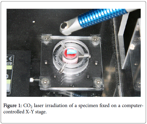
All root surfaces except for a rectangular window (2 mm x 3 mm) on the mesial or distal proximal surface were coated with an acidresistant varnish (Protect varnish, Kuraray Noritake Dental, Tokyo, Japan). The exposed area was irradiated using a CO2 laser (Opelaser 03SIISP, Yoshida, Tokyo, Japan). The irradiation conditions of the CO2 laser were: 10.6 μm wavelength, 0.5 W output power, continuous mode, repeated pulse (5 ms irradiation and a 10 ms pause for groups 1,2 and 3, and 10 ms irradiation and a 10 ms pause for groups 4, 5 and 6) and a defocused beam (1 mm spot size). A computer-controlled X-Y stage (Shot-602, Sigma-koki, Tokyo, Japan) was used to ensure uniform exposure (Figure 1). The root was fixed to the stage, which traversed at 1 mm/s through the laser beam. After each CO2 laser irradiation session, a 2.0% NaF solution was applied to the windows of specimens in groups 1 and 4 for 5 min. Specimens from groups 2 and 5 were immersed into a 0.2% NaF solution for 50 min, and specimens from groups 3 and 6 were immersed into a 0.05% NaF solution for 200 min. Immersions in NaF solution were performed using a room temperature (23°C) bath stirred at 120 rpm.
Demineralization/remineralization test
Specimens were subjected to a 2-day pH-cycling process in which five roots from each group were immersed in a demineralizing solution (pH 4.7, containing 0.05 M acetic acid, 2.2 mM calcium, and 2.2 mM phosphate ions) for 18 hours, then immersed in a remineralizing solution (pH 7.0, containing 0.15 M potassium chloride, 1.5 mM calcium, and 0.9 mM phosphate ions) for 6 hours. The solutions were maintained at 37°C and stirred at 120 rpm. The roots were irrigated with deionized water for 5 minutes during transfers between the solutions and at the completion of the cycling process.
Lesion depth evaluation
The samples were sectioned perpendicular to the root surfaces and through the center of each window using a hard-tissue microtome (Isomet, Buehler, Lake Bluff, IL, USA). Four sections with a thickness of approximately 200 μm were obtained from each window. Each section was ground to a thickness of approximately 100 μm using a whetstone (#2000).
The sections were examined at 200× magnification using a polarized light microscope (PLM, Eclipse LV100POL, Nikon, Tokyo, Japan). Digital photomicrographs were obtained using a CCD camera (DS-L2, Nikon, Tokyo, Japan). The lesion depth in each section was determined by measuring the width between the original root surface and the deepest position of the lesion using the camera’s control software.
Statistical analysis
Data were statistically analyzed with two-way ANOVA and Tukey’s multiple comparison tests for post hoc analysis at a significance level of 0.05. All analyses were performed using a statistical analysis add-in software package for Microsoft Excel (BellCurve for Excel, Social Survey Research Information Co. Ltd, Tokyo, Japan).
Results
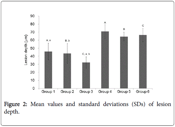
The mean values and standard deviations (SD) of lesion depths are presented in Figure 2. A two-way ANOVA showed that both the laser energy density and the fluoride concentration had significant effects on the lesion depth (p<0.01), and there was a significant interaction between these two factors (p<0.01). A simple main effect analysis and post hoc Tukey’s HSD tests showed that root surfaces treated with laser irradiation at an energy density of 17 J/cm2 had greater acid resistance than those irradiated at an energy density of 25 J/cm2 for all three tested fluoride concentrations (p<0.01). Among the specimens irradiated at an energy density of 17 J/cm2, greater acid resistance was observed with the low-concentration fluoride solution (0.05% NaF applied for 200 minutes) than with medium- and high-concentration solutions (0.2% NaF applied for 50 minutes and 2.0% NaF applied for 5 minutes, respectively; both p<0.01). Acid resistance did not differ between fluoride concentration subgroups for specimens irradiated with an energy density of 25 J/cm2 (p>0.05).
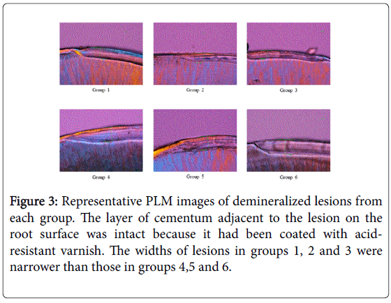
Representative PLM images of demineralized lesions for each group are presented in Figure 3. The layer of cementum adjacent to the lesion on the root surface was intact because it had been coated with acidresistant varnish. Stripes parallel to the bottom of the specimen were observed in some lesions; these indicate remineralization during the pH cycling procedure. The widths of lesions in groups 1,2 and 3 were narrower than those in groups 4,5 and 6.
Discussion
The parameters used for laser irradiation are important factors for evaluating the efficacy of laser irradiation at strengthening the acid resistance of the root surface. In this study, the laser output used was set to employ a continuous series of pulses from a 0.5 W laser beam defocused to a spot 1 mm in diameter. The irradiation cycle consisted of a 10 ms pause and a 5 ms (groups 1,2 and 3) or 10 ms irradiation time (groups 4,5 and 6). The laser beam exposure was ensured to be uniform using a computer-controlled X-Y stage with the specimen fixed to the stage and moved at 1 mm/s throughout laser irradiation. Therefore, the laser energy density in groups 1,2 and 3 was 17 J/cm2 (0.25 J/cm2 per pulse) and that of groups 4,5 and 6 was 25 J/cm2 (0.5 J/cm2 per pulse). These parameters were decided upon to modify the root surface through the heat of laser irradiation, as determined by a previous study in which the effect of CO2 laser irradiation on enamel demineralization was investigated [4-6,8].
Previous studies have shown that laser irradiation followed by fluoride application significantly decreases dentin demineralization compared to a single application of fluoride or a single session of laser irradiation [20-25]. The present study showed that pre-irradiation by a lower energy density (17 J/cm2) laser followed by fluoride application (mean lesion depth: group 1, 45.8 μm; group 2, 43.5 μm; group 3, 32.1 μm) yielded better root surface acid resistance than a higher energy density (25 J/cm2) laser followed by fluoride application (mean lesion depth: group 4, 70.8 μm; group 5, 64.4 μm; group 6, 66.3 μm). Our previous study, in which a laser with an identical energy density to that used in the present study was employed, demonstrated that the mean lesion depths of specimens irradiated by a CO2 laser at 17 J/cm2 and 25 J/cm2 and subjected to the application of 0.05% NaF and 0.2% NaF were 41.0 ± 3.4 μm, 42.3 ± 2.9 μm, 37.9 ± 4.2 μm and 32.6 ± 3.4 μm, respectively [19]. From these data, the combination laser and fluoride treatment investigated in the present study did not significantly decrease the root surface lesion depth compared to a single fluoride or laser treatment as studied our previous work. Therefore, the efficacy of laser irradiation followed by fluoride application for root surfaces may be affected by the energy density of the laser irradiation applied. The laser intensities used in the present study were higher than those in previous studies [20-22]. Several studies have indicated that higher energy densities cause significant thermal damage to irradiated dentin [19,21,23-25]. Therefore, we hypothesize that the synergistic effect of laser irradiation and fluoride application may not be exhibited when laser irradiation with a high energy density is used.
Apatite crystal readily absorbs CO2 and Er:YAG laser beams. At a high CO2 laser intensity, the cementum or dentin is carbonized. The present study showed that a thin polarizing layer of 20-30 μm exists at the intact root surfaces in most specimens, as determined by PLM images, and this layer could be cementum. This layer was not observed in all specimens, which may indicate cementum loss during the root planing process. A stripe parallel to the bottom of the specimen was observed in lesions on all specimens, which may indicate remineralization occurring in the cementum during the pH cycling procedure. As determined by visual observation, substance loss through carbonization occurring on the root surface was not found on specimen surfaces in this study. Although the intensity used in the present study was not sufficiently high to carbonize the tooth surface, the structure of the apatite crystals on the root surface could be altered by laser irradiation. The mechanism of prevention of dentin demineralization by laser treatment is uncertain. Some studies have reported that re-crystallization, melting and fusion occur on enamel and dentin surfaces after CO2 laser irradiation [4-9]. These superficial layers on laser irradiated enamel and dentin are thought to be effective at inhibiting the demineralization of tooth substrates. However, higher intensity laser irradiation would cause significant heat damage to irradiated dentin and induce greater mineral loss during pH cycling. Therefore, laser irradiation at a higher energy density (25 J/cm2) could be too strong to produce a superficial acid-resistance layer and may cause greater mineral loss during an acid challenge. Future studies are needed to confirm the synergistic effect of laser irradiation and fluoride application when using a CO2 laser at a low energy density.
The application of fluoride to root surfaces irradiated at a lower energy density yielded better acid resistance when the lowest concentration of fluoride (0.05% NaF) was applied for an extended time period compared to when the highest concentration of fluoride (2.0% NaF) was applied for a brief period. However, groups irradiated with a higher energy density showed no significant effects with respect to fluoride concentrations. Therefore, the null hypothesis, in which energy density and fluoride concentration in a combination treatment of CO2 laser irradiation and fluoride application does not affect the demineralization/remineralization of root surfaces, was rejected when a lower energy density laser was used. This result suggests that the formation of fluoroapatite on root surfaces irradiated by a lower energy density laser requires a long period of contact between F ions and hydroxyapatite on the superficial cementum or dentin. The synergistic effect of fluoride application and laser irradiation may be related to the efficacy of laser treatment at increasing fluoride uptake to tooth substrates [26,27]. This mechanism could be explained by the formation of micro-spaces on the root surface due to laser irradiation and the enhancement of fluoride uptake into these micro-spaces. This process may require a long period of time, during which F ions penetrate into the micro-spaces formed after laser irradiation at an appropriate intensity. Therefore, the long period of contact between the fluoride solution and the laser-irradiated root surface could be effective at decreasing root surface demineralization even at low fluoride concentrations.
Several studies have investigated the effect of laser irradiation of fluoride-treated tooth surfaces on tooth surface demineralization [28-30]. These studies show that fluoride treatment followed by laser irradiation is effective at decreasing the demineralization of the tooth surface. Based on these previous studies, treatments integrating laser irradiation and fluoride application may be effective at decreasing both enamel and dentin demineralization, regardless of the order of treatment. However, the effect of the order of fluoride application and laser irradiation on the demineralization/remineralization of root surfaces has not been studied. The mechanisms of action by which these treatments increase acid resistance could be different. When fluoride treatment is followed by laser irradiation, the increased acid resistance of tooth surfaces may result from the promotion of F ion penetration into tooth surfaces due to the heat-based effects of laser irradiation. Therefore, a high intensity laser may be more suitable for cases in which fluoride treatment is followed by laser irradiation than a low intensity laser. Future studies are needed to clarify the relationship between the laser energy density and the fluoride concentration when laser and fluoride treatments are pre-applied to improve the acid resistance of root surfaces.
Conclusion
Within the limitations of this study, both the laser energy and the fluoride concentration used in a combination treatment involving CO2 laser irradiation followed by fluoride application showed significant effects on the demineralization/remineralization of root surfaces. The application of fluoride onto laser-irradiated root surfaces yielded better acid resistance when a low concentration of fluoride was applied for an extended time period compared to the use of a high concentration of fluoride applied for a brief period.
References
- Griffin SO, Griffin PM, Swann JL, Zlobin N (2004) Estimating rates of new root caries in older adults. J Dent Res 83: 634-638.
- Kassab MM, Cohen RE (2003) The etiology and prevalence of gingival recession. J Am Dent Assoc 134: 220-225.
- Imazato S, Ikebe K, Nokubi T, Ebisu S, Walls AW (2006) Prevalence of root caries in a selected population of older adults in Japan. J Oral Rehabil 33: 137-143.
- Hsu CY, Jordan TH, Dederich DN, Wefel JS (2000) Effects of low-energy CO2 laser irradiation and the organic matrix on inhibition of enamel demineralization. J Dent Res 79: 1725-1730.
- Featherstone JD, Barrett-Vespone NA, Fried D, Kantorowitz Z, Seka W (2006) CO2 laser inhibitor of artificial caries-like lesion progression in dental enamel. J Dent Res 85: 617-621.
- Hsu DJ, Darling CL, Lachica MM, Fried D (2008) Nondestructive assessment of the inhibition of enamel demineralization by CO2 laser treatment using polarization sensitive optical coherence tomography. J Biomed Opt 13: 054027.
- Can AM, Darling CL, Ho C, Fried D (2008) Non-destructive assessment of inhibition of demineralization in dental enamel irradiated by a lambda = 9.3-microm CO2 laser at ablative irradiation intensities with PS-OCT. Lasers Surg Med 40: 342-349.
- Esteves-Oliveira M, Zezell DM, Meister J, Franzen R, Stanzel S, et al. (2009) CO2 Laser (10.6 micron) parameters for caries prevention in dental enamel. Caries Res 43: 261-268.
- Rechmann P, Fried D, Le CQ, Nelson G, Rapozo-Hilo M, et al. (2011) Caries inhibition in vital teeth using 9.6mm CO2-laser irradiation. J Biomed Opt 16: 071405.
- Westerman GH, Hicks MJ, Flaitz CM, Blankenau RJ, Powell GL, et al. (1994) Argon laser irradiation in root surface caries: in vitro study examines laser’s effects. J Am Dent Assoc 125: 401-407.
- Westerman GH, Hicks MJ, Flaitz CM, Blankenau RJ, Powell GL (1998) Argon laser irradiation effects on sound root surfaces: in vitro scanning electron microscopic observations. J Clin Laser Med Surg 16: 111-115.
- Yamazaki H, Litman A, Margolis HC (2007) Effect of fluoride on artificial caries lesion progression and repair in human enamel: regulation of mineral deposition and dissolution under in vivo-like conditions. Arch Oral Biol 52: 110-120.
- Lynch RJ, Mony U, Ten Cate JM (2006) The effect of fluoride at plaque fluid concentrations on enamel de- and remineralisation at low pH. Caries Res 40: 522-529.
- Schlueter N, Ganss C, Mueller U, Klimek J (2007) Effect of titanium tetrafluoride and sodium fluoride on erosion progression in enamel and dentine in vitro. Caries Res 41: 141-145.
- Toda S, Featherstone JD (2008) Effects of fluoride dentifrices on enamel lesion formation. J Dent Res 87: 224-227.
- Meyer-Lueckel H, Tschoppe P (2010) Effect of fluoride gels and mouth rinses in combination with saliva substitutes on demineralized bovine enamel in vitro. J Dent 38: 641-647.
- Takeshita EM, Exterkate RA, Delbem AC, ten Cate JM (2011) Evaluation of different fluoride concentrations supplemented with trimetaphosphate on enamel de- and remineralization in vitro. Caries Res 45: 494-497.
- Naumova EA, Niemann N, Aretz L, Arnold WH (2012) Effects of different amine fluoride concentrations on enamel remineralization. J Dent 40: 750-755.
- Shinkai K, Suzuki S (2014) Effects of CO2 laser and fluorides application on root demineralization. Asian Pac J Dent 14: 1-6.
- Gao XL, Pan JS, Hsu CY (2006) Laser-fluoride effect on root demineralization. J Dent Res 85: 919-923.
- Geraldo-Martins VR, Lepri CP, Faraoni-Romano JJ, Palma-Dibb RG (2014) The combined use of Er,Cr:YSGG laser and fluoride to prevent root dentin demineralization. J Appl Oral Sci 22: 459-464.
- Esteves-Oliveira M, El-Sayed KF, Dörfer C, Schwendicke F (2017) Impact of combined CO2 laser irradiation and fluoride on enamel and dentin biofilm-induced mineral loss. Clin Oral Investig 21: 1243-1250.
- Geraldo-Martins VR, Tanji EY, Wetter NU, Nogueira RD, Eduardo CP (2005) Intrapulpal temperature during preparation with the Er:YAG laser: An in vitro study. Photomed Laser Surg 23: 182-186.
- Hossain M, Kimura Y, Nakamura Y, Yamada Y, Kinoshita JI, et al. (2001) A study on acquired acid resistance of enamel and dentin irradiated by Er,Cr:YSGG laser. J Clin Laser Med Surg 19: 159-163.
- Hossain M, Nakamura Y, Yamada Y, Kimura Y, Matsumoto N,et al. (1999) Effects of Er,Cr:YSGG laser irradiation in humanenamel and dentin: Ablation and morphological studies. J Clin Laser Med Surg17: 155-159.
- Chin-Ying SH, Xiaoli G, Jisheng P, Wefel JS (2004) Effects of CO2 laser on fluoride uptake in enamel. J Dent 32: 161-167.
- Tepper SA, Zehnder M, Pajarola GF, Schmidlin PR (2004) Increased fluoride uptake and acid resistance by CO2 laser-irradiation through topically applied fluoride on human enamel invitro. J Dent32: 635–641.
- Meurman JH, Hemmerle J, Voegel JC, Rauhamaa-Makinen R, Luomanen M (1997) Transformation of hydroxyapatite to fluorapatite by irradiation with high-energy CO2 laser. Caries Res 31: 397-400.
- Nakagaki S, Iijima M, Endo K, Saito T, Mizoguchi (2015) Effects of CO2 laser irradiation combined with fluoride application on the demineralization, mechanical properties, structure, and composition of enamel. Dent Mater J 34: 287-293.
- Kumar P, Goswami M, Dhillon JK, Rehman F, Thakkar D, et al. (2016) Comparative evaluation of microhardness and morphology of permanent tooth enamel surface after laser irradiation and fluoride treatment - An in vitro study. Laser Ther 25: 201-208.
Relevant Topics
- Cementogenesis
- Coronal Fractures
- Dental Debonding
- Dental Fear
- Dental Implant
- Dental Malocclusion
- Dental Pulp Capping
- Dental Radiography
- Dental Science
- Dental Surgery
- Dental Trauma
- Dentistry
- Emergency Dental Care
- Forensic Dentistry
- Laser Dentistry
- Leukoplakia
- Occlusion
- Oral Cancer
- Oral Precancer
- Osseointegration
- Pulpotomy
- Tooth Replantation
Recommended Journals
Article Tools
Article Usage
- Total views: 2703
- [From(publication date):
August-2017 - Jul 06, 2025] - Breakdown by view type
- HTML page views : 1881
- PDF downloads : 822


