Research Article Open Access
Effect of Ethanolic Extract of Coconut (Cocos nucifera) on Aspirin-induced Gastric Ulcer in Albino Rats
Amjad M* and Tahir MDepartment of Anatomy, Avicenna Medical College, Lahore, Pakistan
- *Corresponding Author:
- Amjad Mcorr
Department of Anatomy, Avicenna Medical College
Phase IX, DHA, Bedian Road, 53100, Lahore, Pakistan
Tel: 042 37167566
Email: dr.madihaamjad@gmail.com
Received date: March 28, 2017; Accepted date: June 20, 2017; Published date: June 27, 2017
Citation: Amjad M, Tahir M (2017) Effect of Ethanolic Extract of Coconut (Cocos nucifera) on Aspirin-induced Gastric Ulcer in Albino Rats. J Gastrointest Dig Syst 7:512. doi: 10.4172/2161-069X.1000512
Copyright: © 2017 Amjad M, et al. This is an open-access article distributed under the terms of the Creative Commons Attribution License, which permits unrestricted use, distribution, and reproduction in any medium, provided the original author and source are credited.
Visit for more related articles at Journal of Gastrointestinal & Digestive System
Abstract
Statement of problem: Aspirin is extensively used as an analgesic, antipyretic, anti-thrombotic and antiinflammatory drug. It is safe in therapeutic doses but it is toxic in over dosage or in chronic injudicious use, when it causes acute or chronic toxicity respectively. Acute toxicity causes gastric ulceration and bleeding. Gastric ulcer developed when the imbalance occurs between aggressive and defensive factors of the stomach and situation favours aggressive factors enough to cause mucosal damage and lead to ulceration. Many antioxidants have been trying to study their protective effects of aspirin and others NSAID’s induced gastric ulcers. Coconut and its products are known for their antioxidant, antibacterial, anti-diabetic, antithrombotic and antiulcer genic effects. Two different studies are documented about the effect of ethanolic extract of coconut on indomethacin induced gastric ulcers. Both are paradoxical, and show controversy at different doses of extract. The present study, therefore, designed in an attempt to observe the effects of high doses of ethanolic extract of coconut on aspirin induced gastric ulcers. This was conducted at University of Health Sciences Lahore. Thirty rats, weighing 175-220 gm, were divided into six groups and were treated with different doses of extract for 14 days and were sacrificed on 15th day of experiment. Methodology involved staining of stomach with H and E and Masson Trichome staining and was observed for desquamation, mucosal congestion, inflammatory Cells and necrosis. Blood samples were drawn for serum analysis of Super Oxide Dismutase (SOD).
Findings: The results showed that ethanolic extract of coconut helps to heal the stomach ulcers and gives best results at 400 mg/kg dose and also helps to reduce oxidative stress which also helps to heal ulcers.
Conclusion and significance: It was concluded that ethanolic extract of coconut provides a very reliable and cost effective adjunctive therapy to be routinely used in patients who take aspirin and aspirin induced gastric ulcers as well.
Keywords
Aspirin; Gastric ulcer; Non-steroidal anti-inflammatory drugs; Super oxide dismutase; Ethanolic extract of coconut; Desquamation; Congestion; Necrosis
Introduction
Andre Robert introduced the term ‘cytoprotection’ first time. He used this term for protection of experimentally induced gastric ulcer by prostaglandins [1]. But later on, this term is broadly used for protection of gastric mucosal injury except injuries caused by gastric acid inhibition or neutralization [2]. Despite intensive research of the last few decades, gastro-protection is incompletely understood, and we are still far away from effectively treating Helicobacter pylori -negative ulcers and preventing non-steroidal anti-inflammatory drugs-caused erosions and ulcers in the upper and lower gastrointestinal tract; hence "gastric cytoprotection" research is still relevant [3]. Aspirin, also known as acetylsalicylic acid, is commonly used as an analgesic, antiinflammatory, antipyretic, antiplatelet and antithrombotic drug. It is also used as a protective drug because it decreases the incidence of transient ischemic attack, unstable angina, coronary artery thrombosis and myocardial infarction and thrombosis after coronary artery bypass grafting. At low doses, it lowers the incidence of colon cancer, probably related to its COX-inhibiting effect [4].
It inhibits the cyclo-oxygenase enzyme (COX) of arachidonic pathway. There are 2 types of enzymes COX-1 and COX-2. Aspirin binds with the active sites of both enzymes in the same manner and leads to irreversible inhibition of both [5]. The main prostaglandin (PG) synthesized in the gastric mucosa is PGI2. PGI2 increases the mucus and bicarbonate secretion and dilates blood vessels, preventing the mucosal injury caused by ischemia. Aspirin decreases the gastric mucus synthesis inhibiting the synthesis of prostaglandins and lead into damage of gastric mucosa [6]. Many antioxidants have been trying to study their protective effects of NSAID’s induced gastric ulcers including coconut. The coconut palm (Cocos nucifera ) is a member of the family Arecaceae (Palm family) is an important and useful fruit tree present in the world. The ethanolic extract of C. nucifera was found to have a vasorelaxant and anti-hypertensive effect via nitric oxide production and endothelial dependent manner through direct activation of nitric oxide, muscarinic receptor stimulation or via the cyclooxygenase pathway [7]. During second world war, in emergency room young coconut water was used as dextrose water in place of sterile dextrose [8].
In 2010, two different studies are documented about effect of ethanolic extract of coconut on indomethacin induced gastric ulcer. Both are paradoxical, and show controversy at 400 mg/kg doses of extract. The present study, therefore, designed in an attempt to observe the effects of high doses of ethanolic extract of coconut on aspirin induced gastric ulcers.
Materials and Methods
Thirty wistar albino male rats 6-8 weeks old, weighing 175-220 gm was procured from University of Health Sciences, Lahore. All the animals were examined thoroughly and weighed before the commencement of the experiment. The rats were housed in the experimental research laboratory of University of Health Sciences, Lahore, under controlled temperature of 23+2°C and humidity 55% ± 5%, and 12 hours light and dark cycle were maintained, and fed on a standard rat diet and tap water ad libitum.
Rats were kept for a week for acclimatization and were handled from time to time to minimize the stress during the experiment. Completely randomized design is adopted for statistical sampling experiment by using the balloting method, where all rats were divided into five groups (A, B, C, D and E) with 6 animals in each group.
Preparation of ethanol extract of coconut
The extract was extracted by the pharmacology laboratory of University of Health Sciences, Lahore. Mature coconut seed was purchased from local markets and broken into tiny bits and were grounded with a mechanical grinder and macerated for 24 hours in absolute ethanol and subsequently filtered with a white cotton cloth and was concentrated into a dry paste at an optimum temperature of 40°C-50°C, by using a rotary evaporator [9].
Doses of ethanol extract of coconut
Doses of ethanol extract of coconut 400, 600 and 800 mg/kg body weight given intraperitoneally in the current study is based on previous studies [9].
Dose of aspirin
Aspirin in powder form was obtained from BDH Limited, Poole, England. Distilled water was used to prepare acetylsalicyclic acid solution and given a single dose of 500 mg/kg body weight orally, to 24 hours fasted rats of group B, C, D, and E.
Histological examination
Desquamation was observed on H&E stained slides. Desquamation was labelled by the presence of shedding or exfoliation of mucosal cells, disrupted mucosal gastric epithelium [10]. Acute gastric mucosal disease is capillary and venular congestion with localized areas of mucosal haemorrhage confined to the lamina propria. Necrosis was defined by the presence of nuclear and cytoplasmic changes in H&E stained histological sections. Necrosis was graded according to the following scale:
Grade 0: No Lesion, Grade 1: Several (≤ 4) focal areas of partialthickness mucosal necrosis, Grade 2: Many (>4) areas of partialthickness mucosal necrosis, Grade 3: Multiple area of necrosis with 1 area of full mucosal thickness necrosis, Grade 4: Extensive confluent full-thickness mucosal necrosis often with sloughing or erosion of necrotic mucosa.
Inflammation was labelled by the presence of inflammatory cells e.g. neutrophils. The inflammation was graded as Grade 0: Normal epithelium, Grade 1: Few inflammatory cells, Grade 2: Moderate amount of inflammatory cells in several layers, Grade 3: High level of inflammation (more than 50 Inflammatory cells) [11].
• Gastric Erosion Severity Score (GESS): The sum of resultant grades of necrosis of each group was the gastric erosion severity score. Mean severity score=GESS/Total number of rats in group
• Gastric Erosion Index (GEI): Gastric Erosion index=Gastric Erosion Severity Score × Incidence of necrosis (Number of rats in a group with gastric necrosis).
Biochemical essay
These blood samples were used to assess the serum SOD enzyme by using specific commercial kits.
Statistical analysis
Statistical analysis was performed by using SPSS 20.0 software. The level of significance was selected at ≤ 0.05.
Results
SOD levels
Table 2a and 2b given below show summary statistics (Mean ± SD) on SOD levels. There is sufficient statistical evidence of p-value=0.014* (<0.05 as α) that Mean Scores of SOD levels are significantly different between the treatment groups including the control group.
| Groups | Intervention and Dosage | Route of Administration | Duration of Administration | Day of Sacrifice |
|---|---|---|---|---|
| Group A | 5ml/kg N/S | Intraperitoneally | Daily for 14 days | 15 |
| Group B | Aspirin 500mg/kg dissolved in D/W | Orally | Single dose, 24 hours after fasting | 15 |
| Group C | Aspirin 500 mg/kg dissolved in D/W | Orally | Single dose, 24 hours after fasting | 15 |
| Ethanolic extract of coconut 400 mg/kg suspended in 5 ml of N/S | Intraperitoneally 4 hours after aspirin administration. | Daily for 14 days | ||
| Group D | Aspirin 500 mg/kg dissolved in D/W | Orally | Single dose, 24 hours after fasting | 15 |
| Ethanolic extract of coconut 600 mg/kg suspended in 5 ml of N/S | Intraperitoneally 4 hours after aspirin administration. | Daily for 14 days | ||
| Group E | Aspirin 500 mg/kg dissolved in D/W | Orally | Single dose, 24 hours after fasting | 15 |
| Ethanolic extract of coconut 800 mg/kg suspended in 5 ml of N/S | Intraperitoneally 4 hours after aspirin administration. | Daily for 14 days |
Table 1: Showing experimental strategy of study.
| Groups | Group A | Group B | Group C | Group D | Group E | p-value |
|---|---|---|---|---|---|---|
| SOD levels | 1.558±0.826 | 0.575±0.214 | 1.048±0.254 | 1.077±0.421 | 1.477±0.470 | 0.014* |
| *Mean differs significantly at the α=0.05 level. | ||||||
Table 2a: Comparison of Super-Oxide Dismutase (SOD) levels among groups (Mean ±SD).
| (I) Group | (J) Group | Mean Difference (I-J) | Std. Error | p-value |
|---|---|---|---|---|
| Group A (1.558) | Group B (0.575) | 0.983* | 0.282 | 0.014* |
| Group C (1.048) | 0.51 | 0.282 | 0.392 | |
| Group D (1.077) | 0.481 | 0.282 | 0.449 | |
| Group E (1.477) | 0.081 | 0.282 | 0.998 | |
| Group B (0.575) | Group C (1.048) | -0.473 | 0.282 | 0.465 |
| Group D (1.077) | -0.502 | 0.282 | 0.407 | |
| Group E (1.477) | -0.902* | 0.282 | 0.028* | |
| Group C (1.048) | Group D (1.077) | -0.029 | 0.282 | 1 |
| Group E (1.477) | -0.429 | 0.282 | 0.558 | |
| Group D (1.077) | Group E (1.477) | -0.401 | 0.282 | 0.621 |
Table 2b: Comparison of Super-Oxide Dismutase (SOD) levels among groups (A, B, C, D, E); Multiplecomparison (TUKEY’s Test).
Microscopic parameters of stomach
Table 3a and 3b shows summary statistics (Mean ± SD) on mucosal congestion presence. There is sufficient statistical evidence of p value= 0.005* (<0.05 as α) that mean scores of mucosal congestion are highly significantly different between the treatment groups including the control group by colour of stomach.
| Presence of Mucosal Congestion |
Group A | Group B | Group C | Group D | Group E | p-value |
| 7.545 ± 1.462 | 15.308 ± 3.569 | 9.463 ± 3.759 | 5.845 ± 0.915 | 3.201 ± 0.976 | 0.005* |
Table 3a: Summary statistics of mucosal congestion of stomach ulcer (Mean ± S.D).
| Tukey HSD Test for Multiple Comparisons | ||||
|---|---|---|---|---|
| Dependent Variable: Mucosal Congestion | Mean Difference (I-J) | Standard Error | p-value | |
| (I) Group | (J) Group | - | - | - |
| Group A (7.545) | Group B (15.308) | -7.763* | 1.433 | 0.000* |
| Group C (9.463) | -1.918 | 1.433 | 0.67 | |
| Group D (5.845) | 1.7 | 1.433 | 0.759 | |
| Group E (3.201) | 4.344* | 1.433 | 0.041* | |
| Group B (15.308) | Group C (9.463) | 5.845* | 1.433 | 0.003* |
| Group D (5.845) | 9.463* | 1.433 | 0.000* | |
| Group E (3.201) | 12.108* | 1.433 | 0.000* | |
| Group C (9.463) | Group D (5.845) | 3.618 | 1.433 | 0.117 |
| Group E (3.201) | 6.263* | 1.433 | 0.002* | |
| Group D (5.845) | Group E (3.201) | 2.644 | 1.433 | 0.371 |
Table 3b: Comparison of mucosal congestionof stomach ulcer (Mean ± S.D).
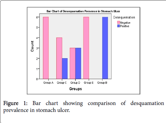
The results showed that the ethanolic extract of coconut doses of 400 mg/kg, 600 mg/kg and 800 mg/kg in groups C, D and E all are significantly effective treatments against ulceration showing the decreasing trend of mucosal congestion. Figure 1 indicates positive strong association of the prevalence of desquamation in stomach ulcer and hence reveal high severity of the prevalence of desquamation in group B. It is concluded that groups C and D show relatively least prevalence of desquamation due to treatment effects of ethanolic extract of coconut 400 mg/kg and 600 mg/kg respectively. However, group E is homogeneous to control group A, and it has better effect on treating desquamation.
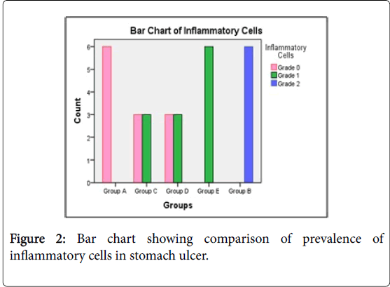
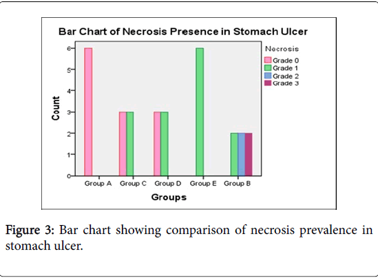
Figure 2 shows the comparison of prevalence of inflammatory cells in stomach ulcer. It is concluded that groups C and D show relatively least prevalence of inflammatory cells due to treatment effects of ethanolic extract of coconut 400 mg/kg and 600 mg/kg respectively. Results of necrosis in stomach ulcer indicate the much stronger positive relationship and hence reveal high severity of the prevalence of necrosis in group E and group B. It is concluded that groups C and D show relatively least prevalence of necrosis due to treatment effects of ethanolic extract of coconut 400 mg/kg and 600 mg/kg respectively.
Healing proportionality against ulceration
Healing proportionality against ulceration=[1 – GEI/(GEI of Group B)] × 100%
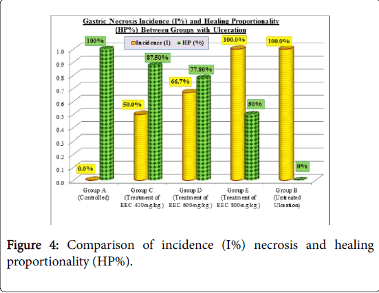
Following Table 4 and Figure 4, shows the incidence, Severity Score, and GEI prevalence of necrosis in ulcerated stomach. It also reveals the Healing Proportionality (HP%) of treatment effects against ulceration in stomach. Control group A shows 100% health because of neither ulceration nor any treatment.
| Gastric Necrosis | ||||
|---|---|---|---|---|
| Groups | Incidence | Severity Score (Mean ± S.D) |
GEI Prevalence in Stomach Ulcer |
Healing Proportionality (%) against Ulceration |
| Group A | 0/6=0 | 0/6=0 | 0 | 100% (=1-0/2) |
| Group B | 6/6=1 | 2 ± 0.894 (=12/6) | 1 × 2=2 | 0% (=1-2/2) |
| Group C | 3/6=0.5 | 0.5 ± 0.548 (=3/6) | 0.5 × 0.5=0.25 | 87.5% (=1-0.25/2) |
| Group D | 4/6=0.67 | 4 ± 0.516 (=4/6) |
0.67 × 0.67=0.444 | 77.80% (=1-0.444/2) |
| Group E | 6/6=1 | 1 (=6/6) | 1 × 1=1 | 50% (=1-1/2) |
Table 4: Shows incidence, SS, GEI and Healing proportionality against ulceration.
Group B is the diseased group with ulceration is the basis to determine HP% of treatment effects against ulceration. Therefore, group C shows 87.5% HP, group D shows 77.8% and group E shows 50% HP against ulceration.
Discussion
NSAID’s had established a defining role as an analgesic, antiinflammatory, antipyretic, antiplatelet, antithrombotic and cytoprotective drug. Beside theses gastrointestinal toxicity associated with NSAIDs has emerged as an important medical problem [12]. The present study was designed to evaluate the possible role of ethanolic extract of coconut on aspirin induced gastric ulcers in wistar albino rats. Coconut (Cocos nucifera ) was reported to protect several organs against oxidative damage activity as an antioxidant due to high content of L-arginine and vitamin C, reduces the free radicals and lipid peroxidation by increasing the level of antioxidant enzymes respectively [13]. The present study stipulates to evaluate the effects of ethanolic extract of coconut on aspirin induced gastric ulcers.
The current study revealed that control group A (control) and group E (treated ulceration) both have homogeneously equal mean scores of SOD levels and are significantly higher SOD levels than that in group B (untreated ulceration, consistent with color of stomach), showing pvalues 0.014* and 0.028* (<α 0.05) respectively. This resultantly means that the ethanolic extract of groups C, D and E is significantly effective treatment against ulceration as compared to the groups B. However, our results support previous findings that SOD activity in rat stomach tissues is decreased by NSAIDs. [14,15]. SOD activity more possibly was inhibited by aspirin (P<0.01), while all doses of ethanolic extract of coconut increased SOD activity near the levels of the control groups.
These results indicate that SOD plays an important role in eliminating gastric damage by partially preventing oxidative damage.
In this study, ulcer inhibition was observed with all the doses of the coconut extract, and was more pronounced in the 400 mg/kg b.w and 600 mg/kg b.w doses of extract. This result is paradoxical, since previous work [9] reported that The ethanolic extracts of coconut increases the gastric erosions induced by indomethacin; rats treated with 100 mg/kg and 200 mg/kg doses of ethanolic extract of coconut are reported to have an ulcer inhibition in 65.4% and 67.9% cases, respectively; whereas 400 mg/kg dose of ethanolic extract of coconut increased the incidence of ulceration when compared to control [9]. In the present study, significant protection against aspirin induced gastric mucosal damage was observed in the animals treated with 400 mg/kg and 600 mg/kg of the coconut extract. This suggests that the extract is anti-ulcerogenic at high doses also, which is similar to results of Chioma et al. [16].
Conclusion
The results showed that ethanolic extract of coconut helps to heal the stomach ulcers at all doses and gives best results at 400 mg/kg dose and also helps to reduce oxidative stress which also helps to heal ulcers. The results of present study suggest that ethanolic extract of coconut is effective in treating ulcers and reducing its size in ethanolic treated stomach of wistar albino rats. This is evident both histopathologically and biochemically by reduction in the inflammation of the stomach. This is helpful in healing ulcer is also evident histopathologically by reduction of desquamation, necrosis and congestion of the stomach. This study also helps us in evolving an inexpensive remedy to heal the ulcers in the wistar albino rats induced by aspirin and other NSAID’s.
Additional research is needed to monitor biochemical parameters like OH (hydroxyl radical), catalase (CAT), and H2O2 (Hydrogen peroxide) should be studied in details.
References
- Robert A (1979) Cytoprotection by prostaglandins. Gastroenterology 77: 761-767.
- D’Souza RS, Dhume VG (1991) Gastric cytoprotection. Indian J PhysiolPharmacol 35: 88-98.
- Szabo S (2014) “Gastric cytoprotection” is still relevant. J GastroenterolHepatol 4: 124-132.
- Katzung BG (2009) Non-steroidal anti-inflammatory drugs. Basic and clinical pharmacology (11th edn) San Francisco: McGraw Hill p: 816.
- Chandrasekharan NV, Dai H, Elton TS, Roos KL, Evanson NK, et al. (2002) COX-3, a cyclooxygenase-1 variant inhibited by acetaminophen and other analgesics/antipyretic drugs. ProcNatlAcadSci USA 99: 13926-13931.
- Camara PRS (2015) Endogenous vasoactive mediators in defense of portal hypertensive gastric mucosa. J ExpIntegr Med 5: 3-15.
- Bankar GR, Nayak PG, Bansal P, Paul P, Pai KS, et al. (2011) Vasorelaxant and antihypertensive effect of Cocosnucifera Linn. endocarp on isolated rat thoracic aorta and DOCA salt-induced hypertensive rats. J Ethnopharmacol 134: 50-54.
- Adodo A (2002) Medicinal value of coconut. A christian approach of herbal medicine. pp: 53-55.
- Anosika CA, Obidoa O (2010) Anti-inflammatory and anti-ulcerogenic effect of ethanol extract of coconut (Cocosnucifera) on experimental rats. AJFAND 10: 4286-4300.
- Salah KM (2015) The Postulated mechanism of protective effect of ginger on the aspirin induced gastric ulcer: Histological and immunohistochemical studies. HistoHistopathol 30:855-864.
- Bennedesen M, Wnag X, Willen R, Wadstrom T, Andersen LP (1999) Treatment of H. pylori infected mice with antioxidant astaxanthin reduces gastric inflammation, bacterial load and modulates cytokine release by splenocytes. ImmunolLett 70: 185-189.
- Sostres C, Gargallo CJ, Arroyo MT, Lanas A (2010) Adverse effects of non-steroidal anti-inflammatory drugs (NSAIDs, aspirin and coxibs) on upper gastrointestinal tract. Best Pract Res ClinGastroenterol 24: 121-132.
- Nevin KG, Rajamohan T (2005) Virgin coconut oil supplemented diet increases the antioxidant status in rats. Food CheM99: 260-266.
- Halici M, Odabasoglu F, Suleyman H (2005) Effects of water extract of Usnealongissima on antioxidant enzyme activity and mucosal damage caused by indomethacin in rats. Phytomedicine 12: 656-662.
- Dengiz GO, Odabasoglu F, Halici Z, Cadirci E, Suleyman H (2007) Gastroprotectiveand antioxidant effects of montelukast on indomethacin-induced gastric ulcer in rats. J PharmacolSci 105: 94-102.
- Chioma A, Obidoa O, Parker E (2010) Anti-ulcerogenic and membrane stabilization effect of ethanol extract of coconut (Cocosnucifera). Research J Pharmacognosy and Phytochemistry2: 85-88.
Relevant Topics
- Constipation
- Digestive Enzymes
- Endoscopy
- Epigastric Pain
- Gall Bladder
- Gastric Cancer
- Gastrointestinal Bleeding
- Gastrointestinal Hormones
- Gastrointestinal Infections
- Gastrointestinal Inflammation
- Gastrointestinal Pathology
- Gastrointestinal Pharmacology
- Gastrointestinal Radiology
- Gastrointestinal Surgery
- Gastrointestinal Tuberculosis
- GIST Sarcoma
- Intestinal Blockage
- Pancreas
- Salivary Glands
- Stomach Bloating
- Stomach Cramps
- Stomach Disorders
- Stomach Ulcer
Recommended Journals
Article Tools
Article Usage
- Total views: 4991
- [From(publication date):
June-2017 - Jul 12, 2025] - Breakdown by view type
- HTML page views : 4002
- PDF downloads : 989


