Make the best use of Scientific Research and information from our 700+ peer reviewed, Open Access Journals that operates with the help of 50,000+ Editorial Board Members and esteemed reviewers and 1000+ Scientific associations in Medical, Clinical, Pharmaceutical, Engineering, Technology and Management Fields.
Meet Inspiring Speakers and Experts at our 3000+ Global Conferenceseries Events with over 600+ Conferences, 1200+ Symposiums and 1200+ Workshops on Medical, Pharma, Engineering, Science, Technology and Business
Case Report Open Access
Eccentric Training for Elbow Hypermobility
| Kaux JF1*, Forthomme B1, Foidart-Dessalle M1, Delvaux F1, Debray FG2, Crielaard JM1and Croisier JL1 | |
| 1Physical Medicine and Rehabilitation Department, University Hospital of Liège and Motor Science Department, University of Liège, 4000 Liège, Belgium | |
| 2Human Genetics Department, University Hospital of Liège, University of Liège, Belgium | |
| Corresponding Author : | Jean-François Kaux Physical Medicine and Sport Traumatology Service University Hospital of Liège, Avenue de l’Hôpital B35, 4000 Liège, Belgium Tel: 32-4366-8241 Fax: 32-4366-7230 E-mail: jfkaux@chu.ulg.ac.be |
| Received July 23, 2013; Accepted August 23, 2013; Published August 26, 2013 | |
| Citation: Kaux JF, Forthomme B, Foidart-Dessalle M, Debray FG, Crielaard JM , et al. (2013) Eccentric Training for Elbow Hypermobility. J Nov Physiother 3: 180. doi:10.4172/2165-7025.1000180 | |
| Copyright: © 2013 Kaux JF, et al. This is an open-access article distributed under the terms of the Creative Commons Attribution License, which permits unrestricted use, distribution, and reproduction in any medium, provided the original author and source are credited. | |
Visit for more related articles at Journal of Novel Physiotherapies
| Introduction |
| The hypermobility can be defined as an increase in range of motion compared to normal amplitudes for the same age, sex and ethnic group [1]. It is also due to ligament laxity determined by coding genes for fibrous proteins such as collagen, elastin, fibrillin and tenascin and can be affected by mutations [2,3]. |
| Conventionally, these people with an exclusively joint hypermobility are considered solely hypermobile and those with musculoskeletal symptoms due to their abnormal joint mobility associated do fall into are contained in the ��?hypermobility syndrome� [3]. |
| Benign Joint Hypermobility Syndrome (BJHS) affects between 5 and 10% of the Caucasian population (women predominantly) but it also concerns rare hereditary dystrophies with abnormal collagen structure or metabolism (i.e.: Ehlers-Danlos Syndrome (EDS) and Marfan or Osteogenesis imperfecta) [4]. The hypermobility also can be hardly and reversibly acquired after repeated stretching just as for ballet dancers and gymnasts. The acquired generalized hypermobility is also found sometimes in certain pathologies such as acromegaly, hyperparathyroidism, chronic alcoholism, or systemic lupus erythematosus [5]. |
| Beighton criteria [6], used clinically to detect a joint hypermobility in a patient include: |
| - - Passive dorsiflexion of little fingers up to 90°. |
| - Passive opposition of thumbs on the forearm. |
| - Both hands flat on the ground with stretched legs. |
| - Hyperextension of the elbow up to 10°. |
| - A recurvatum of the knees up to 10°. |
| More recently, these criteria have been included into a more complex classification that of Brighton [7], divided into major and minor criteria (Table 1). |
| Patients with joint hypermobility complain regularly of acute osteoarticular such as shoulder luxation or ankle sprains but also chronic joint pain from many causes: high frequency of subluxation, repeated tendon or ligament pathologies, peripheral neurological irritation, multiple operations [1,8-12]. Generally, a classic rehabilitation causes little positive effects on both pain and quality of life. The rehabilitative content should be adapted individually by prioritizing techniques directly focused on joint hypermobility [3]. Muscle reinforcement for active stabilization of the involved joint is therefore preferred, especially in eccentric mode to develop the suppressive and protective role of the trained muscle group, as already used in the rehabilitation of instabilities of traumatic origin [13]. The use of an isokinetic dynamometer allows to train safely in eccentric mode. Indeed, this device permits us working gradually, at controlled speed, within a limited range of joint motion in flexion and extension to avoid risk of luxation. |
| We illustrate this concept by the description of a clinical case. |
| Clinical Case |
| A 16 year old girl, diagnosed as EDS type III, consulted us for pain in the elbow and right wrist (dominant side) when playing tennis [14]. |
| She had particularly: |
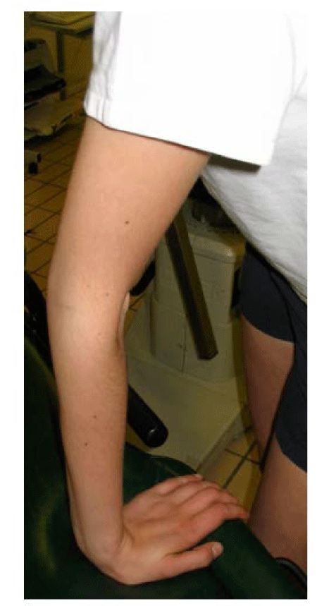
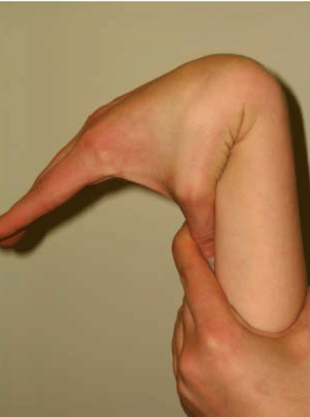
| - A significant known hypermobility (1 major criteria and 2 minor criteria according Brighton) (Figures 1 and 2). |
| - A hypermobile type EDS according to clinical and immunohistochemical studies. |
| - A myopia. |
| - Regular epistaxis. |
| - Multiple ankle and knee sprains when playing soccer. |
| Her bone density was within the standards for her age. At the family level, several subjects had hypermobility, heart problems or significant scoliosis. Her twin brother died prematurely, after 4 months of gestation (recognized sign in certain types of EDS) [15]. |
| You may recall that the EDS is a rare hereditary dystrophy affecting the synthesis or metabolism of collagen (I, III or V) or due to an enzymatic deficiency (lysyl hydroxylase and procollagen peptidase) involved in the synthesis of connective tissue [2,11,15]. The last classification called Villefranche classification, based on partially identified genetic and biochemical alterations and clinical presentations, recognizes 6 types [5,11,12,15] (Table 2). All have two major criteria, expressed at different levels: joint hyper mobility and skin hyper elasticity (+ possibly dystrophic scars) [5,16]. |
| The immuno-histological analyzes make it possible to detect pathologies of the connective tissue but in all cases are not capable of a precise diagnosis. The appropriate course of action is, in the case of strong suspicion of EDS (mainly classic and vascular forms), to resort to the research of biochemical abnormalities of collagen produced by a skin fibroblast lines under culture: quantitative or qualitative (electrophoretic) abnormalities of type I and V collagens (classic or previous I and II type EDS) or of type III procollagen (EDS vascular or previous IV) [15]. Based on this analysis, we can afterwards try to find a direct mutation in the corresponding gene (Table 2). However, in the hypermobile form (previous type III), the incriminated gene remains unknown at the present time and the electrophoretic analysis is generally normal in most cases [9,15,17]. The diagnosis of hypermobile type EDS is still clinical at present and can be confused with SHB [12]. |
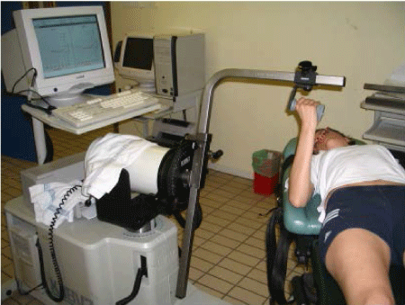
| A rehabilitation program, 3 times a week, during 6 weeks, aimed in particular at eccentric strengthening of flexor and extensor muscles of the elbow together with prosupinators. Indeed, a prior isokinetic test had noted a deficit in muscle strength on the affected side compared to the opposite side of -15 to -35% at slow (60�?°/s) and fast speed (180�?°/s) speeds, with a prevailing impairment on flexors (Table 3). In order to protect joints and maintain movements within a reasonable range of motion (avoiding the hyperextension of the elbow), eccentric strengthening exercises at slow speeds, were carried out on an isokinetic dynamometer (Cybex Norm®) (Figure 3). Thus, she was trained to move her wrist at increasing speeds [30�?°/s, 60�?°/s, and 90�?°/s] within a limited range of motion in flexion and extension. All the more, as our purpose was to improve the gesture control, she started a submaximal eccentric program, using slow speeds, very gradually intensified (3 to 5 sets of 15 repetitions), in a safe range of motion in order to promote protective braking action of the joint. |
| A proprioceptive rehabilitation has also been undertaken in order to improve voluntary control of extreme ranges of motion. The wearing of a semi-rigid ortheosis to limit the wrist range of motion when playing tennis was also recommended. Rapidly, the patient noted a significant decrease in pain (assessed using a 10-point visual analogue scale which dropped from 7/10 to 1/10, and quality of life scale MOS SF-36 which went from 70 to 100) and an increase in the stability of the elbow when playing tennis, as reported by the patient and the objectivation of a better control of the positioning of the elbow in space. An isokinetic control test at the end of 18 sessions noted a 20 to 25% increase in the time of maximum strength in the right elbow muscles and a recovery of the equilibrium of studied values in relation to the left elbow, with the exception of high speed concentric flexors. It would have been appropriate to continue with the isokinetic strengthening in order to hope for the complete normalization of muscular profile, however, the young patient, satisfied, could not continue with the program treatment given the remote home location. At the end of the sessions in rehabilitation center, the patient was encouraged to continue with proprioception and isometric strengthening exercises at home as instructed in physiotherapy (for example, self-mobilization of the wrist and elbow in predefined range of motion, with eyes open and then closed; isometric contractions in different positions of the wrist and elbow, especially close to extreme positions). |
| Discussion |
| The patients suffering from hypermobility regularly develop associated proprioception disorders. However, it is still not clearly defined whether or not this deficit is present from birth or developed during childhood. Indeed, some hypermobile patients develop symptoms only after puberty or in adulthood. These proprioceptive disorders may therefore appear gradually (over several years) due to repeated (micro) traumas related to this joint hypermobility and potentially magnified by a specific sporting activity [18]. |
| The Panjabi model [19] shows that joint stability depends on three subsystems which are functionally interdependent: |
| - The passive musculoskeletal system, including bones, joint surfaces, ligaments, joint capsules and passive mechanical properties of muscles; |
| - The active musculoskeletal system, including muscles and tendons surrounding the joint; |
| - The control (or neural) system and the feedback, which include transducers of strength and movement variations located in the ligaments, tendons, muscles and the peripheral and central nervous system. |
| These concepts provide an explanation to some symptoms in hypermobile patients and can form the basis of their rehabilitation to increase the joint stability by muscle control. |
| In addition, these hypermobile patients frequently have specific muscle weakness compared to healthy subjects. The origin of this weakness would probably be multifactorial: increased muscle and ligament elasticity, lack of proprioception, joint instability, associated pain [20]. It is therefore important to undertake a specific rehabilitation for muscle strengthening (concentric motor role and protective suppressive eccentric function) and fresh proprioceptive training. |
| The goal of rehabilitation, based on the Panjabi model [19], entailed avoiding the joints hypermobility of the elbow and wrist, painful when playing tennis, by using the suppressive and stabilizer role of this arm��?s muscles. We thought appropriate to emphasize the importance of measuring muscle performances, in order to quantify possible shortfalls and objectivize the benefits of rehabilitation [20,21]. Regarding the selected procedures in following our patient progress, only the concentric mode was appraised. Indeed, by definition, the purpose of the appraisal is to obtain the maximum intensities that can be developed by the tested muscle group. Knowing that the tension developed in the eccentric mode is higher compared to the concentric mode, we preferred to avoid the taking of risk, especially in a young patient. |
| However, the eccentric rehabilitation began in accordance with submaximal procedures, very gradually intensified [21]. The objective here clearly aimed to ��?motion control� and proprioceptive in relation to a possible objective of pure muscle strengthening. Although eccentric exercises against manual resistance are possible, delivering the eccentric exercise using an isokinetic dynamometer has some advantages in terms of security that cannot be possible with conventional strengthening techniques: the speed control, a fixed amplitude managed by electronic stops, the control of the developed force level (graphics and display values). In addition, during the exercises, in case of pain and stoppage of muscle contraction, the isokinetic device stops, the movement is imposed on the basis of a minimum tension to develop and not as an ��?blind� motor, if the patient reaches a level of strength above the fixed limit, the device stops (��?torque limiter�) [21]. |
| Stretching exercises have of course been banned because in one hand, the patient is already hypermobile and in the other, risks of fracture have been reported [22]. In addition, a semiflexible ortheosis should have been worn when playing tennis [23]. |
| In conclusion, the rehabilitation of hypermobile patients with joints pain should be individually rehabilitated. Exercises must include muscle eccentric strengthening in order to obtain a voluntary limitation of the involved joint range and in the other hand, a proprioceptive retraining compensating the deficit in proprioception in these patients [18,20]. The contribution of isokinetic dynamometry acknowledges two axes: at the evaluative and rehabilitative level [21]. Indeed, this device allows directing the management of the patient according to deficits objectivized by a pre-test, to ensure the followup and measure the effectiveness of the treatment by a post-test. In addition, the rehabilitation can be performing on it safely by avoiding excessive loads and greater range of motion. |
References |
|
Tables and Figures at a glance
| Table 1 | Table 2 | Table 3 |
Figures at a glance
 |
 |
 |
| Figure 1 | Figure 2 | Figure 3 |
Post your comment
Relevant Topics
- Electrical stimulation
- High Intensity Exercise
- Muscle Movements
- Musculoskeletal Physical Therapy
- Musculoskeletal Physiotherapy
- Neurophysiotherapy
- Neuroplasticity
- Neuropsychiatric drugs
- Physical Activity
- Physical Fitness
- Physical Medicine
- Physical Therapy
- Precision Rehabilitation
- Scapular Mobilization
- Sleep Disorders
- Sports and Physical Activity
- Sports Physical Therapy
Recommended Journals
Article Tools
Article Usage
- Total views: 17059
- [From(publication date):
December-2013 - Apr 01, 2025] - Breakdown by view type
- HTML page views : 12442
- PDF downloads : 4617
