Case Report Open Access
Early Intervention with a Tactile Discrimination Task for Phantom Limb Pain that is Related to Superficial Pain: Two Case Reports
| Michihiro Osumi1*, Hideki Nakano1,2, Masahiko Kusaba3 and Shu Morioka1 | |
| 1Department of Neurorehabilitation, Graduate School of Health Science, Kio University, Japan | |
| 2Japan Society for the Promotion of Science, Japan | |
| 3Osaka medical college hospital, Takatsuki, Osaka, Japan | |
| Corresponding Author : | Michihiro Osumi Department of Neurorehabilitation Graduate School of Health Science Kio University, 4-2-2 Umami-naka, Koryo-tyo Kitakatsuragi-gun, Nara 635-0832, Japan Tel: +81-745-54-1601 Fax: +81-745-54-1600 E-mail: p0511109@univ.kio.ac.jp |
| Received August 17, 2012; Accepted September 22, 2012; Published September 25, 2012 | |
| Citation: Osumi M, Nakano H, Kusaba M, Morioka S (2012) Early Intervention with a Tactile Discrimination Task for Phantom Limb Pain that is Related to Superficial Pain: Two Case Reports. J Nov Physiother S1:003. doi:10.4172/2165-7025.S1-003 | |
| Copyright: © 2012 Osumi M, et al. This is an open-access article distributed under the terms of the Creative Commons Attribution License, which permits unrestricted use, distribution, and reproduction in any medium, provided the original author and source are credited. | |
Visit for more related articles at Journal of Novel Physiotherapies
Abstract
We report the outcome of an early intervention with a tactile discrimination task in 2 patients with phantom limb pain related to superficial pain, after qualitative assessment. Two patients experienced phantom limb pain related to superficial pain after amputation performed because of diabetic gangrene. The 2 patients were asked to discriminate the region of tactile sensation when a cushion was held to the amputation stump. At the beginning of the discrimination task, the patients could not discriminate the region of tactile sensation accurately. However, after the discrimination task, the subjects could discriminate the region of tactile sensation, and the phantom limb pain improved. This result shows the importance of an immediate decision regarding intervention, after the qualitative assessment of phantom limb pain.
| Keywords |
| Amputation; Phantom limb pain; Tactile discrimination task |
| Introduction |
| Phantom limb pain (PLP) is pain that occurs in a part of the body that was lost due to amputation. Approximately 60-80% of amputees experience PLP [1] and it becomes chronic in 25% of amputees [2]. Chronic PLP is difficult to treat [3]. PLP may become chronic because of plastic changes in the central nervous system (CNS) [4]. McCabe et al. [5] hypothesized that the incongruence between motor intention and sensory feedback causes PLP. There have been some studies on interventions designed to resolve this incongruence. Examples of these interventions include mirror therapy [6], motor illusion [7], motor imagery [8], and action observation [9]. These interventions effectively reduce PLP, provided that patients learn to move their phantom limb in their imaginations, control involuntary movements of their phantom limb [10], and resolve the sensory–motor incongruence [11]. |
| However, Sumitani et al. [12] reported that while resolving sensory–motor incongruence is effective for PLP that is categorized as deep pain (e.g., twisting, clenching, or cramp-like pain), it has little effect on PLP categorized as superficial pain (e.g., knife-like, electric shock-like, or stinging pain). Sumitani et al. [12] speculated that these differences in the effectiveness of the intervention occur because deep pain is derived from a higher-order cognitive process of sensory–motor integration and movement representation in the CNS, but superficial pain is derived from hyperexcitability of the pain pathways and abnormal firing pattern of neurons within them. The hyperexcitability of neurons in pain pathways is related to patients’ long-term experience of pain prior to amputation [13]. Furthermore, hyperexcitability of pain pathways evokes abnormal remapping of somatotopic representations in the primary somatosensory area (S1) after amputation, causing PLP [13,14]. To resolve this change in S1 and reduce PLP, appropriate remapping of the somatotopic representation in S1 can be accomplished by interventions in which patients learn to discriminate different frequencies and locations of high intensity non-painful electric stimuli applied to the stump [15,16]. |
| We hypothesized that coordinating sensory–motor incongruence provides effective relief of deep PLP, and remapping the appropriate somatotopic representation in S1 (e.g., discrimination of tactile stimuli applied to the stump) provides effective relief of superficial pain. Therefore, it is important in clinical practice to understand the quality and source of patient PLP and to determine the appropriate therapeutic intervention. This has rarely been done as part of past PLP rehabilitation programs. We report 2 patients with superficial PLP who were treated using a stump tactile and pressure discrimination task soon after amputation to prevent PLP from becoming chronic. |
| Patient Characteristics |
| Patient M. Y. was a 56-year-old woman. M. Y. had diabetic gangrene that required amputation of her right lower leg. For a few years prior to the amputation, she was unable to walk because of pain in her right heel that was due to diabetic disturbances of peripheral circulation. One week after amputation, she described a vivid phantom lower leg that was normal in size and length. She further described PLP that felt like electricity shooting through her phantom heel. The intensity of the PLP was moderate (Numerical Rating Scale score of 5/10). M. Y said “I felt pain in my heel when it touched the bed I was lying on before the amputation, but I still feel the same pain after amputation.” |
| Patient S. F. was a 42-year-old man. He had diabetic gangrene that required amputation of his left lower leg. For a few years prior to amputation, he experienced pain in his left toe that was due to diabetic disturbances of peripheral circulation. However, he was able to work as a courier and perform normal daily activities. One week after amputation, he had a vivid phantom limb below his ankle that was normal in size and length. He reported a tingly PLP in his left phantom toe. The PLP intensity was slight (Numerical Rating Scale score of 3/10). S. F. said “I felt pain in my toe when I woke up in the morning before the amputation, and I get the same pain after the amputation”. He actually had felt PLP at the only time of awaking. |
| The location, quality, and timing of PLP in both patients corresponded to the pain that arose from diabetic disturbances in peripheral circulation before amputation. Neither patient experienced any involuntary movement of the phantom limb. Both patients provided consent for the details of their cases to be published. |
| Intervention |
| We categorized the PLP experienced by both patients as superficial because the quality of the PLP was “like shooting electricity” or “tingly” and because neither patient experienced involuntary movement of the phantom limb. As the PLP experienced by both patients was superficial and identical to that experienced before amputation, we chose to treat the PLP by using a stump tactile discrimination task. |
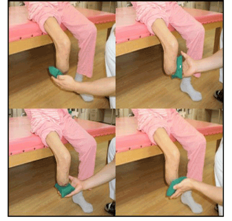
| We modified the intervention method described by Flor et al. [14], so that it could be easily used in a clinical setting. We used a soft square cushion to apply tactile stimuli and pressure to the patients’ stumps by the physical therapist. The hardness of this cushion is 107.9 N as measured by an automatic hardness tester (type JIS K6400, Asker JA). The physical therapist applied tactile stimuli to the front side, back side, left side, right side, or basal side of their stump (Figure 1). Patients were asked to indicate the location of each stimulus, and patients answered orally where the tactile stimuli applied, after which they looked to see whether their response was correct. The physical therapist kept on applying the tactile stimuli until the patients could answer. Patients performed this task about 30 times within 30 min each day, considering their tiredness. We began performing this therapeutic intervention after postoperative stump pain had subsided (M. Y. started 14 days after amputation; S. F. started 10 days after amputation). Both patients also participated in standard physical therapy consisting of range of motion exercises and gait exercises using parallel bars. |
| Result of Intervention |
| During the first session, M. Y. incorrectly located 5/10 stimuli, while S. F incorrectly located 6/10 stimuli. Neither patient was able to determine the location of tactile nor pressure stimuli applied to the stump. |
| After 18 days of discrimination task therapy, M. Y. experienced no PLP and identified the location of all tactile and pressure stimuli correctly. After 9 days of discrimination-task therapy, S. F. experienced no PLP and incorrectly identified only 2/10 stimuli. In addition, neither patient continued to experience any phantom limb phenomena and both came to feel that their leg existed only as far as the stump. Both patients were eventually able to walk independently after being fitted with a prosthetic limb and neither has experienced recurrence of PLP. |
| Discussion |
| After qualitatively assessing the PLP of 2 patients, we determined that early intervention in the form of a tactile stimulus and pressure discrimination task resulted in the elimination of the PLP. |
| The experience of pain prior to amputation increases the excitability of S1 and establishes the memory of pain [13]. Somatotopic representation in S1 may also overlap the amputated representation in S1, increasing pain signals, resulting in chronic PLP [13,17]. In the cases presented here, the patients experienced pain for a long time (years) before amputation, and the quality of their PLP was similar to the pain they experienced before amputation. We hypothesized that there was hyperexcitability of S1 pain pathways due to the long-term experience of pain prior to amputation. Inappropriate remapping in the S1 somatotopic representations resulted in the superficial PLP reported by the patients. We successfully prevented the PLP experienced by the 2 patients described here from becoming chronic by engaging them in a tactile and pressure stimulus discrimination task soon after surgery. |
| MEG and fMRI studies indicate that tactile stimulus discrimination tasks bring about reorganization in the S1 [18,19]. Additionally, the ability of patients to determine the location of tactile stimuli is correlated with S1 reorganization [19]. In both the cases described here, there was a simultaneous improvement in the perception of the stump and a reduction in PLP. The phantom limb eventually disappeared, and both patients came to feel that their leg existed only as far as their stump. We therefore believe that the S1 somatotopic representation was successfully re-mapped in these patients. Since this occurred soon after amputation, this prevented overlapping of the amputated region with adjacent regions. |
| The results of past studies on PLP treatments are highly variable. This may be due to the indiscriminate selection of PLP treatments. It is important to customize the treatment for each patient [20]. In the cases described here, we selected the method of intervention after assessing the quality the PLP experienced by the patients, and determining its mechanism. This enabled us to prevent the PLP from becoming chronic, as the intervention was tailored to the PLP pathology. This suggests that it is necessary to determine PLP pathology through qualitative assessment prior to selecting a method of intervention. However, this remains a matter of speculation, because there are few quantifiable PLP assessment tools and we did not monitor changes in S1 with fMRI or MEG. |
| This study is further limited by a lack of control over the rate of correctly identified tactile and pressure stimuli locations and the assessment of the relationship between the time-dependent change in the rate of correct answers and the time-dependent change in PLP. In order to more thoroughly investigate the effectiveness of discrimination task therapy, the number of tasks must be controlled, and a method of qualitative PLP assessment must be developed that is correlated to functional brain imaging results. |
References
- Nikolajsen L, Jensen TS (2001) Phantom limb pain. Br J Anaesth 87: 107-116.
- Ramachandran VS, Hirstein W (1998) The perception of phantom limbs. The D. O. Hebb lecture. Brain 121 : 1603-1630.
- Flor H (2002) Phantom-limb pain: characteristics, causes, and treatment. Lancet Neurol 1: 182-189.
- Melzack R (1990) Phantom limbs and the concept of a neuromatrix. Trends Neurosci 13: 88-92.
- McCabe CS, Haigh RC, Halligan PW, Blake DR (2005) Simulating sensory-motor incongruence in healthy volunteers: implications for a cortical model of pain. Rheumatology (Oxford) 44: 509-516.
- Ramachandran VS, Rogers-Ramachandran D (1996) Synaesthesia in phantom limbs induced with mirrors. Proc Biol Sci 263: 377-386.
- Mercier C, Sirigu A (2009) Training with virtual visual feedback to alleviate phantom limb pain. Neurorehabil Neural Repair 23: 587-594.
- MacIver K, Lloyd DM, Kelly S, Roberts N, Nurmikko T (2008) Phantom limb pain, cortical reorganization and the therapeutic effect of mental imagery. Brain 131: 2181-2191.
- Beaumont G, Mercier C, Michon PE, Malouin F, Jackson PL (2011) Decreasing phantom limb pain through observation of action and imagery: a case series. Pain Med 12: 289-299.
- Roux FE, Ibarrola D, Lazorthes Y, Berry I (2001) Virtual movements activate primary sensorimotor areas in amputees: report of three cases. Neurosurgery 49: 736-741.
- Ramachandran VS, Altschuler EL (2009) The use of visual feedback, in particular mirror visual feedback, in restoring brain function. Brain 132: 1693-1710.
- Sumitani M, Miyauchi S, McCabe CS, Shibata M, Maeda L, et al. (2008) Mirror visual feedback alleviates deafferentation pain, depending on qualitative aspects of the pain: a preliminary report. Rheumatology (Oxford) 47: 1038-1043.
- Flor H (2008) Maladaptive plasticity, memory for pain and phantom limb pain: review and suggestions for new therapies. Expert Rev Neurother 8: 809-818.
- Flor H, Nikolajsen L, Staehelin Jensen T (2006) Phantom limb pain: a case of maladaptive CNS plasticity? Nat Rev Neurosci 7: 873-881.
- Flor H, Denke C, Schaefer M, Grüsser S (2001) Effect of sensory discrimination training on cortical reorganisation and phantom limb pain. Lancet 357: 1763-1764.
- Huse E, Preissl H, Larbig W, Birbaumer N (2001) Phantom limb pain. Lancet 358: 1015.
- Ramachandran VS (2005) Plasticity and functional recovery in neurology. Clin Med 5: 368-373.
- Godde B, Ehrhardt J, Braun C (2003) Behavioral significance of input-dependent plasticity of human somatosensory cortex. Neuroreport 14: 543-546.
- Pilz K, Veit R, Braun C, Godde B (2004) Effects of co-activation on cortical organization and discrimination performance. Neuroreport 15: 2669-2672.
- McAvinue LP, Robertson IH (2011) Individual differences in response to phantom limb movement therapy. Disabil Rehabil 33: 2186-2195.
Figures at a glance
 |
| Figure 1 |
Relevant Topics
- Electrical stimulation
- High Intensity Exercise
- Muscle Movements
- Musculoskeletal Physical Therapy
- Musculoskeletal Physiotherapy
- Neurophysiotherapy
- Neuroplasticity
- Neuropsychiatric drugs
- Physical Activity
- Physical Fitness
- Physical Medicine
- Physical Therapy
- Precision Rehabilitation
- Scapular Mobilization
- Sleep Disorders
- Sports and Physical Activity
- Sports Physical Therapy
Recommended Journals
Article Tools
Article Usage
- Total views: 7543
- [From(publication date):
specialissue-2012 - Jul 02, 2025] - Breakdown by view type
- HTML page views : 2963
- PDF downloads : 4580
