Case Report Open Access
Early Diagnosis of Necrotizing Fasciitis of Unknown Origin: A Challenge to Prevent the Delay of Surgical Treatment
| Borja Apellaniz Aguirre*, Elena Gomez Garcia, Jose Luis Cebrian Carretero and Miguel Burgueño Garcia | ||
| University Hospital La PAZ, Madrid, Spain | ||
| Corresponding Author : | Borja Apellaniz University Hospital La PAZ, Madrid, Spain Tel: 629015310 E-mail: borja_apellaniz@hotmail.com |
|
| Received September 06, 2014; Accepted October 16, 2014; Published October 28, 2014 | ||
| Citation: Aguirre BA, Garcia EG, Cebrian Carretero JL, Garcia MB (2014) Early Diagnosis of Necrotizing Fasciitis of Unknown Origin: A Challenge to Prevent the Delay of Surgical Treatment. J Infect Dis Ther 2:174. doi:10.4172/2332-0877.1000174 | ||
| Copyright: © 2014 Aguirre BA, et al. This is an open-access article distributed under the terms of the Creative Commons Attribution License, which permits unrestricted use, distribution, and reproduction in any medium, provided the original author and source are credited. | ||
Related article at Pubmed Pubmed  Scholar Google Scholar Google |
||
Visit for more related articles at Journal of Infectious Diseases & Therapy
Abstract
Cervical Necrotizing Fasciitis is an uncommon infection. It is characterised by a rapidly progressive polymicrobial infection that spreads along the deep fascial planes of the neck. Its fulminant course and high morality makes this disease a diagnostic challenge for the maxillofacial surgeon. Therefore, when clear clinical signs and suspicious images are available, the patient must be taken to the operating room for surgical treatment. Although it is considered a rare entity, misdiagnosis is not acceptable due to its fatal outcome. Close physical examination with a thorough medical history may set the alarm. Further medical test will help to achieve the diagnosis, but never should delay the start of the appropriate treatment. We present a case of cervical necrotizing fasciitis of unknown origin successfully managed in our department. Odontogenic infection and less frequently tonsilar infections are the origin of this infection. Nevertheless, just an auricular wound could be identified as a possible source of this cervical necrotizing fasciitis. We believe that the aggressive surgical treatment combined with broad-spectrum antibiotic and intensive medical care was the key of the early recovery and few after-effects. The aim of this article is to insist in the weigh of the maxillofacial surgeon on the early surgical treatment of the cervical necrotizing fasciitis.
| Keywords |
| Cervical necrotizing fasciitis; Cervico-facial; Surgical debridement; Infection |
| Introduction |
| The Necrotizing Fasciitis (NF) is a real surgical emergency; an uncommon life-threatening infection that affects to the superficial fascia. Although head and cervical NF is rare, due to its odontogenic common origin and its need of surgical treatment to be correctly treated, the maxillofacial surgeon must be aware of this entity and his implication in the therapeutic sequence. NF should never be underestimated since its mortality can reach 30% [1]. |
| Local and systemic signs become more evident as the infection spreads. It mainly results from a polymicrobial or mixed aerobic-anaerobic infection, with a prevalence of Streptococcus species and anaerobic Bacteroides in synergistic coexistence [2]. The immune-compromising diseases such as diabetes predispose to suffer NF. Although healthy adults and children can be affected, this is relatively rare (13-31%) [3]. Therefore, clinical history will help identify predisposing factors. Blood tests, radiological imaging and surgical exploration will help to confirm the diagnosis. |
| Its fulminant course and fatal outcome makes early diagnosis and surgical treatment vital to ensure the best possibilities of survival and minimize the morbidity. The use of antibiotic without proper surgical debridement is associated with mortality close to the 100% [4]. |
| Intensive care treatment and broad-spectrum empiric antibiotic associated with the surgery are the mainstay of the treatment. Once the infection has been resolved and the patient stabilized, the reconstructive treatment can be performed. |
| Case Report |
| A 30-year-old Asian woman was derived from her primary care centre doctor due to a history of 3-day fever and preauricular erythema. No previous medical or surgical conditions were referred by the patient. She also denied any toxic habit. |
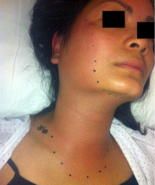
| On admission to hospital the woman was found febrile and prostrate. Right preauricular swelling and erythema, which extended over right hemifacial and high right hemicervical region (Figure 1). No odontogenic origin was identified or any other infectious source except for a 0.5 cm wound on the right ear helix with phlogotic signs. |
| The patient’s blood pressure (BP) was 90/60 mmHg and heart rate (HR) 100 beats per minute (bpm). The white blood cell count (WBCC) was 15.0x10³/µL with an 89.4% of neutrophils; C reactive protein (CRP) was 183 mg/L; the glucose 80 mg/dL, potassium 2,7 mmol/L and creatinine 0.79 mg/dl. The creatine kinase (CK) levels were in normal range (33 UI/L). |
| Empiric antibiotic therapy was started with Amoxicillin-Clavulanic acid. Within 4 hours since the admission, the clinical state of the patient worsened. The erythema reached the supraclavicular space and extended across the middle line. The BP had fallen to 80/55 mmHg and a 113 bpm HR was measured. New blood tests were taken with a WBCC of 24x103/µL with a 94.5% of neutrophils, CRP195 mg/L and creatinine of 0.67 mg/dl. CK levels stayed normal. |
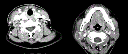
| Therefore blood cultures were obtained and CT scan was performed. Right hemicervical superficial fascial thickening was observed with trabeculation of the adjacent fat. The number of right cervical lymphatic nodes was significantly increased, but with no pathological appearance. No subcutaneous emphysema or fluid collection was noticed (Figure 2). |
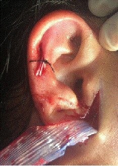
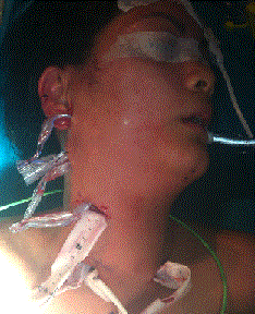
| With clinical criteria of septic shock, the patient was taken to the operating room (OR). Orotracheal intubation to preserve the airway was performed. Under general anaesthesia, several skin incisions where performed in the preauricular (Figure 3) and cervical area; followed by blunt dissection in a subplatismal plane (Figure 4). Although no necrosis was identified, a cloudy serous liquid was observed. The microbiology department cultured samples taken from the surgical field. No specimen for pathology examination was send. Drainages were placed and secured. No tracheostomy was performed because of the early state, no airway involvement and the aggressive surgical treatment applied. The patient was transferred intubated to a surgical intensive care unit where triple empiric antibiotic therapy was started with Meropenem, Linezolid and Clindamycin, as clinical guides recommended. After 48 hours the vasoactive drugs were suspended and she was extubated. No sign of skin necrosis was detected. 24 hours later, the patient was transferred to the maxillofacial hospitalization floor were she kept stable. |
| The infectious disease unit decided to discontinue the Linezolid on day 7th post surgery, since no previous contact with hospital environment was detected neither Methicillin Resistant Staphylococcus aureus (MRSA) colonization. All microbiological cultures were sterile. Clinically the patient was stable and the blood test showed tendency to normal ranges after 14 days of hospitalization. The drainages were removed and the wounds healed by second intention. On the 15th day the PCR was 8.65 mg/L and 8.5x10³/µL WBCC with a 61% of neutrophils. Clindamycine and Meropenem were suspended and oral Amoxicillin-Clavulanic acid was started because of its suitability against orofacial pathogens. On the 18th day the blood test showed a WBCC 5.6x10³/µL with a 56.4% of neutrophils. The RCP was in normal ranges (<2.9 mg/L). So, the patient was discharged with oral Amoxiclin-Clavulanic acid for 1 more week and kept under close follow up the weeks after with no complication. |
| Discussion |
| The necrotizing fasciitis (NF) is an acute soft tissue infection characterized by its fulminant course and high mortality. Necrosis of the superficial fascia takes place while clinical signs of septic shock become more evident. Therefore, early diagnosis becomes the key to provide proper surgical and medical treatment. |
| This clinical entity has been known with different names including gangrenous ulcer, gangrenous erysipelas, necrotizing cellulitis, phagedenis ulcer, Fournier’s gangrene, hospital gangrene and Melaney’s gangrene. But it was not until 1952 when Wilson first used the term NF [5]. |
| Most commonly the NF affects the abdominal, perineum and limbs. Less than 2, 6-5% of the cases involve the cervicofacial region known as cervicofacial necrotizing fasciitis (CNF) [6]. In these cases, a precipitant event can be identified. The odontogenic infection is the most common cause in the published series [7,8]. Other types of head and neck infections have been reported as possible causes: tonsilar abscess, parotiditis, otitis media trauma or surgery of the head and neck [9,10]. |
| Bacteria cause tissue necrosis along less-vascular planes such as muscle fascia and dissection proceeds rapidly along the potential spaces of the neck [11]. The NF is comprised of three different bacteriologic entities. Type I consist of an aerobic and anaerobic polymicrobial infection. Usually anaerobic species such as Bacteroides, Clostridium or Peptostreptococcus are isolated with one or more facultative anaerobic Streptococci (other than group A) and members of the Enterobacteriaceae. When we focus on the CNF, mouth anaerobes such as Fusobacteria, anaerobic Streptococci, Bacteroides and Spirochetes can be found. Type II is considered a group A Streptococcal monomicrobial infection. But it can be found in combination with other species, most commonly, S. aureus. Type III is a rare type due to a marine vibrio. The CNF commonly is associated with oral or pharyngeal mixed aerobic-anaerobic flora (Streptococcus and anaerobic Bacteroides). Methicillin resistant S. aureus (MRSA) has been isolated in 39% on NF [12]. Therefore, MRSA directed antibiotics should be considered promptly. |
| The host commonly presents certain disorders that can make him more susceptible to this infection. Up to 89,4% in some series had systemic diseases [13]. The immune-compromised patients have a higher risk of developing NF. Systemic diseases such as diabetes, AIDS, cirrhosis, lymphoma, renal failure or toxic habits such us drug use, alcoholism or tobacco abuse are risk factors to be consider due to micro vascular alteration. |
| NF can progress from a small odontogenic infection to an extensive skin necrosis associated to septic shock and fatal outcome. Bacteria cause tissue necrosis along less-vascular planes such as muscle fascia, and dissection proceeds rapidly along the potential spaces of the neck. Due to its fulminant evolution, maxillofacial surgeons must be alert to identify real NF from routine cellulitis. |
| Usually, the systemic signs and symptoms are disproportioned to the skin necrosis. Local infection is associated to phlogotic signs and sever pain. But it is not until the spread through the fascial plane takes place when the patient starts to become clinically ill, with septic shock signs such as hypotension, tachycardia and malaise. The onset is gradual, but subsequently may spread rapidly. |
| Skin emphysema can be associated when gas-forming organisms are involved. If no treatment is started, skin necrosis will start and lead to organ failure (acute respiratory distress syndrome, renal or haematological). The infection typically spreads along the muscle fascia because of its relatively poor blood supply. That is why overlying skin can be little affected, and surgical intervention is mandatory to confirm the NF diagnosis. Early in the infection, necrosis and liquefaction of the fascia and fat occur, followed by venous thrombosis, inflammatory cell infiltration (abscess), and arterial compromise due to endarteritis obliterans of the nutrient vessels [14]. |
| Wang et al. proposed a clinical staging based on the skin signs. Stage I involves inflammatory signs of erythema, tenderness, heat and swelling. Stage II is considered when blistering and bullae appears in the surface. Final stage III includes crepitus, skin anaesthesia and skin necrosis [15]. |
| In early stages care must be taken to differentiate from erysipelas, where the superficial layer of the skin and lymphatic are affected or cellulitis where the deeper subcutaneous tissue is involved. None of those has the fascia altered. |
| Radiographic imaging can be handful to reach a NF diagnosis. Nevertheless, this test should never delay when clinical evidence of soft tissue infection progression or crepitus is observed. Soft tissue X-ray will be helpful to detect gas. CT scan will be able to detect constant features such as: diffuse thickening and infiltration of the cutis and subcutis, diffuse enhancement and/or thickening of the superficial and deep cervical fascia, enhancement and thickening of platysma, sternomastoid and strap muscles and fluid collections in multiple neck compartment [16]. If anaerobic bacteria are involved, gas collection may be visible. The presence of rapidly progressive infection and systemic response should not exclude prompt surgical exploration based on a CT with negative findings regarding the presence of gas [17]. But a normal CT does not rule out NF [2]. |
| The first step of management involves prompt resuscitation of the patient, followed by aggressive surgery as the next step to be done. Once the first surgery has been performed, critical care support with adjuvant antibiotics will help to resolve the infection. Finally the reconstructive treatment can be planned once the infection has been stopped. |
| The patient needs fast resuscitation treatment allowing the patient to be taken to the OR as soon as possible. A combination of fluids, vasoactive drugs, insulin and electrolyte correction will make the surgery possible. Usually these patients need several surgeries and long stay under sedative drugs; therefore, early tracheotomy is recommended [18]. Antibiotic can be initiated at this time, but never delaying surgical treatment. |
| Upper airway management is essential in the resuscitation stage. Obtaining secure airway may be difficult due to neck rigidity, trismus, pooled secretions and possible discharge of pus and fluid collection. Therefore, an awake fiberoptic intubation is the safest way to intubate these patients trachea. A blind nasal intubation with the patient breathing spontaneously is an acceptable alternative [18]. Multiple surgeries may be expected, therefore, early tracheostomy has been advocated for obtaining a secure airway, that also helps to ease the postoperative care in the ICU [19]. But as in our case, when prompt surgical treatment is performed with little airway compromise and a short ICU stay is expected, tracheostomy may be avoided. |
| Surgery is the corner stone of the treatment [3]. All the authors agree that early surgical treatment is essential [9,20]. Once the CNF is evident such us in stages II and III the surgery must be performed as soon as possible. Stage I could be unnoticed. But when skin inflammatory sings and deep fascial involvement with systemic affectation is detected, surgical exploration should be done to confirm the diagnosis . The sooner the surgical debridement is undertaken, the better the prognosis [9]. The main goal is to remove all the necrotic tissue. The debridement must be performed until viable tissue is exposed. Samples for gram staining and culture should be obtained. All the liquid collections must be drained. Reluctance to aggressive surgery often leads to under treatment. So, bilateral cervicotomy should be performed allowing complete excision and debridement of the fascia, subcutaneous and all devitalized tissues [14]. |
| Drainages should be placed so no secretion stays in the fascial planes and may provide bacterial culture medium. Delay of debridement and insufficient drainage are the primary causes of high mortality [21]. |
| Besides surgery, broad-spectrum empiric antibiotics should be started as soon as possible. As we mentioned before, most commonly involved microorganism are group A Streptococcus and anaerobes. We should never forget the MRSA increase in our medium. Therefore, the most suitable antibiotic therapy should include a Carbapenem or beta-lactamic+beta lactamase inhibitor together with Clindamycin for its antitoxin effects against toxin elaborating strains of Streptococci and Staphylococci. An agent against MRSA such as Linezolid, Vancomycin or Daptomycin is highly recommended. Treatment duration has not been defined, but it should be adjusted according to the microbiological cultures and clinical evolution. The literature recommends keep it until further debridement is not necessary and clinically the patient remains stable. |
| Conclusion |
| Early diagnosis of CNF can be a challenge in the emergency department. Its incipient signs and symptoms can lead to misjudge this infection. But once it is established, its fast spread and systemic repercussion can make it more difficult to handle it increasing the mortality alarmingly. |
| Medical treatment is essential to treat the infection. But when applied on an isolated treatment design it becomes useless. Surgical treatment always must be part of the therapeutic plan. It’s aggressive an early performance is demonstrated to increase the survival. |
| We believe that our patient hospital-stay and single surgery was due to the fast onset of the surgical and medical treatment. Early drainage avoided the progression of the necrosis and the following consequences. When the patient was taken to the OR was in stage I, but no tracheostomy was performed because of the thorough debridement performed that avoided more reoperations. |
References
- Malik V, Gadepalli C, Agrawal S, Inkster C, Lobo C (2010) An algorithm for early diagnosis of cervicofacial necrotising fasciitis. EurArch Otorhinolaryngol 267: 1169-1177
- Leyva P, Herrero M, Eslava JM, Acero J (2013) Cervical necrotizing fasciitis and diabetic ketoacidosis: literature review and case report. IntJ Oral MaxillofacSurg 42: 1592-1595.
- Sahoo NK, Tomar K (2014) Necrotizing fasciitis of the cervico-facial region due to odontogenic infection. J Oral Maxillofac Surgery, Med Pathol 26: 39-44.
- Anaya DA, Dellinger EP (2007) Necrotizing soft-tissue infection: diagnosis and management. Clin Infect Dis 44: 705-710.
- Wilson B (1952) Necrotizing fasciitis. Am Surg 18: 416-431
- Wong C-H, Chang H-C, Pasupathy S, Khin L-W, Tan J-L, et al. (2003) Necrotizing fasciitis: clinical presentation, microbiology, and determinants of mortality. J Bone Joint Surg Am85-A:1454-1460.
- Whitesides L, Cotto-Cumba C, Myers RA (2000) Cervical necrotizing fasciitis of odontogenic origin: a case report and review of 12 cases. J Oral MaxillofacSurg 58: 144-1451
- Tung-Yiu W, Jehn-Shyun H, Ching-Hung C, Hung-An C (2000) Cervical necrotizing fasciitis of odontogenic origin: a report of 11 cases. J Oral MaxillofacSurg 58: 1347-1352
- Ord R, Coletti D (2009) Cervico-facial necrotizing fasciitis. Oral Dis 15: 133–141.
- Antunes AA, Avelar RL, de Melo WM, Pereira-Santos D, Frota R (2013) Extensive cervical necrotizing fasciitis of odontogenic origin. J CraniofacSurg 24: e594-597.
- Sarna T, Sengupta T, Miloro M, Kolokythas A (2012) Cervical necrotizing fasciitis with descending mediastinitis: literature review and case report. J Oral MaxillofacSurg 70: 1342-1350.
- Lee TC, Carrick MM, Scott BG, Hodges JC, Pham HQ (2007) Incidence and clinical characteristics of methicillin-resistant Staphylococcus aureusnecrotizing fasciitis in a large urban hospital. Am J Surg 194: 80-812.
- Lin C, Yeh FL, Lin JT, Ma H, Hwang CH, et al. (2001) Necrotizing fasciitis of the head and neck: an analysis of 47 cases. PlastReconstr Surg. 107: 1684–1693.
- Cruz Toro P, Callejo Castillo A, TorneroSaltó J, González Compta X,Farré A, et al. (2014) Cervical necrotizing fasciitis: Report of 6 cases and review of literature. Eur Ann Otorhinolaryngol Head NeckDis
- Wang YS, Wong CH, Tay YK (2007) Staging of necrotizing fasciitis based on the evolving cutaneous features. Int J Dermatol 46: 1036-1041.
- Becker M, Zbären P, Hermans R, Becker CD, Marchal F, et al (1997) Necrotizing fasciitis of the head and neck: role of CT in diagnosis and management. Radiology 202: 471-476.
- McMahon J, Lowe T, Koppel DA (2003) Necrotizing soft tissue infections of the head and neck: case reports and literature review. Oral Surg Oral Med Oral Pathol Oral RadiolEndod95: 30-37.
- Durrani MA, Mansfield JF(2003) Anesthetic implications of cervicofacial necrotizing fasciitis. J ClinAnesth 15: 378-381.
- Kantu S, Har-El G (1997) Cervical necrotizing fasciitis. Ann OtolRhinolLaryngol 106: 965-970.
- Martínez AY, McHenry CR, MenesesRivadeneira L (2014) Fasceítisnecrosantecervicofacial: unainfecciónseveraquerequieretratamientoquirúrgicotemprano. Rev Española Cirugía Oral y Maxilofa.
- Sandner A, Börgermann J, Kösling S, Silber RE, Bloching MB (2007) Descending necrotizing mediastinitis: early detection and radical surgery are crucial. J Oral MaxillofacSurg 65: 794-800.
Figures at a glance
 |
 |
 |
 |
|||
| Figure 1 | Figure 2 | Figure 3 | Figure 4 |
Relevant Topics
- Advanced Therapies
- Chicken Pox
- Ciprofloxacin
- Colon Infection
- Conjunctivitis
- Herpes Virus
- HIV and AIDS Research
- Human Papilloma Virus
- Infection
- Infection in Blood
- Infections Prevention
- Infectious Diseases in Children
- Influenza
- Liver Diseases
- Respiratory Tract Infections
- T Cell Lymphomatic Virus
- Treatment for Infectious Diseases
- Viral Encephalitis
- Yeast Infection
Recommended Journals
Article Tools
Article Usage
- Total views: 17042
- [From(publication date):
December-2014 - Nov 21, 2024] - Breakdown by view type
- HTML page views : 12562
- PDF downloads : 4480
