Case Report Open Access
Early Diagnosis and Management of Extracranial Carotid Vasculopathy in Mitigating Neurological Complications of Sickle Cell Disease
| Sri Hari Sundararajan* | |
| Rutgers Robert Wood Johnson Medical School, New Brunswick, USA | |
| Corresponding Author : | Sri Hari Sundararajan Rutgers Robert Wood Johnson Medical School of Radiology MEB 404, New Brunswick, NJ 08093, USA Tel: 6469155621 E-mail: ssundararajan@univrad.com |
| Received: July 02, 2015 Accepted: July 16, 2015 Published: July 20, 2015 | |
| Citation: Sundararajan SH (2015) Early Diagnosis and Management of Extracranial Carotid Vasculopathy in Mitigating Neurological Complications of Sickle Cell Disease. OMICS J Radiol 4:195. doi:10.4172/2167-7964.1000195 | |
| Copyright: © 2015 Sundararajan SH. This is an open-access article distributed under the terms of the Creative Commons Attribution License, which permits unrestricted use, distribution, and reproduction in any medium, provided the original author and source are credited. | |
Visit for more related articles at Journal of Radiology
Abstract
While cerebral vasculopathy is a well-known early complication of sickle cell disease that has warranted transcranial doppler surveillance, little has been documented regarding the role of cervical circulation in the development of cerebrovascular events and transient ischemic attacks amongst sickle cell patients. We present the unique case of a symptomatic 18-year-old male with sickle cell disease found to have critical cervical carotid stenosis in the absence of significant intracranial vasculopathy. Increased awareness of the potential role of the extracranial circulation in the development of sickle cell related cerebrovascular events is warranted.
|
Abstract
While cerebral vasculopathy is a well-known early complication of sickle cell disease that has warranted transcranial doppler surveillance, little has been documented regarding the role of cervical circulation in the development of cerebrovascular events and transient ischemic attacks amongst sickle cell patients. We present the unique case of a symptomatic 18-year-old male with sickle cell disease found to have critical cervical carotid stenosis in the absence of significant intracranial vasculopathy. Increased awareness of the potential role of the extracranial circulation in the development of sickle cell related cerebrovascular events is warranted.
Introduction
The neurologic manifestations of sickle cell disease include early onset of cerebral ischemia, transient ischemic attacks, and stroke [1]. The most common secondary diagnosis associated with transient ischemic attack development in children is sickle cell disease, with other associated diagnoses including congenital heart disease, migraine, moyamoya disease, and stroke from other thromboembolic phenomena [2]. The peak incidence of overt stroke in sickle cell disease is 1.02 per 100 patient years in patients between the ages 2 and 5 [3]. Silent cerebral infarctions are, however, more common than overt stroke, occurring in up to 37% of patients with either heterozygote or homozygous expression of the sickle cell gene [3]. It is recommended that all sickle cell patients undergo annual transcranial doppler for evaluation of dynamic or fixed cerebral arterial stenoses from age 2 to 16 years. This has become the standard of care after results of the Stroke Prevention Trial in Sickle Cell Anemia study demonstrated regular blood transfusions reduce the risk of stroke by 90% in children with elevated intracranial blood velocities [3]. However, assessment of the cervical vasculature is not yet part of routine management of sickle-cell disease patients. We present the case of a young male patient with sickle-cell disease, who despite prior intracranial imaging demonstrating stable changes, was found to have a critical left internal carotid arterial stenosis. The patient provided consent for use of the case history and imaging for publication purposes.
Case Presentation
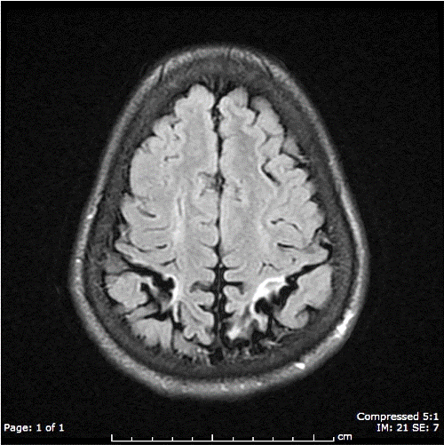
This is an 18-year-old African-American male with homozygous sickle cell disease on chronic packed red blood cell transfusion given mildly elevated cerebral artery velocities noted in 2002 during transcranial doppler surveillance. Follow-up imaging since then have reconfirmed stable silent bilateral chronic parietal lobe infarcts and volume loss out of proportion to patient’s age (Figure 1). Given the long-term stability of repeat transcranial doppler and MRI brain exam results, an MRA carotid exam was ordered to establish the patient’s baseline carotid anatomy.
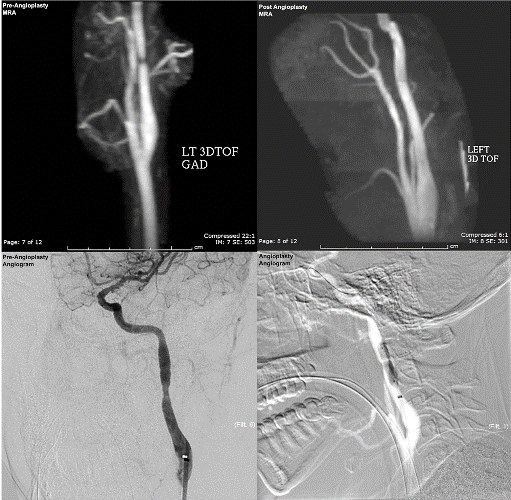
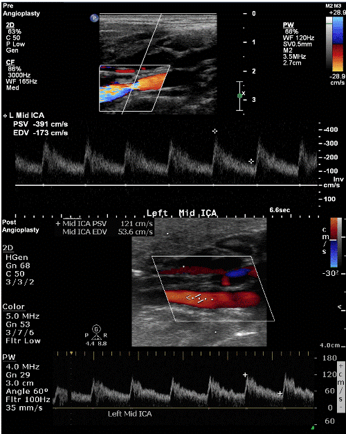
Surprisingly, the carotid MRA exam demonstrated a >80% stenosis within the mid left cervical ICA (Figure 2). Carotid duplex interrogation confirmed an 80-99% concentric stenosis in the mid left ICA, with focal fibrosis of the vessel wall and elevated peak-systolic and diastolic velocities (Figure 3). The patient was deemed high risk for irreversible carotid occlusion due to the severity of the left carotid stenosis. Baseline platelet function was qualified with the VerifyNow System platelet reactivity test (Accumetrics®, California, USA) before Plavix 75 mg daily was initiated alongside Aspirin 81 mg daily. Endovascular evaluation and treatment was offered to minimize risk of debilitating stroke. A focal band with change in vessel caliber in the mid left ICA at C3-C4 was identified during angiography (Figure 2). While central lucency suggestive of webbing within the stenotic lumen was appreciated, the degree of stenosis was apparently less severe than expected based on carotid duplex and MRA examinations. Despite its less impressive angiographic appearance, we pursued left carotid angioplasty given parameters corresponding to severe stenosis on carotid duplex and MRA. Using roadmap technique, a 5-mm diameter Emboshield NAV6 distal protection device (Abbott Vascular, Illinois, USA) was maneuvered through the left ICA stenosis and opened in the high cervical ICA (Figure 2). There was notable decrease in the left ICA severe stenosis following angioplasty with Viatrac 5 × 20 and 6 × 20 mm balloons (Abbott Vascular, Illinois, USA). Given this improvement, we felt that placement of a stent would not be necessary. No complications occurred during the procedure. Post-angioplasty carotid duplex and MRA exams demonstrated significant improvement in left ICA hemodynamic parameters and resolution of the high-grade stenosis (Figures 2 and 3). Discussion
The presence of extracranial internal carotid stenosis has been associated with clinical stroke. Elevated extracranial internal carotid velocities in sickle cell patients have been previously correlated with younger age, higher rates of hemolysis, elevated white blood cell count, severe extracranial internal carotid stenoses, and even dissections. Such extracranial stenoses may explain why some strokes occur without an immediately evident intracranial vasculopathy [3,4].
Our patient’s carotid duplex and MRA examinations demonstrated severe left mid internal carotid artery stenosis. While only a moderate change in vessel caliber was appreciated on catheter-based angiography, post angioplasty carotid duplex demonstrated the peak systolic velocity of the mid left ICA decreased by 69% of its original value. This was reaffirmed on post-procedural MRA, which showed resolution of the high-grade stenosis. The degree of stenosis during catheter-based angiography was likely not as striking given the radiolucency of the underlying webbing. This case uniquely demonstrates how carotid duplex and MRA examination allowed for a more accurate assessment of improvement in our patient’s internal carotid artery stenosis. The pathogenesis of cerebral vasculopathy in sickle cell disease is multifactorial. Contributory mechanisms include shearing intimal injury from deformed red blood cells, decreased nitric oxide availability, and up regulation of fibroproliferative and inflammatory cytokines related to hypoxia. The end result of these mechanisms is the creation of progressive hemodynamically significant stenosis from intimal damage and reactive tubular intimal fibroplasia [5]. We find it noteworthy that the vascular changes described in sickle cell patients appear similar to those described in fibromuscular dysplasia of the intimal layer. Fibromuscular dysplasia is an uncommon non-atherosclerotic disorder that results in variable degrees of flow impedance dependent on the layer and severity of abnormal collagen proliferative deposition. Medial and perimedial FMD are the most common cervicocephalic subtypes accounting for 75-80% and 10-15% of cases respectively. Their string of beads appearance in which the beading diameter is either larger (medial) or small (perimedial) than the involved artery differentiates them. Intimal fibromuscular dysplasia is a rare subtype that accounts for <10% of cases, creating variable degrees of tubular stenosis akin to the narrowing demonstrated in our patient’s angiography [6,7]. Cervical internal carotid dissections have also been reported in sickle-cell patients [4]. Given our patient’s short segment high-grade stenosis, it is likely the left ICA stenosis was further propagated by focal arterial dissection. Perhaps the irregularly shaped red-blood cells flowing through such vessels instigate the progressive intimal fibroproliferative changes of intimal fibromuscular dysplasia that, in time, lead to arterial dissection and critical stenosis development. Conclusion
Pediatric patients with sickle cell disease may benefit from not only cranial imaging, but also cervical vasculature imaging via doppler and MRA examination. We conclude by proposing that carotid duplex be performed at the time of transcranial doppler screening. This allows for previously unrecognized sickle cell disease patients with hemodynamically significant extracranial carotid stenosis to benefit from optimal anticoagulation and interventions such as angioplasty. These measures were thought to improve our patient’s quality of life and reduce future neurological episodes. Prospective trials are needed to establish the medical validity of implementing great vessel imaging in the diagnostic paradigm for stratifying stroke risk in sickle cell disease patients.
References
|
Figures at a glance
 |
 |
 |
| Figure 1 | Figure 2 | Figure 3 |
Relevant Topics
- Abdominal Radiology
- AI in Radiology
- Breast Imaging
- Cardiovascular Radiology
- Chest Radiology
- Clinical Radiology
- CT Imaging
- Diagnostic Radiology
- Emergency Radiology
- Fluoroscopy Radiology
- General Radiology
- Genitourinary Radiology
- Interventional Radiology Techniques
- Mammography
- Minimal Invasive surgery
- Musculoskeletal Radiology
- Neuroradiology
- Neuroradiology Advances
- Oral and Maxillofacial Radiology
- Radiography
- Radiology Imaging
- Surgical Radiology
- Tele Radiology
- Therapeutic Radiology
Recommended Journals
Article Tools
Article Usage
- Total views: 14091
- [From(publication date):
August-2015 - Mar 29, 2025] - Breakdown by view type
- HTML page views : 9565
- PDF downloads : 4526
