Case Report Open Access
Duplication of Gall bladder: Review of Literature and Report of a Case
Hoda Salah Darwish*
Department of radio-diagnosis, Faculty of medicine. Suez Canal University, Egypt
- *Corresponding Author:
- Hoda Salah Dariwsh
Department of radio-diagnosis
Faculty of medicine. Suez Canal University, Egypt
Tel: 00966 0558858648
E-mail: darwish.hoda.@yahoo.com
Received date: June 03, 2016; Accepted date: June 27, 2016; Published date: June 30, 2016
Citation: Darwish HS (2016) Duplication of Gall bladder: Review of Literature and Report of a Case. OMICS J Radiol 5:226. doi:10.4172/2167-7964.1000226
Copyright: © 2016 Darwish HS. et al. This is an open-access article distributed under the terms of the Creative Commons Attribution License, which permits unrestricted use, distribution, and reproduction in any medium, provided the original author and source are credited.
Visit for more related articles at Journal of Radiology
Abstract
The gallbladder duplication consider a rare anatomic malformation, it can now be detected by preoperative multiple imaging study. The preoperative diagnosis is very important to avoid potential damage to the duct system during surgery. We preferred Ultrasound which is modality of imaging. Surgical treatment consists of the removal of both gallbladders to prevent later complications. We report one case of congenital double gall-bladder as it is very rare which were demonstrated radio logically and proved at surgery.
Keywords
Double gallbladder; Gallbladder duplication
Introduction
The gallbladder is one of the most our body organs subject to anatomical variations; that may be related to number, shape and position and may also affect the cystic duct and cystic artery [1]. Duplication of the gallbladder is a rare congenital malformation, occurring in about one per 4000 births [1]. It associated with an increased risk of complications after laparoscopic cholecystectomy [2]. Preoperative diagnostic imaging should be helpful. The diagnosis easily missed intraoperatively, especially laparoscopically, if there is no prior knowledge of the exact abnormality [3]. Ultrasound has a high sensitivity and specificity and it is the preferred modality of imaging [4] . CT scan and MRI are the nonvasive modality and can be used to delineate the anatomy [5].
Case Report
A 26 years old man presented as an emergency with acute right hypocondrium pain. He was complaining of anorexia and weight loss. The routine blood test, revealed high bilirubin level which was raised at 44 umol/l.
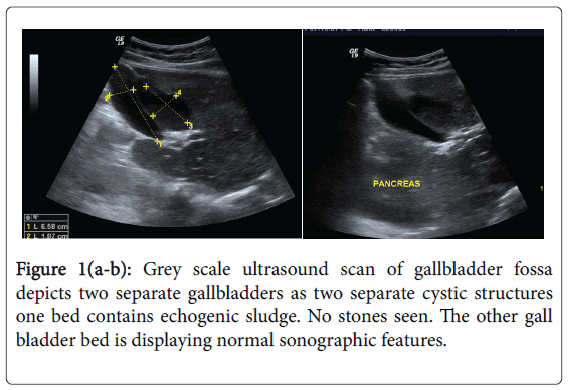
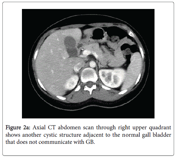
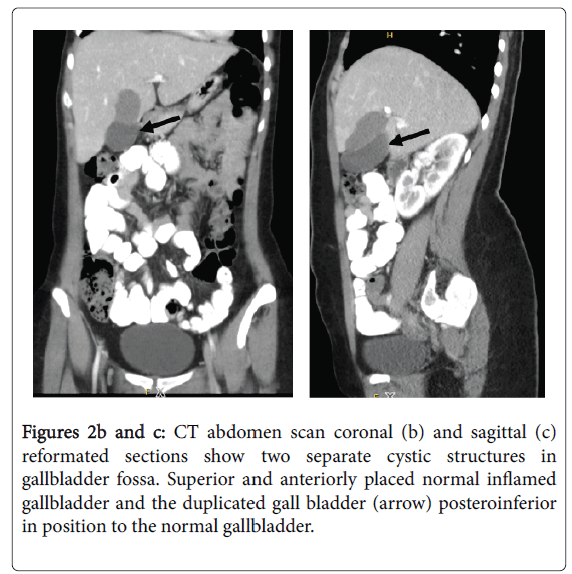
Abdominal Ultrasound scan demonstrated no stones with small amount of echogenic sludge within the gallbladder (Figure 1). Normal gallbladder wall thickness and there was no evidence of intra or extra hepatic biliary tree dilatation. Another cystic structure was seen with normal wall thickness and clear content adjacent to it. CT abdomen (Figures 2a, 2b and c) and the intraoperative findings confirm the diagnosis of gall bladder duplication with inflammation of one bed and clear the bed. The report of histolopathology confirmed the specimen to be a chronically mild inflamed gallbladder with no stones.
Discussion
Double gallbladder is a rare congenital anomaly with an incidence of 1:4000 patients [1]. There are several literature reported double and also triple gallbladder [3]. Several classifications have been proposed according to anatomic or embryologic development of the gallbladder. Double gallbladders are classified according to the Boyden's classification [1]. The two main types of duplications are vesica fellea divisa (bilobed gallbladder) and, vesica fellea duplex (true duplication) which is more common, with two different cystic ducts. However the true duplication is sub classified into H shaped type which entails two separate gallbladders and cystic ducts entering separately into the common bile duct; and another Y shaped type, where the two cystic ducts unites before entering the common bile duct [3].
The accessory gallbladder of ductular type may be adjacent to the normal organ or may be intrahepatic, subhepatic or within the gastrohepatic ligament. The true duplication is more common, occurs during the 5th and early 6th week of embryonic life due to bifurcation of gallbladder primodium [1,5-7]
In our patient we found a complete duplication of the gallbladder with its own cystic artery and duct entering the common bile duct at lower level than normal [3]. The differential diagnosis of double gallbladder include the following gallbladder fold, focal adenomyomatosis phrygian cap, intraperitoneal fibrous (Ladd's) bands, choledochal cyst, pericholecystic fluid and gallbladder diverticulum [8].
Diagnosis includes various modalities like ultrasound, Oral cholecystogram (OCG), scintigraphy, ERCP, PTC, CT scan and MRI. OCG and scintigraphy depend on certain conditions such as hepatobiliary uptake and excretion with a patent cystic duct. ERCP and PTC are the invasive procedures and will be used rarely. Ultrasound imaging is the modality of choice, with a high sensitivity and specificity. CT scan and MRI are the nonvasive modality and can be used to delineate the anatomy [5]. Congenital duplication of gall bladder tends to lead to biliary complications, such as cholelithiasis and acute cholecystitis of both gallbladders. The clinical features are usually RUQ pain and tenderness and sometimes jaundice [9]. In symptomatic patient, cholecystectomy is recommended with the excision of both is recommended [5].
Conclusion
Double gallbladder is a rare congenital abnormality. Pre-operative diagnosis is very important not to miss the second gallbladder. We are reporting a case of double gallbladder as it is very rare.
References
- Boyden EA (1926) The accessory gallbladder: an embryological and comparative study of aberrant biliary vesicles occuringin man and the domestic mammals. Am J Anat 38: 177-231.
- Gigot J, Van Beers B, Goncette L, Etienne J, Collard A, et al. (1997) Laparoscopic treatment of gallbladder duplication: a plea for removal of both gallbladders.Surg Endosc 11: 479-482.
- Lefemine V, Lazim TR (2009) Neuroma of a double gallbladder: a case report. Cases J 2:11
- Tiwary SK, Agarwal S, Singh S, Khanna AK (2006) Double Gallbladder. The Internet Journal of Gastroenterology.
- Mazziotti S, Minutoli F, Blandino A, Vinci S, Salamone I, et al. (2001) Gallbladder duplication: MR cholangiography demonstration. Abdom Imaging 26: 287-289.
- Ozgen A, Akata D, Arta A, Demirkazik FB, Ozmen MN, et al.(1999) Gallbladder duplication: imaging findings and differential considerations. Abdom Imaging 24: 285-288.
- Gray SW, Skandalphia JE (1974) Embryology for Surgeons. Obstetrics & Gynecology 44: 786.
- Goiney RC, Schoenecker SA, Cyr DR, Shuman WP, Peters MJ, et al. (1985) Sonography of gallbladder duplication and differential consideration AJR Am J Roentgenol 145: 241-243.
- Hishinuma M, Isogai Y, Matsuura Y, Kodaira M, Oi S, Ichikawa N, et al. (2004) Double gallbladder. J Gastroenterol Hepatol 19: 233-235.
--
Relevant Topics
- Abdominal Radiology
- AI in Radiology
- Breast Imaging
- Cardiovascular Radiology
- Chest Radiology
- Clinical Radiology
- CT Imaging
- Diagnostic Radiology
- Emergency Radiology
- Fluoroscopy Radiology
- General Radiology
- Genitourinary Radiology
- Interventional Radiology Techniques
- Mammography
- Minimal Invasive surgery
- Musculoskeletal Radiology
- Neuroradiology
- Neuroradiology Advances
- Oral and Maxillofacial Radiology
- Radiography
- Radiology Imaging
- Surgical Radiology
- Tele Radiology
- Therapeutic Radiology
Recommended Journals
Article Tools
Article Usage
- Total views: 15722
- [From(publication date):
June-2016 - Mar 29, 2025] - Breakdown by view type
- HTML page views : 14732
- PDF downloads : 990


