Case Report Open Access
Duodenal Hookworm- An Endoscopic Diagnosis
| Vikas Singla, Varun Gupta*, Ashish Kumar, Praveen Sharma, Naresh Bansal and Anil Arora | |
| Department of Gastroenterology and Hepatology, Sir Ganga Ram Hospital, New Delhi, India | |
| *Corresponding Author : | Varun Gupta Department of Gastroenterology and Hepatology Sir Ganga Ram Hospital, New Delhi, India E-mail: varun_doc@yahoo.co.in |
| Received: January 07, 2016 Accepted: February 17, 2016 Published: February 22, 2016 | |
| Citation: Singla V, Gupta V, Kumar A, Sharma P, Bansal N, et al. (2016) Duodenal Hookworm- An Endoscopic Diagnosis. J Infect Dis Ther 4:268. doi:10.4172/2332-0877.1000268 | |
| Copyright: © 2016 Singla V, et al. This is an open-access article distributed under the terms of the Creative Commons Attribution License, which permits unrestricted use, distribution, and reproduction in any medium, provided the original author and source are credited. | |
Visit for more related articles at Journal of Infectious Diseases & Therapy
Abstract
Hookworm infections are not an uncommon cause of anemia and can affect any age group. Usually a diagnosis is made by identifying eggs in the stool samples. In this case of 61 years old male, who was suffering from anemia for last one year without any identifiable cause we were able to identify hookworm infestations in the duodenum by performing an upper gastrointestinal endoscopy. Patient recovered completely by giving a single stat dose of albendazole. Gastrointestinal endoscopy can be useful in diagnosing easily treatable hookworm infection if other modalities are inconclusive.
| Keywords |
| Hookworm; Endoscopy; Albendazole |
| Case Report |
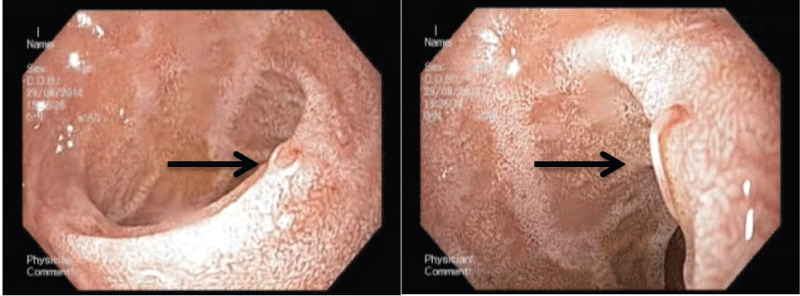
| A 61 years old male presented with generalized weakness and easy fatigability for last one year. He was evaluated and was diagnosed to have anemia, for which he was being managed symptomatically with iron and folic acid supplements along with three units of packed cell transfusions in last one year. There was no history of hematemesis, melena, or blood loss from any other site. At our center, his investigation revealed iron deficiency anemia (Hemoglobin 9.2 gm/ dL). Stool for occult blood was positive on three separate samples but stool for ova and cyst was normal at all the three occasions. On suspicion of carcinoma colon, colonoscopy examination was done which was unremarkable. Subsequently, an upper GI endoscopy was done which revealed multiple hookworms buried partially in the wall of bulb and second part of duodenum and the sucked blood could be appreciated in the cavity (pseudocoelom) of hookworms. Antihelminthic (Albendazole 400 mg, as a single dose) and iron supplements (for 3 months) were prescribed to the patient. On follow up, he improved clinically within 4 weeks, and his hemoglobin increased spontaneously to 13 gm/dl after 3 months. |
| Ancylostoma duodenale and Necator americanus are the hookworm species affecting the human beings. An Italian physician, Dubini, first described hookworm in 1838, after an autopsy. Hookworm is transmitted through penetration of intact skin by the third stage larva, though the swallowed larva of A. duodenale can also develop in full adult. After skin penetration, larva reaches heart and enters lung capillaries and then is swallowed to the proximal intestine, where it develops in adult worms, which are about one cm in length. The most common disease in humans is iron deficiency anemia, which occurs due to mechanical sucking of blood and due to blood loss secondary to release of anticoagulants by the worm. |
| Diagnosis is usually made by evaluation of stool for the eggs. Recently, newer small bowel imaging modalities like capsule endoscopy and double balloon enteroscopy has identified hookworm as a cause of obscure gastrointestinal bleed [1,2], but it is rare to find hookworm during upper GI endoscopy (Figure 1). Multiple worms can be identified feeding on the mucosa of the small intestine and they appear red in color or they can be identified in the luminal cavity and appear white in color. It is important to prevent the infection by avoiding barefoot walking and maintenance of food hygiene [3,4]. Treatment with antihelminthic, albendazole (400 mg as a single dose) or mebendazole (100 mg twice daily for 3 days) is highly effective and simple. Clinician should not miss this treatable cause of anemia, and even if the stool samples are normal one can use GI endoscopic modalities to identify undiagnosed cases without any other apparent proven cause of anemia. |
| Conflict of Interests |
| Authors declare that there are no conflicts of interests. |
References
- Reyes-Leiva BA, De León JL, Castellanos F, Sandoval E, García-Martínez I (2015) Obscure overt gastrointestinal bleeding due to Necatoramericanus diagnosed by double-balloon enteroscopy. Endoscopy 47: E57.
- Ghoshal UC, Venkitaramanan A, Verma A, Misra A, Saraswat VA (2015) Hookworm infestation is not an uncommon cause of obscure occult and overt gastrointestinal bleeding in an endemic area: A study using capsule endoscopy. Indian J Gastroenterol 34: 463-7.
- Hotez PJ, Brooker S, Bethony JM, Bottazzi ME, Loukas A, et al. (2004) Hookworm infection. N Engl J Med 351: 799-807.
- Hotez PJ, Pritchard DI (1995) Hookworm infection. Sci Am 272: 68-74.
Figures at a glance
 |
| Figure 1 |
Relevant Topics
- Advanced Therapies
- Chicken Pox
- Ciprofloxacin
- Colon Infection
- Conjunctivitis
- Herpes Virus
- HIV and AIDS Research
- Human Papilloma Virus
- Infection
- Infection in Blood
- Infections Prevention
- Infectious Diseases in Children
- Influenza
- Liver Diseases
- Respiratory Tract Infections
- T Cell Lymphomatic Virus
- Treatment for Infectious Diseases
- Viral Encephalitis
- Yeast Infection
Recommended Journals
Article Tools
Article Usage
- Total views: 12532
- [From(publication date):
February-2016 - Apr 05, 2025] - Breakdown by view type
- HTML page views : 11624
- PDF downloads : 908
