Dual Malignancy of Pancreas: A Rare Case Report and Review of Literature
Received: 07-Jan-2022 / Manuscript No. JCD-22-51319 / Editor assigned: 10-Jan-2022 / PreQC No. JCD-22-51319(PQ) / Reviewed: 24-Jan-2022 / QC No. JCD-22-51319 / Revised: 28-Jan-2022 / Manuscript No. JCD-22-51319(R) / Accepted Date: 31-Jan-2022 / Published Date: 04-Feb-2022 DOI: 10.4172/2476-2253.1000136
Abstract
Multiple primary neoplasms (MPNs) are found in approximately 5 to 10% of the population which can present within the same organ (infrequent) or can occur in different organs (much more frequent). The presence of MPNs within the pancreas is exceptionally rare, ranging only from 0.06 to 0.2% of all pancreatic tumors. They are also termed as collision tumors, which are comprised of at least two types of cancers within the same anatomical organ. Majority of the available literature on double primary pancreatic tumors is focused on pancreatic duct adenocarcinoma (PDA), either derived from or concomitantly with an intraductal papillary mucinous neoplasm (IPMN) or association between pancreatic neuroendocrine tumor (NET) and IPMN. Simultaneous PDA with NET is extremely rare. We herein, present a similar case of PDA and NET of pancreas.
Case Report
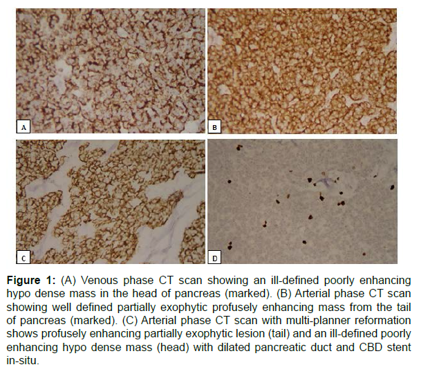
A 76-year-old man with multiple co-morbidities was admitted for evaluation of progressive jaundice for the last 4 months with anorexia, weight loss and intermittent low-grade fever. Patient had already undergone ERCP stenting. On laboratory examination, he was found to have direct hyperbilirubinemia (direct bilirubin 6.8 mg/ dl) prior to stenting with elevated CA 19-9 level (1092 U/mL; normal Figure 1). Positron Emission Tomography - Computed Tomography (PET/CT) showed a space occupying lesion in head of pancreas which was non-FDG avid. Endoscopic ultrasound (EUS) was suggestive of a 2.3 × 2.3 cm hypo echoic mass in head of pancreas with enlarged peril-pancreatic, portocaval and Para-aortic lymph nodes. Fine needle aspiration biopsy (FNAB) for pancreatic head mass was suggestive of ductal adenocarcinoma while biopsy of Para-aortic lymph node biopsy suggested reactive lymphadenopathy. The patient underwent diagnostic laparoscopy, on which neither liver nor peritoneal metastasis were found, a 2 × 2 cm solid irregular mass in head of pancreas was found to be just abutting the portal vein and 2 × 2 cm firm, well circumscribed homogenous lesion with cystic component was identified at the junction of body and tail region. Open Whipple’s operation for the pancreatic head mass along with enucleation of pancreatic tail lesion under the impression of ductal adenocarcinoma with NET was performed. The postoperative period was uneventful [1].
Figure 1: (A) Venous phase CT scan showing an ill-defined poorly enhancing hypo dense mass in the head of pancreas (marked). (B) Arterial phase CT scan showing well defined partially exophytic profusely enhancing mass from the tail of pancreas (marked). (C) Arterial phase CT scan with multi-planner reformation shows profusely enhancing partially exophytic lesion (tail) and an ill-defined poorly enhancing hypo dense mass (head) with dilated pancreatic duct and CBD stent in-situ.
Pathology
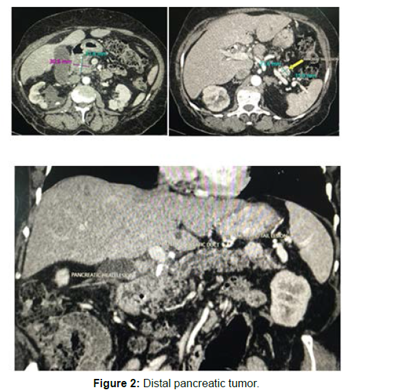
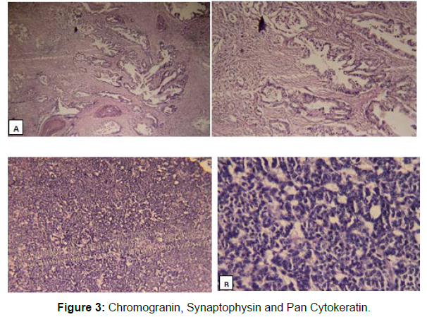
Pancreatic head tumor was 2.5 × 2 × 2 cm, moderately differentiated ductal adenocarcinoma with metastasis to 5 out of 12 lymph nodes. There was no lymphovascular or per neural invasion (p T2N2 Lv0 Pn0). Distal pancreatic tumor was 1.5 ×1.5 ×1.4 cm, univocal; grade 2 NET with mitotic rate of 3 mitosis / 50 HPF (High-Power-Field) (Figure 2). It was a non-functional tumor with neither necrosis nor lymphovascular and per neural invasion (pT1N0). On immunohistochemistry (IHC), tumor cells from the tail lesion were positive for Chromogranin, Synaptophysin and Pan Cytokeratin respectively with an intermediate proliferation rate by Ki67 of 5% (Figure 3).
Discussion
We report an extremely rare presentation of dual malignancy of pancreas constituted by PDA in head of pancreas and NET in the tail of pancreas. Pre-operative work-up was favorable for adenocarcinoma with NET. Eventually the provisional diagnosis was confirmed by histopathology and IHC. According to the World Health Organization (WHO), collision tumors can be histologically classified as those with at least two different malignant components, without mixed or transitional area [2]. They can occur in any organ of the body with stomach and esophagus being the most commonly involved organs. Pancreas is a very unusual site for such presentation.
As per the available literature, sporadic cases comprising of PDA with IPMN, IPMN with NET, solid pseudo papillary neoplasm with NET and cancer of bile duct with pancreas have been reported [3,4]. Chang et al. [5] reported one such case of solitary concomitant NET and PDA in a 58-year-old lady and provided a simple classification for such neoplasms. Similarly, a case of pancreatic collision tumor composed of NET and concurrent PDA has also been reported [6].
Pre-operative diagnosis of dual malignancy of pancreas can be complex at times. Serafini et al. [1] presented a case of collision of PDA with pancreatic NET, in which pre-operative radiology suggested pancreatic IPMN. Whereas, histopathology of totally pancreatectomy specimen revealed an altogether different picture. Likewise, a very rare case of double malignancy of pancreas (PDA and NET) has been reported in remnant pancreas which was diagnosed on 4th year of follow-up [7].
From the above evidences it is believed the co-existence of dual malignancy is an incidental diagnosis. It is almost impossible to diagnose them pre-operatively [8]. However, in our case, the diagnosis was well established preoperatively perhaps because of location of both tumors on extreme ends. Surgical resection is deemed to be treatment of choice in operable cases but they still have a poor prognosis [6]. Our patient underwent surgical resection followed by adjuvant chemotherapy, and did well for follow-ups with disease free 15 months after surgery before having a natural death.
Conclusion
We present a rarest subtype of pancreatic dual malignancy (PDA with NET). A preoperative diagnose is mostly challenging. Hence, the coexistence of multiple primary malignancies should always be considered in case of atypical radiology. Surgical resection is the treatment of choice for operable tumors but the prognosis is still uncertain.
Acknowledgement
A. Gajdhar wrote the manuscript. Balakrishnan S and A. Vijai edited the manuscript. P. Nalankilli and C. Palanivelu performed the surgery and reviewed the manuscript. Informed consent was obtained for this case report.
References
- Serafini S, Dalt GD, Pozza G, Blandamura S, Valmasoni M, et al. (2017) Collision of ductal adenocarcinoma and neuroendocrine tumor of the pancreas: A case report and review of the literature. World J Surg Oncol 15.
- Hamilton SR, Aaltonen LA (2000) World Health Organization Classification of Tumours: Pathology and Genetics of Tumours of the Digestive System. IARC.
- Yan SX, Adair CF, Balani J, Mansour JC, Gokaslan ST (2015) Solid pseudopapillary neoplasm collides with a well-differentiated pancreatic endocrine neoplasm in an adult man: Case report and review of histogenesis. Am J Clin Pathol 143:243-247.
- Izumi H, Furukawa D, Yazawa N, Masuoka Y, Yamada M, et al. (2015) A case study of a collision tumor composed of cancers of the bile duct and pancreas. Surg Case Reports 1:40
- Chang SM, Yan ST, Wei CK, Lin CW, Tseng CE (2010) Solitary concomitant endocrine tumor and ductal adenocarcinoma of pancreas. World J Gastroenterol 16:2692-2697.
- Wang Y, Gandhi S, Basu A, Ijeli A, Kovarik P, et al. (2018) Pancreatic Collision Tumor of Ductal Adenocarcinoma and Neuroendocrine Tumor. ACG Case Reports J 5:e39.
- Kim HJ, Park MH, Shin B (2018) Double primary tumors of the pancreas: A case report. Med 97:e13616.
- Kim HJ, Choi BG, Kim CY, Cho CK, Kim JW, et al. (2013) Collision tumor of the ampulla of Vater - Coexistence of neuroendocrine carcinoma and adenocarcinoma: report of a case. Korean J Hepatobiliary Pancreat Surg 17:186-190.
Indexed at, Google Scholar, Crossref
Indexed at, Google Scholar, Crossref
Indexed at, Google Scholar, Crossref
Indexed at, Google Scholar, Crossref
Citation: Gajdhar A, Balakrishnan S, Vijai A, Nalankilli P, Srikanth B, et al. (2022) Dual Malignancy of Pancreas: A Rare Case Report and Review of Literature. J Cancer Diagn 6: 136. DOI: 10.4172/2476-2253.1000136
Copyright: © 2022 Gajdhar A, et al. This is an open-access article distributed under the terms of the Creative Commons Attribution License, which permits unrestricted use, distribution, and reproduction in any medium, provided the original author and source are credited.
Share This Article
Recommended Journals
Open Access Journals
Article Tools
Article Usage
- Total views: 1988
- [From(publication date): 0-2022 - Apr 05, 2025]
- Breakdown by view type
- HTML page views: 1482
- PDF downloads: 506


