Dramatic Morphological Changes of Astrocytes in Influenza Associated Encephalopathy
Received: 03-Mar-2022 / Manuscript No. JIDT-22-55994 / Editor assigned: 07-Mar-2022 / PreQC No. JIDT-22-55994 (PQ) / Reviewed: 21-Mar-2022 / QC No. JIDT-22-55994 / Revised: 28-Mar-2022 / Manuscript No. JIDT-22-55994 (R) / Published Date: 04-Apr-2022 DOI: 10.4172/2332-0877.1000496
About the Study
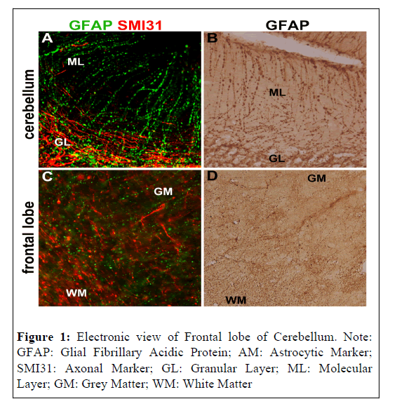
Clasmatodendrosis is an abnormal morphological change of astrocytes characterized by fragmentation of distal processes and vacuolation of cell bodies. Clasmatodendrosis has been found in postmortem brain tissues in diseases such as dementia, head trauma, Neuro Myelitis Optica, and infectious encephalopathies, including influenza-associated encephalopathy [1,2]. The figure shows immunohistochemical stainings of Glial Fibrillary Acidic Protein (GFAP; astrocytic marker) in the postmortem brains of patients with IAE; A and B show the molecular and granular layers of the cerebellum, and C and D are the cortico-medullary border areas of the frontal lobe (Figure 1). Beaded, truncated astrocytic endfeet were observed in the cerebellar molecular layer and deep in the cortex. Such changes in astrocyte morphology were detected in various areas of IAE brains. Although the mechanism of clasmatodendrosis remains unclear, cytokines such as TNF-α are thought to cause such morphological changes in astrocytes. Clasmatodendrosis may be one of the causes of irreversible defects of neurological function after IAE.
References
- Tachibana M, Mohri I, Hirata I, Kuwada A, Kimura S, et al. (2019) Clasmatodendorosis is associated with dendritic spines and does not represent autophagic astrocyte death in influenza-associated encephalopathy. Brain Dev 41:85-95.
[Crossref] [Google Scholar] [PubMed]
- Balaban D, Miyawaki ED, Bhattacharyya S, Torre M (2021) The phenomenon of clasmatodendrosis. Heliyon 7: e07605.
[Crossref] [Google Scholar] [PubMed]
Citation: Tachibana M (2022) Dramatic Morphological Changes of Astrocytes in Influenza Associated Encephalopathy. J Infect Dis Ther 10: 496. DOI: 10.4172/2332-0877.1000496
Copyright: © 2022 Tachibana M. This is an open-access article distributed under the terms of the Creative Commons Attribution License, which permits unrestricted use, distribution, and reproduction in any medium, provided the original author and source are credited.
Share This Article
Recommended Journals
Open Access Journals
Article Tools
Article Usage
- Total views: 1688
- [From(publication date): 0-2022 - Nov 21, 2024]
- Breakdown by view type
- HTML page views: 1339
- PDF downloads: 349

