Different Bone injury on Bone Scan in a Case of Unsuspected Child Abuse
Received: 13-Aug-2021 / Accepted Date: 27-Aug-2021 / Published Date: 03-Sep-2021 DOI: 10.4172/2167-7964.1000337
Keywords:
Introduction
Radiological examinations are significant while evaluating youngsters who might have been exposed to actual maltreatment. These examinations assume a critical part in kid misuse cases in light of the fact that the set of experiences gave is frequently fragmented or misdirecting and the actual assessment may not promptly recognize mysterious wounds, particularly in babies and small kids.
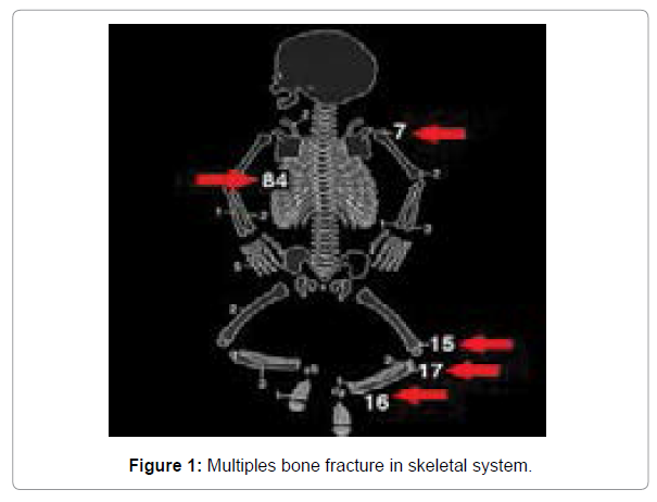
The battered youngster disorder comprises of a star grouping of signs that might be either obvious or incognito. Many examples of injury have been depicted in the kid misuse condition. Bone scintigraphy is a significant imaging methodology in the assessment of the battered youngster. It is normal used to assess skeletal injury and recognize breaks which beforehand would have been disregarded [1-5] (Figure 1).
Case Report
An eleven-month-old new-born child weighted 1780 g with an untimely birth age of 34 weeks was in the escalated care for seizure assault, goal pneumonia, and subdural and subarachnoid drain. He created reformist lower appendages enlarging and his left elbow skeletal distortion following a fall. To secure the protection of our patients, their full records are not straightforwardly accessible. The radionuclide bone output with 185 MBq (5 mCi) Tc-99m MDP was performed for an assessment of dubious munnion break, bone contamination, or prior ailments in light of the fact that metabolic problems and bone illnesses might make a kid’s bones more defenseless against crack [6]. Different expanded radioactive foci all through the entire body (Figure 1) were out of the blue found. There was a solid likelihood of youngster misuse. A progression of plain film radiographs showed calvarias break lines at left temporoparietal locale, hard oddity of the spine, numerous old cracks with callus arrangement including back part of left tenth, eleventh ribs, right proximal humerus, two-sided proximal femurs, and metaphysis of tibias the above discoveries were likewise steady with youngster misuse. Radiographic skeletal review and radionuclide pictures.
Conversation
The assessed occurrence of revealed youngster misuse has expanded from 3% in 1985 to 4.5% in 1992. The rate of skeletal injury in these kids is roughly 20% and is more normal among those under 1 year old enough. Kids more established than 3 years old will in general have transcendently delicate tissue injury. Cerebral injury is normal at whatever stage in life. The cracks are normally numerous, including the long bones, skull, vertebrae, ribs, and facial bones as well as often showing various phases of recuperating. Bone scintigraphy is a significant imaging methodology in the assessment of these small kids, particularly in recognizing injury in ribs, cost vertebral intersections, hands, feet, spine, and diaphysis of long bones [2]. Youngster misuse ought to be viewed as when diagnosing expanded solitary bone take-up on bone scintigraphy, which might show no accidental injury. The blend of bone output and X-beam with experienced hands can lessen the bogus negative rate from 12.3% to 0.8%. Albeit the bone output might be positive as ahead of schedule as 7 hours after injury, the youngster is typically brought to a clinic so late that the bone mending has started the picture modalities assume a vital part in the examination and documentation of the battered youngster disorder. The essential indicative imaging concentrate in presumed kid misuse is either a bone output and X-beam series or a total radiographic skeletal study by X-beam series in children and babies. Skeletal review and bone scintigraphy are corresponding examinations in the assessment of no accidental injury and ought to both be acted in instances of suspected youngster misuse Further investigations ought to be embraced in the present situation to look for existing together wounds, particularly as the set of experiences and system of injury may regularly be muddled. Bone output might require sedation, and this methodology is presently less regularly utilized, particularly in the developing setting In any case, in situations where kids are possibly being lost to follow-up, this will help the analysis of most of cracks during the underlying appraisal and, along these lines, assist with guaranteeing the wellbeing of the kid [3]. Irreconcilable circumstances the creators pronounce they have no irreconcilable situations in distributing this contextual investigation. Creators.
References
- Howard JL, Barron B J, Smith GG (1990) “Bone scintigraphy in the evaluation of extra skeletal injuries from child abuse, radiographic 10:67-81.
- Smith FW, Gilday DL, Ash JM, Green MD (1980) Unsuspected custom-vertebral fractures demonstrated by bone scanning in the child abuse syndrome, Pediatr Radiol 10(2):103-106.
- Bainbridge JK, Huey BM, Harrison SK (2015) Should bone scintigraphy be used as a routine adjunct to skeletal survey in the imaging of non-accidental injury? A 10 year review of reports in a single centre. Clin Radiol 70: 83-89.
- Worlock P, Stower M, Barbor P (1986) Patterns of fractures in accidental and non-accidental injury in children a comparative study. British Med J 293: 100-102.
- Van Rijn RR, Perez-Rossello JM, Kleinman PK (2009) “Skeletal imaging of child abuse (non-accidental injury).†Paediatr Radiol 39(5):461-470.
- Flaherty EG, Perez-Rossello JM, Â Levine MA, (2014) Evaluating children with fractures for child physical abuse. Paediatrics133: 477-489.
Citation: Nivedh S (2021) Different Bone injury on Bone Scan in a Case of Unsuspected Child Abuse. OMICS J Radiol 10: 337. DOI: 10.4172/2167-7964.1000337
Copyright: © 2021 Nivedh S. This is an open-access article distributed under the terms of the Creative Commons Attribution License, which permits unrestricted use, distribution, and reproduction in any medium, provided the original author and source are credited.
Select your language of interest to view the total content in your interested language
Share This Article
Open Access Journals
Article Tools
Article Usage
- Total views: 2672
- [From(publication date): 0-2021 - Dec 05, 2025]
- Breakdown by view type
- HTML page views: 1933
- PDF downloads: 739

