Dieulafoy-Like Lesion at the Brim of a Gastric Diverticulum: A Very Rare Cause of Gastrointestinal Bleeding
Received: 30-Jun-2018 / Accepted Date: 17-Jul-2018 / Published Date: 23-Jul-2018 DOI: 10.4172/2161-069X.1000572
Introduction
Gastric diverticulum (GD) is the rarest form of gastrointestinal diverticula with a reported prevalence of 0.01%-0.11% [1]. There is no gender predilection and it usually presents in the fifth or sixth decade of life. More than 70% of congenital GD are mostly located in the posterior wall of the fundus, 2 cm below the oesophagogastric junction and 3 cm from the lesser curve. Acquired GD are typically located in the antrum and usually occur due to chronic inflammatory disease, malignancy, surgery, or gastric outlet obstruction. GD are mostly asymptomatic and when symptoms occur, they vary from vague upper abdominal pain, vomiting, dysphagia, halitosis and eructation.
Description
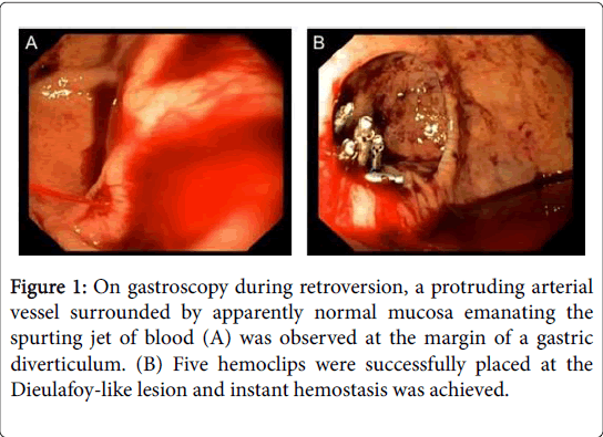
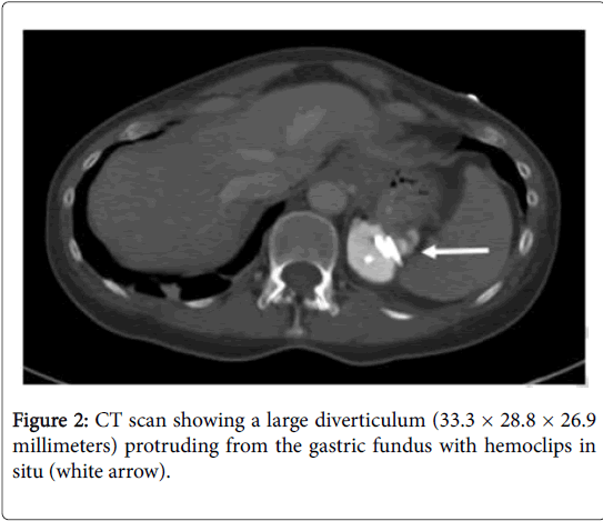
A 55-year-old female presented to our hospital with sudden hematemesis and epigastric pain. The hemodynamics were stable and physical examination was unremarkable. Blood tests revealed a hemoglobin level of 8.7 g/dl. The patient’s history offered no preliminary symptoms and no preexisting diseases, surgery or medication. An esophagogastroduodenoscopy was performed immediately and visualized an active arterial hemorrhage from a Dieulafoy-like lesion at the brim of a GD located in the posterior wall of fundus (Figure 1A). The bleeding was successfully stopped endoscopically (Figure 1B). A CT scan confirmed a large GD (Figure 2).
Figure 1: On gastroscopy during retroversion, a protruding arterial vessel surrounded by apparently normal mucosa emanating the spurting jet of blood (A) was observed at the margin of a gastric diverticulum. (B) Five hemoclips were successfully placed at the Dieulafoy-like lesion and instant hemostasis was achieved.
The usual bleeding site is an abberant vessel in the dome of diverticulum. In the present case, the diagnosis is consistent with Dieulafoy's disease for the following reasons: no bleeding history, the sudden onset, a bleeding spot at the marginal GD surface and a protruding vessel surrounded by apparently normal mucosa. An association of Dieulafoy-like lesion with gastrointestinal diverticula has been described [2]. The hemoclip application is considered the most appropriate for endoscopic treatment [3].
Discussion
However, when the diverticulum is >4 cm in diameter, patients are still symptomatic after PPIs administration, or perforation occur, surgical diverticulectomy is the recommended approach [1].
References
- Rashid F, Aber A, Iftikhar SY (2012) A review on gastric diverticulum. World J Emerg Surg 7: 1.
- de Benito Sanz M, Roman MC, Yuste RT (2018) A Dieulafoy's lesion in a duodenal diverticulum: An infrequent cause of UGIB. Rev Esp Enferm Dig 110: 266-267.
- Nojkov B, Cappell MS (2015) Gastrointestinal bleeding from Dieulafoy's lesion: Clinical presentation, endoscopic findings, and endoscopic therapy. World J Gastrointest Endosc 7: 295-307.
Share This Article
Recommended Journals
Open Access Journals
Article Tools
Article Usage
- Total views: 3655
- [From(publication date): 0-2018 - Nov 21, 2024]
- Breakdown by view type
- HTML page views: 3018
- PDF downloads: 637


