Diagnosis and Management of an Adolescent with a Recurrent Chondromyxoid Fibroma in the Metatarsals: A Case Report
Received: 13-Jul-2021 / Accepted Date: 27-Jul-2021 / Published Date: 03-Aug-2021 DOI: 10.4172/2472-016X.1000150
Abstract
Chondromyxoid Fibroma (CMF) is a rare benign aggressive cartilaginous tumor that represents less than 0.5% of all bone tumors. It most commonly arises from the metaphysis of long bones. However, CMF can rarely arise from other short bones as well as the hand and foot bones. Moreover, it is characterized by some radiological findings, most described as a well circumscribed lytic lesion with scalloped and sclerotic margins. Histopathology is required to confirm the diagnosis of CMF since this tumor is known for its unique histopathological characteristic findings as well. The mainstay treatment is surgical excision via curettage with or without bone grafting or cementing. In this article, we report the diagnosis and management of an 18-year-old male with a painful CMF arising from the 5th metatarsal bone of the right foot that has recurred after curettage and bone grafting surgery, ultimately managed with marginal excision of the tumor and resection of half of the 5th metatarsal bone followed by reconstruction.
Keywords: Chondromyxoid fibroma; CMF; Metatarsal bone; Recurrence; Management
Introduction
Chondromyxoid fibroma (CMF) is defined as a rare benign aggressive neoplasm of the chondroid, myxoid and fibrous tissue [1]. Most common site of this disease is the metaphysis of long bones, especially those around the knee joint [2]. It comprises about 0.5% of all bone tumors, 2% of benign bone tumors, 8% in the bones of the foot, and 71% of the cases affecting the long bones of the lower extremities’ epiphyseal aspects, most commonly involving the knee joint [3]. However, one study reports that CMF represents less than 5% of all bone tumors, and its overall incidence in the craniofacial bones is between 2% to 5%, while another study reports its occurrence in the craniofacial bones to be only 2% with the commonest sites being the maxilla and the mandible [2,3]. Interestingly, CMF can arise from the orbital bones which is extremely rare [4]. The main treatment method of CMF is through curettage with or without bone grafting or cementing which will pose a 20%-25% recurrence risk [5]. To the best of our knowledge, only 15 cases of CMF arising from the metatarsal bones were reported. Therefore, we present a case of recurrent CMF that has originated from the right foot 5th metatarsal head along with how it was diagnosed and managed.
Case Presentation
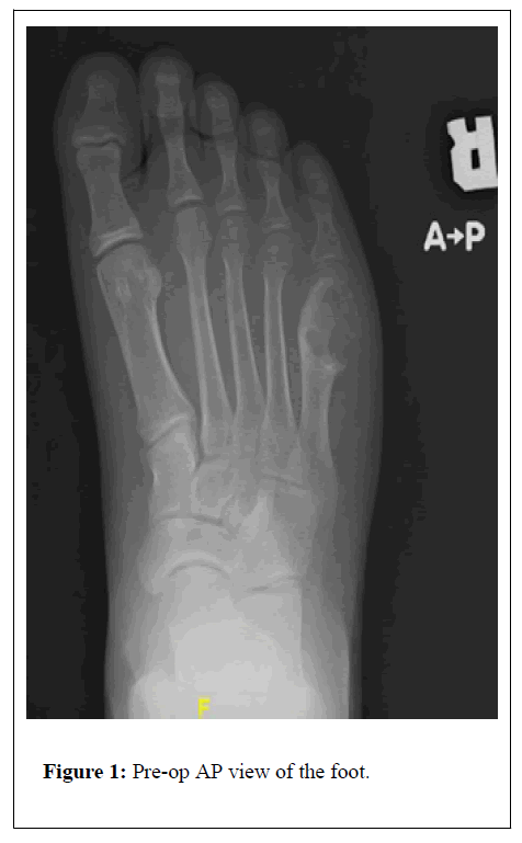
This is a case of an 18-year-old male who is medically and surgically free presented to the clinic complaining of right foot pain on the lateral side associated with a swelling for one month prior to presentation. Physical examination, clinical and radiological investigations were carried out and accordingly the diagnosis of right foot metatarsal head CMF was proposed in Figures 1 and 2. Thereafter, the patient was opted for intralesional curettage and bone grafting, which was performed on December 5th, 2018, and an intraoperative tissue biopsy was taken, which eventually confirmed the diagnosis.
Post-operatively the patient was doing well and was discharged, he attended his regular follow-up visits in which he was having no complaints. A few months later, he started complaining of mild pain at the same site of the lesion, for which X-rays of the foot were performed showing recurrence of the lesion. One year after the 1st operation, he was planned for marginal excision of the lesion with resection of half of the 5th metatarsal bone and reconstruction procedure on December 25, 2019 was observed in Figures 3-5. Surgery was uneventful, and the patient was kept on backslap for pain and swelling control, sutures were removed 10 days later. Fortunately, the patient started bearing weight fully one-month post-operation.
Patient came to the clinic four months later for a follow-up visit, in which he was walking without pain or any other complaints. He, thankfully, came for a follow-up appointment one year later, he was doing very well, walking normally with no issues at all, and all his follow-up X-rays were unremarkable was observed in Figures 6 and 7.
Results and Discussion
CMF is an aggressive benign bone tumor that is most seen in the metaphysis of long bones, especially the bones surrounding the knee joint followed by the foot bones [3]. The onset of the disease is usually in the 2nd and 3rd decades of life with male predominance [5,6]. The symptoms and clinical manifestations of the disease vary based on the location and size of the tumor, however, the most common manifestation to be presented with is chronic pain, which was found in a study involving 10 cases with CMF as most of them presented with chronic pain [5]. This is consistent with what happened in the present case since the patient started complaining of chronic pain at the site of the lesion for a month as well as swelling prior to presentation.
Radiologically, the characteristic description of CMF is a well circumscribed, lytic lesion with scalloped and sclerotic margins like a metaphyseal fibrous defect [7]. CMF is usually confined to the metaphysis but occasionally it may involve the epiphysis as well, which was reported in their case [6]. Additionally, computed tomography (CT) is helpful to define cortical integrity and to confirm no mineralization of the matrix [8]. However, magnetic resonance imaging (MRI) is the preferred modality to determine the extent of the disease [1]. On MRI, CMF lesion is usually hypointense or isointense on T1-weighted images and hyperintense on T2-weighted images [1,8]. The diagnosis can only be made definite through histopathology which typically consists of lobules of spindle-shaped or stellate cells with abundant myxoid or chondroid intercellular material with varying numbers of multinucleated giant cells [3,6,9].
In the presented case, all radiological studies were in line with the diagnosis being CMF, but other differentials cannot be excluded, and the diagnosis cannot be definitively confirmed until biopsy was done. Following histopathological confirmation, the management plan was started.
The differential diagnosis of CMF includes a list of chondroid lesions such as chondroblastoma, which is a benign cartilaginous tumor, that has a similar presentation to CMF [2,10]. Chondroblastoma can be differentiated from CMF histologically, as it lacks stellate cells which are characteristic for CMF [2,10]. Furthermore, chondrosarcoma is another important differential that is occasionally confused with CMF since, as reported in the literature, 22% of CMF cases were misdiagnosed as chondrosarcoma [2]. This is usually pertained to the difficulty in distinguishing between them radiologically, for example calcifications can be seen sometimes in CMF though they are more prominent in chondrosarcoma [1]. Histologically, chondrosarcoma is characterized by nuclear pleomorphism and atypia, which are rarely seen in CMF, and they lack the bands of mitochondrion tissue [1]. In fact, according to two of the ten cases that were initially involved in their study had the primary diagnosis of pelvic CMF, but the final diagnosis turned out to be chondrosarcoma [5]. Another differential diagnosis of this disease is chondroma, which is distinguished histologically by its pathognomonic cells known as physaliferous cells described as “vacuolated or bubbly eosinophilic cytoplasm” [1].
As for the treatment of CMF, the mainstay is surgical excision through intralesional curettage alone, curettage with bone grafting or cementing, en-bloc excision, or amputation [6,11]. The recurrence rate after a simple curettage is 12.5%-25%, hence the importance of bone grafting or cementing in reducing the risk of recurrence [3]. The recurrence rate in a study involving the management of 22 CMF cases is reported to be 9.1%, all patients were treated with intralesional curettage except four cases [11]. This low risk is pertained to the adjuvant bone cementing in four of the cases, which is beneficial in reducing the risk of future fractures by providing more stability and destroying residual tumor cells [11]. As for en-bloc resection, it was performed as the main treatment in a case diagnosed with CMF in the frontal bone, and fortunately the disease has not recurred in a follow-up period of 20 months [12].
As for the presented case, it was managed through intralesional curettage and bone grafting, since intralesional curettage is the most used treatment modality, and bone grafting decreases the risk of recurrence [13]. However, after a few months, recurrence was confirmed radiologically after the patient complained of pain at the site of the lesion. Therefore, he underwent marginal excision of the recurrent lesion with resection of half of the 5th metatarsal bone as well as reconstruction, which was successful, and the patient had no recurrence for the follow-up period of 12 months. The same treatment plan, which is comprised of curettage and bone grafting, was used in another reported CMF case of the metatarsal bone, in which the surgery was uneventful and there was no recurrence in the 10-month period of follow-up [13].
Conclusion
In conclusion, CMF is an extremely rare benign tumor that most commonly involves the metaphysis of long bones, and it represents less than 0.5% of all bone tumors. In the present article, we report a case of CMF originating from the right 5th metatarsal bone that has recurred in a few months as well as elaborating more on its diagnosis and management. The clinical manifestations of the disease are dependent upon the location and size of the lesion, which was chronic pain on the lateral side of the foot in the present case. Moreover, radiological studies usually are characteristic for CMF, however, histopathological confirmation through biopsy is essential to make the diagnosis definite and exclude other differential diagnosis that may be confused with the disease such as chondrosarcoma and chondroblastoma. The standard treatment of CMF is surgical excision via intralesional curettage with or without bone grafting or cementing.
References
- El Kouri N, Elghouche A, Chen S, Shipchandler T, Ting J (2019) Sinonasal chondromyxoid fibroma: Case report and literature review. Cureus 5: 11.
- Elsamanody A, Van den Aardweg M, Smits A, Willems S, Topsakal V (2020) Chondromyxoid fibroma of the mastoid: A rare entity with comprehensive literature review. J Int Adv Otol 16: 117-122.
- Liu T, Yao J, Li X, Qi X, Zhao P (2020) Chondromyxoid fibroma of the temporal bone. Medicine (Baltimore) 99: e19487.
- Grewal A, Singh M, Vishwajeet V, Thakur U, Das A (2019) Primary chondromyxoid fibroma of the orbit: An orbital mass with calcification. Indian J Ophthalmol 67: 2110.
- Jamshidi K, Mazhar FN, Jafari D (2015) Chondromyxoid fibroma of pelvis, surgical management of 8 cases. Arch Iran Med 18: 367-370.
- Vasudeva N, Shyam Kumar C, Ayyappa Naidu CR (2020) Chondromyxoid fibroma of distal phalanx of the great toe: A rare clinical entity. Cureus 12: 1-16.
- Shen S, Chen M, Jug R, Yu CQ, Zhang WL (2017) Radiological presentation of chondromyxoid fibroma in the sellar region. Medicine (Baltimore) 96: e9049.
- Sharma H, Jane MJ, Reid R (2006) Chondromyxoid fibroma of the foot and ankle: 40 years’ Scottish bone tumour registry experience. Int Orthop 30: 205-209.
- Zheng YM, Wang HX, Dong C (2018) Chondromyxoid fibroma of the temporal bone: A case report and review of the literature. World J Clin Cases 6: 1210-1216.
- Siddiqui B, Habib Faridi S, Faizan M, Sherwani RK (2016) Cytodiagnosis of chondromyxoid fibroma of the metatarsal head: A case report. Iran J Pathol 11: 272-275.
- Bhamra JS, Al-Khateeb H, Dhinsa BS, Gikas PD, Tirabosco R (2014) Chondromyxoid fibroma management: A single institution experience of 22 cases. World J Surg Oncol 12: 283.
- Yerleflen FK (2008) Chondromyxoid fibroma of frontal bone: A case report frontal kemikte yerleflen kondromiksoid fibroma: Olgu Sunumu ve Literaturun Gozden. World J Surg Oncol 8: 249-253.
- Dey B, Deshpande A, Brar R, Ray A (2018) Chondromyxoid fibroma of the metatarsal bone: A diagnosis using fine needle aspiration biopsy. J Cytol 35: 67.
Citation: Almousa N, Alshaya O (2021) Diagnosis and Management of an Adolescent with a Recurrent Chondromyxoid Fibroma in the Metatarsals: A Case Report. J Orthop Oncol 7:150. DOI: 10.4172/2472-016X.1000150
Copyright: © 2021 Almousa N, et al This is an open-access article distributed under the terms of the Creative Commons Attribution License, which permits unrestricted use, distribution, and reproduction in any medium, provided the original author and source are credited.
Share This Article
Recommended Journals
Open Access Journals
Article Tools
Article Usage
- Total views: 1908
- [From(publication date): 0-2021 - Nov 28, 2024]
- Breakdown by view type
- HTML page views: 1362
- PDF downloads: 546


