Diagnosing Stroke Cases Using Unenhanced Helical Computed Tomography in Recently Brain Stroked Patients in Duhok City
Received: 04-Apr-2021 / Accepted Date: 13-Apr-2021 / Published Date: 20-Apr-2021 DOI: 10.4172/2167-7964.1000324
Abstract
Background: Cerebral Stroke has been recorded as a major health problem in the world, it accounts the second most common cause of personal disability in the worldwide. This study was undertaken for the use of unenhanced computed tomography (CT) imaging findings in the persons who were clinically diagnosed and referred to the radiological units as a cerebrally stroked patients,aiming to detect their overall number (incidence) in the city, taking into consideration the most common period of onset and duration of their relevant radiological unenhanced CT findings regarding both haemorrhagic, ischemic changes and their subtypes, looking for the most commonly affected vascular territories in different stroke types considering the site and side of the vessel affected, detecting also the most common brain lobes involved in different strokes in detailed disruption with their mass effects in addition to checking the incidence of normal (-ive) CT scan findings among those clinically diagnosed and referred to as stroked patients in addition to finding out the sensitivity of the unenhanced CT in detecting early stroked cases compared to the clinical diagnosis and to find out the most common related risk factor in each kind of stroke.
Results: The overall number of these patients was 104 case study, the male patients constitute 60.6% of the total number and the female patients were 39.4%, the mean period of onset of their relevant radiological appearances on the unenhanced CT scan in overall acute cases regardless of the kind of stroke was 6.9 hours from the onset of the patients symptoms, the incidence of ischemic stroke was more than haemorrhagic stroke as it was 33.7% of the overall cases while the haemorrhagic stroke constituted 23.1%. Among all 104 patients, the middle cerebral artery (MCA) was the most affected vessel in both ischemic and haemorrhagic strokes constituting about half of the cases followed by the posterior cerebral artery (PCA) which accounts 17% in ischemic and about 21% of haemorrhagic cases, the right side of both of these vessels was commonly affected, the parietal lobe was most commonly affected in ischemic stroke cases followed by the basal ganglia, while in haemorrhagic cases the opposite was seen, 43.3% of the cases showed negative CT scan findings. The sensitivity of unenhanced CT scan radiological findings in confirming the diagnosis was 56.7% compared to the clinical diagnosis as reference standard. Regarding the risk factor, the age was the most common factor followed by hypertension and Diabetes Mellitus, more mass effects seen in haemorrhagic than ischemic stroke, the mean time of appearance of radiological changes as seen in CT scan was 5.8 hrs in haemorrhagic type compared with 7.5 hrs. in ischemic stroke.
Conclusion: In overall cases of stroke, Male were affected more than the female, 6.9 hours was the mean period of onset of appearance of their unenhanced CT findings, ischemic kind of stroke exceeds haemorrhagic one by about 10%, the right MCA was the most affected vessel in both types of stroke followed by the PCA, the parietal lobe was the most common lobe affected in ischemic stroke while the basal ganglia were most commonly affected in haemorrhagic stroke, negative unenhanced CT findings account for about one third of the clinically diagnosed cases, the unenhanced CT scan detection and disease confirmation sensitivity was more than half compared with the reference clinical diagnosis, age increment was the most common risk factor for overall stroke followed by diabetes mellitus. The use of unenhanced CT scan in the patients presented and clinically confirmed as having stroke, imply an early assessment, giving proper estimation, has a major role in differentiating ischemic from haemorrhagic types of stroke which is considered the major dilemma, in addition it can be performed rapidly, a feature that help recognize early signs of stroke as soon as 6 hours from starting symptoms
Keywords: Unenhanced helical computed, Brain parenchyma, ischemic stroke, Haemorrhagic stroke, Hounsfield unit, Cerebrovascular accident
Background
Cerebral Stroke has been regarded as the main etiology of mortality and morbidity in the world. The prevalence rate for stroke ranged between 508 and 777 per 100,000 populations in the Middle East in its two major types (ischemic and haemorrhagic). Ischemic stroke was the most reported type followed by intracerebral haemorrhage and subarachnoid haemorrhage [1]. There is sufficient analysis demonstrating association between the use of unenhanced Computed Tomography (CT) and early management with better outcomes of these patients with recent stroke in its hyper acute and acute states which have found to be the most effective cause of patient dilemma. Therefore, their early detection is an important factor for proper management of these patients. The objectives of an imaging assessment for recent stroke are to build up a diagnosis as soon as possible and to get accurate and early knowledge of the types of stroke, any intracranial abnormal vasculature and brain parenchymal perfusion state helping to choose the proper treatment to avoid uncontrollable complications.
An early assessment may be performed with a native computed tomography (CT), which has major role in differentiating ischemic from haemorrhagic stroke and it can be performed rapidly, which can help recognize early signs of stroke as soon as 6 hours from starting symptoms [2]. The native head CT can recognize the early signs of stroke, but above all can exclude intracerebral haemorrhage and lesions that might resemble acute ischemic stroke like tumour,abscess,contusion, and demyelinating disease [3].
Methods
This study is a hospital based cross sectional study of patients who were clinically diagnosed with a new onset of (cerebrovascular accident) CVA and referred to radiological department at Azadi Teaching and Zakho general hospitals and in coordination with emergency department at Azadi teaching hospital for doing unenhanced Brain CT scan image to these patients living in Duhok city. The data were collected for a period of eight months (1st January to 30th of August, 2019), a total of one hundred four cases with a clinically diagnosed of CVA who were referred to radiological departments were scanned by native CT, patients mean age was 56 years (±SD 15, range 21-96 years old), the number of male was 63 and female was 41 have been included in this study. Unenhanced CT scanning step was the first modality imaging to differentiate the ischemic and haemorrhagic CVA,and to exclude other abnormality causing CVA like symptoms form normal CT scan .The author developed a questionnaire (Appendix I) according to the evidence-based documents of the current literature to obtain general information of each patient and record the CT findings, all the CT imaging of the patients were performed by the researcher under supervision of a specialized radiologist.
CT Scan Machine and Parameters
The CT scan machines used for the study are Toshiba and Philips, 64 slices. Patients examined without preliminary preparation, putted in supine position, multiple axial, coronal and sagittal sections were taken for each patients, in addition to 3D reformatted images and using the following parameters: 140 Kv, 170 mA, 5 mm thick slices with 5 mm imaging spacing, 0.8 second rotation time, 22-cm field-of view. The windows which are used including slandered window with slandered setting (35 HU centre -100 HU width level), bone window brain window and stroke window
Data Analysis
The study is cross sectional prospective,the sample of the study consists of 104 cases and these cases are collected after unenhanced CT scan done for them, all the cases in the data have been analysed for the incidence of overall stroke among patients, mean age, gender, time of doing examination from starting symptoms, findings of unenhanced CT scan regarding Ischemic, haemorrhagic and normal findings, lesion side and vascular territories, associated parenchymal mass effect for each subtypes of stroke, and most associated risk factors, then detailed description of all data in tables and figures were done.
Statistical Analysis
Data were analysed by means of SPSS 26 (IBM, 2019). Data were first described using frequency and frequency percentages, and mean, range and standard deviation (SD). Then, categorical variables were analysed by Chi square test, and numerical variables by unpaired t-test or one-way analysis of variance (ANOVA) with Sidak post-hoc (intergroup comparison) test. Pearson correlation (r) was used to test the association between density and duration. A P value less than 0.05 was considered statistically significant. Sensitivity of CT scan was calculated as the number of patients with positive CT findings divided by the number of referred stroke patients.
Results
The Sensitivity of CT scan, with clinical diagnosis as reference standard = (59 / 104) X 100 = 56.7%.
The sample of the study is 104 patients, the sample characteristic are shown in Table 1. Patients were examined within 3-14 hrs of their symptoms (mean)=6.9 hrs. ± SD 2.6 hrs. The mean age of patients was 56 years, ranging between (21-96) years ± SD 15, 63 of them were males so they constitute 60.6%, and 41 were females, the percentage is 39.4%. About the CT scan findings, no abnormality was the commonest and of these 104 patient 45 (43.3%) had no abnormality, followed by ischemic stroke was 35 (33.6%), and the least CT scan findings among these referred patients were haemorrhagic type, they constitute 24 (23.1%) Table 2.
| Characteristic or finding | Mean | Range | SD | No. | % | ||
|---|---|---|---|---|---|---|---|
| Age (years) | 56 | 21 - 96 | 15 | ||||
| Sex | Male | 63 | 60.6 | ||||
| Female | 41 | 39.4 | |||||
| CT findings | Ischemic | 35 | 33.6 | ||||
| Haemorrhagic | 24 | 23.1 | |||||
| No abnormality | 45 | 43.3 | |||||
| Duration (hours) | 6.9 | 3.0-14.0 | 2.6 | ||||
| Total | 104 | 100.0 | |||||
Table 1: Characteristics and main CT finding of all referred stroke patients (n=104).
| Territory | Ischemic | Haemorrhagic | Total | |||
|---|---|---|---|---|---|---|
| No. | % | No. | % | No. | % | |
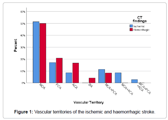
| Middle Cerebra Artery(MCA) | 18 | 51.4 | 12 | 50.0 | 30 | 50.8 |
| Posterior Cerebral Artery(PCA) | 6 | 17.1 | 5 | 20.8 | 11 | 18.6 |
| Anterior Cerebral Artery(ACA) | 3 | 8.6 | 4 | 16.7 | 7 | 11.9 |
| Basilar Artery (BA) | 1 | 4.2 | 1 | 1.7 | ||
| Middle Cerebral Artery (MCA)+Posterior Cerebral Artery(PCA) | 4 | 11.4 | 2 | 8.3 | 6 | 10.2 |
| Middle Cerebral Artery (MCA)+ Anterior Cerebral Artery(ACA) | 3 | 8.6 | 3 | 5.1 | ||
| Middle Cerebral Artery (MCA)+ Posterior Cerebral Artery (PCA)+ Anterior Cerebral Artery (ACA) | 1 | 2.9 | 1 | 1.7 | ||
| Total | 35 | 100.0 | 24 | 100.0 | 59 | 100.0 |
Table 2: Vascular territories of the ischemic and haemorrhagic strokes.
Regarding the territory of ischemic and haemorrhagic stroke,in the present study the most common one involved in both stroke subtypes was MCA, in ischemic was 51.4% (N=18) and in haemorrhagic was 50%(N=30). The second most involved territory was PCA, in ischemic was 17.1%(N=6) and in haemorrhagic was 20.8% (N=5). As shown in Figure 1 and Table 3.
| Territory | Rt. | Lt. | Bilateral | Total | ||||
|---|---|---|---|---|---|---|---|---|
| No. | % | No. | % | No. | % | No. | % | |
| Middle Cerebra Artery(MCA) | 16 | 57.1 | 13 | 50.0 | 1 | 20.0 | 30 | 50.8 |
| Posterior Cerebral Artery(PCA) | 6 | 21.4 | 4 | 15.4 | 1 | 20.0 | 11 | 18.6 |
| Anterior Cerebral Artery(ACA) | 1 | 3.6 | 6 | 23.1 | 7 | 11.9 | ||
| Basilar Artery (BA) | 1 | 3.6 | 1 | 1.7 | ||||
| Middle Cerebral Artery (MCA)+Posterior Cerebral Artery(PCA) | 4 | 14.3 | 1 | 3.8 | 1 | 20.0 | 6 | 10.2 |
| Middle Cerebral Artery (MCA)+ Anterior Cerebral Artery(ACA) | 2 | 7.7 | 1 | 20.0 | 3 | 5.1 | ||
| Middle Cerebral Artery (MCA)+ Posterior Cerebral Artery (PCA)+ Anterior Cerebral Artery (ACA) | 1 | 20.0 | 1 | 1.7 | ||||
| Total | 28 | 100.0 | 26 | 100.0 | 5 | 100.0 | 59 | 100.0 |
Table 3: Vascular territories of the (ischemic and haemorrhagic) strokes, by side.
About the side of stroke lesions, the right side was the most common in patients were the MCA and PCA are involved, in MCA the percentage was 57.1% (N=16) at the right side and 50%(N=13) at the left side, followed by the PCA, was constitute 21.4(N=6) at right side and 15.4(N=4) at left side, and in cases of bilateral side, the MCA and PCA are equally involved, they constitute 20%,while in ACA, the left side was affected more, the percentage is 23.1% (N=6), in comparison to right side the percentage was 3.6% (N=1). As shown in Table 3.
Risk factors in all referred stroke patients
Regarding the risk factors, the aging factor was regarded significant (P value less than 0.05) in comparison to the young ages (20-34 years), middle age (35-49 years) and older age (50- 60 years and more). In both ischemic and haemorrhagic strokes, as increased age, the incidence of both stroke subtypes also increase, in the ischemic stroke between the age (20-34 years) the percentage was 0 (N=0), while in haemorrhagic stroke in the same age group the percentage was 8.3% (N=7), followed by 17.1% (N=6) in ischemic and 29.2% (N=7) in haemorrhagic between the age (35-49 years), while in older age (50-64 years and more) patients, the incidence was 34.3% (N=12) -48.6% (N=17) in ischemic stroke and in haemorrhagic stroke the incidence was 45.8% (N=11) -16.7 (N=4) in same age groups. The other risk factor follow the age was hypertension as had significant P value, the incidence of both ischemic and haemorrhagic strokes were increase in hypertensive patients, 85.7% (N=30) and 70.8% (N=17) were hypertensive patients in comparison to non-hypertensive patients, the incidence was 14.3 (N=5), 29.2 (N=7) in both stroke subtypes. This is shown in Tables 4 and 5.
| Risk factors | CT findings | P | Total | |||||||||
|---|---|---|---|---|---|---|---|---|---|---|---|---|
| Ischemic | Haemorrhagic | No abnormality | ||||||||||
| No. | % | No. | % | No. | % | * | No. | % | ||||
| Age(years) | 20 – 34 | 0 | 0.0 | 2 | 8.3 | 7 | 15.6 | 0.038 | 9 | 8.7 | 31.8% | |
| 35 – 49 | 6 | 17.1 | 7 | 29.2 | 11 | 24.4 | 24 | 23.1 | ||||
| 50 – 64 | 12 | 34.3 | 11 | 45.8 | 17 | 37.8 | 40 | 38.4 | 68.2% | |||
| 65+ | 17 | 48.6 | 4 | 16.7 | 10 | 22.2 | 31 | 29.8 | ||||
| Sex | Male | 18 | 51.4 | 13 | 54.2 | 32 | 71.1 | 0.155 | 63 | 60.6 | ||
| Female | 17 | 48.6 | 11 | 45.8 | 13 | 28.9 | 41 | 39.4 | ||||
| Hypertension | Yes | 30 | 85.7 | 17 | 70.8 | 26 | 57.8 | 0.025 | 73 | 70.2 | ||
| No | 5 | 14.3 | 7 | 29.2 | 19 | 42.2 | 31 | 29.8 | ||||
| Diabetes mellitus | Yes | 15 | 42.9 | 6 | 25.0 | 17 | 37.8 | 0.366 | 38 | 36.5 | ||
| No | 20 | 57.1 | 18 | 75.0 | 28 | 62.2 | 66 | 63.5 | ||||
| Heart disease | Yes | 15 | 42.9 | 8 | 33.3 | 15 | 33.3 | 0.635 | 38 | 36.5 | ||
| No | 20 | 57.1 | 16 | 66.7 | 30 | 66.7 | 66 | 63.5 | ||||
| Smoking | Yes | 11 | 31.4 | 7 | 29.2 | 10 | 22.2 | 0.629 | 28 | 26.9 | ||
| No | 24 | 68.6 | 17 | 70.8 | 35 | 77.8 | 76 | 73.1 | ||||
| Total | 35 | 100.0 | 24 | 100.0 | 45 | 100.0 | 104 | 100.0 | ||||
Table 4: Risk factors in all referred stroke patients.
| Affected area | Ischemic | Haemorrhagic | Total | |||
|---|---|---|---|---|---|---|
| No. | % | No. | % | No. | % | |
| Parietal | 8 | 22.9 | 1 | 4.2 | 9 | 15.3 |
| Basal ganglia | 6 | 17.1 | 3 | 12.5 | 9 | 15.3 |
| Frontal | 6 | 17.1 | 6 | 10.2 | ||
| Occipital | 3 | 8.6 | 3 | 12.5 | 6 | 10.2 |
| Occipital-parietal | 4 | 11.4 | 1 | 4.2 | 5 | 8.5 |
| Front-parietal | 3 | 12.5 | 3 | 5.1 | ||
| Thalamic | 3 | 8.6 | 3 | 5.1 | ||
| Centrum semi vale | 2 | 5.7 | 1 | 4.2 | 3 | 5.1 |
| Temporal | 2 | 5.7 | 2 | 3.4 | ||
| Temporo-parietal | 2 | 8.3 | 2 | 3.4 | ||
| SAH + IVH | 2 | 8.3 | 2 | 3.4 | ||
| Epidural | 2 | 8.3 | 2 | 3.4 | ||
| Basal ganglia + IVH | 2 | 8.3 | 2 | 3.4 | ||
| Parietal+ basal ganglia | 1 | 4.2 | 1 | 1.7 | ||
| Internal capsule | 1 | 4.2 | 1 | 1.7 | ||
| Cerebellum | 1 | 4.2 | 1 | 1.7 | ||
| Both lobes(diffuse) | 1 | 2.9 | 1 | 1.7 | ||
| Bi-thalamic | 1 | 4.2 | 1 | 1.7 | ||
| Total | 35 | 100.0 | 24 | 100.0 | 59 | 100.0 |
Table 5: Area affected by ischemic or haemorrhagic lesions.
Regarding the most affected brain area, the parietal lobe was mostly affected in ischemic stroke, the percentage is 22.9% (N=8) followed by basal ganglia 17.1% (N=6), while in haemorrhagic stroke, the basal ganglia is the commonest, the percentage is 12.5% (N=3) followed by parietal lobe, the percentage is 4.2% (N= 1). As shown in Table 5.
The surrounding features of stroke by type of lesion
Regarding the mass effect, the haemorrhagic stroke had more mass effect than ischemic stroke,the haemorrhagic stroke constitutes 79.1% (N=19), while ischemic stroke constitutes 11.4% (N=4), in comparison to just an edema, the ischemic stroke had edema without mass effect, they constitute 88.6% (N=30), while in haemorrhagic stroke they constitute 20.8% (N=5), so the P value was significant. As shown in Table 6.
| Surrounding features | Ischemic | Haemorrhagic | Total | |||
|---|---|---|---|---|---|---|
| No. | % | No. | % | No. | % | |
| Edema | 31 | 88.6 | 5 | 20.8 | 36 | 61 |
| Edema+ mass effect | 4 | 11.4 | 19 | 79.2 | 23 | 39 |
| Total | 35 | 100.0 | 24 | 100.0 | 59 | 100.0 |
P< 0.001 (based on Chi square test)
Table 6: The surrounding features of stroke by type of lesion.
Duration of strokes
Regarding the duration,the Haemorrhagic stroke duration time of occurrence of stroke according to CT criteria was ranging between (3- 12 hrs ± SD 2.3), while in ischemic stroke duration time was raging between (3-13 hrs ± SD 2.3), as a result the mean time of haemorrhagic stroke (mean=5.8 hrs ) is less than ischemic stroke (mean=7.5 hrs). This is shown in Table 7.
| Type of stroke | Duration (hours) | P* | |||
|---|---|---|---|---|---|
| No. | Mean | Range | SD | ||
| Ischemic | 35 | 7.5 | 3.0-13.0 | 2.3 | 0.038 |
| Haemorrhagic | 24 | 5.8 | 3.0-12.0 | 2.3 | |
| No abnormality | 45 | 7.1 | 3.0-14.0 | 2.9 | |
| Total | 104 | 6.9 | 3.0-14.0 | 2.6 | |
* Based on one-way ANOVA. Sidak intergroup tests showed a
Table 7: Duration of strokes.
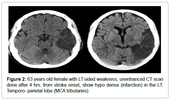
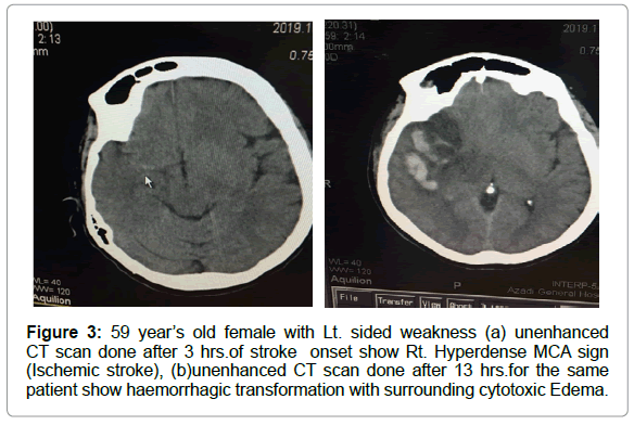
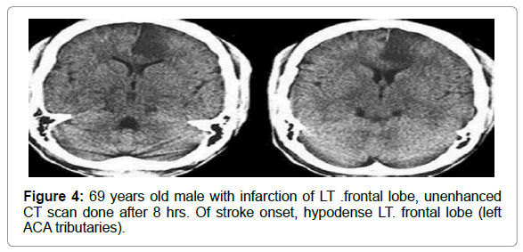
Significant difference between ischemic and haemorrhagic strokes (P = 0.037) (Figures 2-4).
Discussion
In the current study the sensitivity of unenhanced CT scan compared to clinical diagnosis that was taken as a reference standard was 56.7%, the mean time of occurrence (onset) according to CT criteria was 6.9 hrs. of stroke onset as seen in Table 1. This finding is comparable to the study done by [4-6] who found the sensitivity of unenhanced CT the first 8 hrs. was 58%, while the sensitivity of unenhanced CT scan was 41.8% in the study done by, [7] This sensitivity was less than our current study, this is most probably due to time lag between the clinical symptoms of patients and their unenhanced CT performance. Theshortest period of time lag (1.2 hrs) between symptoms onset and a negative unenhanced CT especially in ischemic stroke In current study, as shown in Tables 2 & 3, the main stem of MCA was the mostly affected vessel in both ischemic and haemorrhagic strokes, its percentage with each type was 51.4% and 50% respectively, followed by PCA and VBS main stem, their percentages was 17.1% and 20.8% in both type of stroke respectively. This was scientifically comparable to other studies like in the study conducted by [8], the percentage of main MCA involvement was 49.6%, while the VBS and PCA had been less affected, their percentage was 33.8%, also this finding was similar to the study done by the [9]; their related to main MCA was (48.26%) and the PCA was (20.57%). Regarding the risk factor, in the current study and as shown in Table 4, the incidence of stroke was higher in the older age, the age increment was the most common followed by hypertension, the percentage was 31.8% between the age of (20-49) years, while between the age of ( 50-64years and more), the percentage was 68.3%,this was similar to the study done in the united states by [10], the percentage was 70% above the age of 65 years. In the current study, the risk of aging process followed by the hypertension and DM, the percentage of the stroke was high in the hypertensive patients, the percentage was 73% and in DM was 38%. The study done by [11-13], hypertension was about 67% followed by DM 36%. Regarding the neighbouring regional parenchymal effects.
Regarding the neighbouring regional parenchymal effects, in the present study and as shown in Table 5, the edema and mass effects were seen more in haemorrhagic stroke than in ischemic stroke, the percentage in haemorrhagic stroke was 79.1% and ischemic was(20.8%). This is similar to the study done by who found mass effect was seen in 88% in haemorrhagic stroke and 22% in ischemic sroke in the present study and as shown in Table 6, the edema and mass effects were seen more in haemorrhagic stroke than in ischemic stroke, the percentage in haemorrhagic stroke was 79.1% and ischemic was (20.8%). This is similar to the study done by [14,16,17] who found mass effect was seen in 88% in haemorrhagic stroke and 22% in ischemic stroke. In the current study and as shown in Table 7, the mean time of earliest radiological findings seen in haemorrhagic stroke as detected by unenhanced CT scan was less than ischemic stroke, in haemorrhagic stroke was 5.8 hrs, while in ischemic was 7.5 hrs. this was due to the limitation of nonenhanced CT scan to identify early ischemic changes in the brain, this study also approved by the[17-19],who found the haemorrhagic stroke can be detected earlier than ischemic stroke by unenhanced CT scan, as the ischemic stroke changes need more time to be noticed in unenhanced CT scan.[20].
Conclusions
Unenhanced CT scan showed sensitivity of 56.7% relative to the clinical diagnosis as a referred standard, it can help differentiating ischemic from haemorrhagic stroke confidentially with adjacent or surrounding local brain effects, also it can detect the exact brain area vascular territories regarding both ischemic and haemorrhagic strokes, meanwhile, the negative unenhanced CT scan doesn’t mean normal brain parenchyma especially in early onset of symptoms, as it need time to visualize the brain imaging changes especially in hyper acute ischemic brain stroke, unenhanced CT scan can detect haemorrhagic stroke earlier than ischemic one as the hyper acute stroke needs time to appear on this imaging device.
References
- Abbas SG, Kumar S (2018) Importance of Computed Tomography scans in patients with. Cerebrovascular Accidents 6(11):48-50
- Michael K, Abraham M, Chang W (2016) Subarachnoid Hemorrhage. Emergency Medicine Clinics of North America 34(4): pp.901-916.
- Adams Adams HP, Bendixen B, Kappelle L, Biller J, Love B, et.al (2001) Classification of subtype of acute ischemic stroke. Definitions for use in a multicenter clinical trial. TOAST. Trial of Org 10172 in Acute Stroke Treatment. Stroke 24(1): pp.35-41.
- Allen L, Hasso A, Handwerker J, Farid H (2012) Sequence-specific MR Imaging Findings That Are Useful in Dating Ischemic Stroke. Radiographic 32(5): pp.1285-1297.
- Allmendinger A, Tang E, Lui Y, Spektor V (2012) Imaging of Stroke: Part 1, Perfusion CT???Overview of Imaging Technique, Interpretation Pearls, and Common Pitfalls. American J Radiology 198(1): pp.52-62.
- American College of Radiology (2009) ACR Appropriateness Criteria. “Cerebrovascular Disease.†ACR. Reston, VA. www.acr.org/ac
- AngelesFernandez-Gil M, Palacios-Bote R., Leo-Barahona M, Mora-Encinas J (2010). Anatomy of the Brainstem: A Gaze into the Stem of Life. Seminars in Ultrasound, CT and MRI, 31(3); pp.196-219.
- Arenillas J, Ãlvarez-SabÃn J (2005) Basic Mechanisms in Intracranial Large-Artery Atherosclerosis: Advances and Challenges. Cerebrovascular Diseases 20(2):  pp.75-83.
- Ariesen M, Claus S, Rinkel G, Algra A (2003) Risk Factors for Intracerebral Hemorrhage in the General Population. Stroke 34(8): pp.2060-2065.
- Assarzadegan F, Tabesh H, Shoghli A, Ghafoori Yazdi M, Tabesh H et al. (2015) Relation of Stroke Risk Factors with Specific Stroke Subtypes and Territories. Iran J Public Health 44: p.48.
- Banerjee T, Mukherjee C, Sarkhel A (2001) Stroke in the Urban Population 20 pp: 201 to 207
- Biller J, Love B, Gordon D, Marsh E (2001) Classification of subtype of acute ischemic stroke Definitions for use in a multicenter clinical trial. TOAST. Trial of Org 10172 in Acute Stroke Treatment. Stroke 24(1): pp.35-41.
- Birenbaum D (2010) Emergency neurological care of strokes and Bleeds. J  Emergencies, Trauma Shock 3(1): p.52.
- Brant W, Helms C (2012) Fundamentals of diagnostic radiology. 4th ed. Philadelphia Wolters Kluwer/Lippincott Williams & Wilkins Health.
- Brouwers H, Biffi A, Ayres A, Schwab K, Cortellini L, et al. (2012) Apolipoprotein E Genotype Predicts Hematoma Expansion in Lobar Intracerebral Hemorrhage. Stroke 43(6): pp.1490-1495.
-  Brown MM, Pereira AC (2001) Cerebrovascular Disease: Experience Within Large Multicenter Trials. Clinical Trials in Neurology pp. 229-249.
- Thorsten M Buzug (2008)Â Introduction to Computed Tomography (1st Edition). Dordrecht: Springer p.475
- Carter R (2014)Â The Human Brain Anatomy. [Online] Verywell Mind. Available at: https://www.verywellmind.com/the-anatomy-of-the-brain-2794895 [Accessed 26 Jul. 2019].
- Cha S, Castillo M, Naidich T, Smirniotopoulos J (2013)Â Imaging of the Brain. Elsevier Health Sciences, pp.167-169.
- Zazulia AR, Diringer MN, Derdeyn CP, Powers WJ (1999) Progression of mass effect after intracerebral hemorrhage. Pub Med 30(6):1167-1173
Citation: Saeed M (2021) Diagnosing Stroke Cases Using Unenhanced Helical Computed Tomography in Recently Brain Stroked Patients in Duhok City. OMICS J Radiol 10: 324. DOI: 10.4172/2167-7964.1000324
Copyright: © 2021 Saeed M. This is an open-access article distributed under the terms of the Creative Commons Attribution License, which permits unrestricted use, distribution, and reproduction in any medium, provided the original author and source are credited.
Select your language of interest to view the total content in your interested language
Share This Article
Open Access Journals
Article Tools
Article Usage
- Total views: 2707
- [From(publication date): 0-2021 - Dec 06, 2025]
- Breakdown by view type
- HTML page views: 1860
- PDF downloads: 847


