Developmental Conduction in Aphasia after the Neonatal Stroke
Received: 02-Jun-2022 / Manuscript No. nnp-22-67698 / Editor assigned: 04-Jun-2022 / PreQC No. nnp-22-67698(PQ) / Reviewed: 18-Jun-2022 / QC No. nnp-22-67698 / Revised: 22-Jun-2022 / Manuscript No. nnp-22-67698(R) / Published Date: 29-Jun-2022 DOI: 10.4172/2572-4983.1000242
Abstract
Impairment of speech repetition following injury to the dorsal language stream is a feature of conduction aphasia, a well-described “disconnection syndrome” in adults. The impact of similar lesions sustained in infancy has not been established.
Introduction
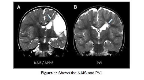
Conduction aphasia was first described by Carl Wernicke1 and was characterized by relatively intact comprehension, with paraphasic expressions and wordfinding problems [1]. The notion of impaired repetition was introduced by Lichtheim and is now a core part of modern diagnostic criteria. Conduction aphasia was regarded as a “disconnection syndrome” by Geschwind,representing an interruption of connections between the anterior and posterior language areas.This early connectionist model was expanded by recent imaging studies into the dual stream model of language. This distinguishes a “dorsal language stream,” of which the arcuate fasciculus (AF) is a key element, from an anatomically distinct “ventral stream.” [2]. The dorsal stream connects speech regions in the temporal lobe with the inferiorparietal cortex and posterior frontal lobe. These regions are involved in mapping speech sounds to articulatory representations, which is critical for speech repetition, especially when repeating and learning novel words. In contrast, the ventral stream projects rostrally via the anterior temporal lobe toward the inferior frontal lobe and involves the extreme capsule fiber system and uncinate fasciculus (UF). This stream serves “sound to meaning mapping” by associating speech with semantic and conceptual representations in the anterior temporal lobe, a system important for meaningful speech [3] (Figure 1).
The roles of the streams during language development continues to be debated, and the differential impact of early dorsal versus ventral stream lesions is not known. Nevertheless, it is generally accepted that damageto perisylvian language areas in infancy or childhood does not result in aphasia syndromes analogous to those in adults, presumably as a result of cerebral plasticity [4]. A critical question is whether the left dorsal stream, and in particular the AF, fulfils specific functional roles that cannot be compensated for by the ventral stream or the right cerebral hemisphere. This question is of direct clinical relevance in view of the comparatively high incidence of stroke in the neonatal period which commonly occurs in the territory of the left middle cerebral artery, supplying most of the dorsal stream regions [5]. Given the association between adult dorsal stream injury and conduction aphasia, we sought to determine whether repetition deficits might be present following similar lesions in a cohort of term-born children who sustained a stroke at birth. We chose a range of repetition tasks, involving digit strings, nonwords, and sentences of increasing complexity, separating participants into dorsal and nondorsal (including ventral stream) lesion groups. Speech repetition problems are thought to impact on language learning and are established predictors ofimpaired literacy and language development in childhood.Also, in view of the presumed functional role of the dorsal stream in language learning we assessed speech repetition in the context of other abilities, such as vocabulary, comprehension, reading, and spelling [6]. Language lateralization was assessed using functional magnetic resonance imaging (fMRI), given that repetition deficits might be modified by interhemispheric reorganization.4 Patients and Methods Study Population We recruited 30 term-born English-speaking individuals (age 5 7–18 years) with MRI-confirmed ischemic/hemorrhagic stroke in the neonatal period. Participants were drawn from a prospective cohort of neurologically symptomatic infants, born at or referred to the Hammersmith Hospital, London. All had early MRI, which was reviewed for the present study, and all were followed-up regularly. A term-born control group (n 5 40) was also recruited, group-matched for age (range 5 6–18 years), sex, and maternal education [7]. A total of 13 individuals were bilingual with native English proficiency (stroke cohortEthical Approval and Patient Consent Ethical approval for the study was obtained from institutional research ethics committee, and written informed consent was obtained from all participants or their parents.
Discussion Our key findings are that at the group level, neonatal left dorsal language stream injury can lead to long-lasting impairment in verbatim sentence repetition, which is similar to conduction aphasia in adults; and this function is relatively preserved in cases with right hemisphere language dominance [8]. In keeping with earlier studies, we confirm that injury to left hemisphere eloquent regions is associated with language decrements independent of lesion size and that many affected cases do not show a language shift to the right hemisphere. In contrast to previous work, we propose the existence of a developmental form of with severely impaired repetition had a lesion involving the left anterior AF, leaving posterior area Spt intact. This supports the notion that the necessary and sufficient basis for the speech repetition deficit is a “disconnection” between posterior and anterior association cortices [9]. Children with injury to other regions of the left or right cerebral hemispheres, including the left ventral language stream, did not show such impairments, confirming the anatomical specificity of this finding. Acquired fluent aphasia is described in childhood, and in some cases conduction aphasia has been documented. However, apart from a single case report following a stroke at 3 years of age, repetition deficits are generally reported to be transient, presumably attributable to lesion resolution or functional reorganization [10]. In contrast, our study provides evidence for longlasting repetition impairment despite the early timing of the injuries, when the potential for cerebral plasticity is presumably at a maximum [11,12].
Conclusions
This is the first study to examine the impact of early injury to the dorsal language stream and identifies adevelopmental form of conduction aphasia with anatomical lesion distribution and common neuropsychological features similar to the classical “disconnection syndrome” described in adults. Although the absence of paraphasia in our cohort is noted, a milder phenotype is not uncommon in developmental variants and may reflect the inherent plasticity of the immature brain. In those children who did not show interhemispheric transfer, the demonstration of long-lasting repetition deficits (and more subtle additional language problems) provides intriguing evidence that there are inherent limitations to cerebral plasticity, even when damage occurs in the very young. This finding is particularly important given the association with subsequent academic difficulties in this population and underscores the need for early identification and remediation. It also offers a neuroanatomical basis for a key behavioral marker of specific language impairmentby showing that nonword and sentence repetition is highly dependent on the integrity of the left dorsal language stream.
References
- Bratteby, L.E.Garby, L.Groth, T.Schneider ,W.Wadman B. (1968)Studies on erythro-kinetics in infancy. XIII. The mean life-span and the life-span frequency function of red blood cells formed during foetal life. ActPaediat. Scand 57-311.
- OskiF,A.Naiman J.L, (1966).Hematologic problems of the newborn. W. B. Saunders Company,Baltimore19-66.
- Smith CA (1959)The physiology of the newborn infant 3.Charles C Thomas, Publisher,Philadelphia.
- Guest GM, Brown E.W (1957) Erythrocytes and hemoglobin of the blood in infancy and childhood. III. Factors in variability, statistical studies. mer. J. Dis. Child.93-486.
- Gairdner, D. Marks ,J.Roscoe. (1952) Blood formation in infancy. Part I. The normal bone marrow. Arch. Dis. Child 27-128.
- Sturgeon P. (1950) Volumetric and microscopic pattern of bone marrow in normal infants and children. Pediatrics 7-577.
- Sturgeon P (1951) Volumetric and microscopic pattern of bone marrow in normal infants and children. Pediatrics 7-642.
- Sturgeon P (1951)Volumetric and microscopic pattern of bone marrow in normal infants and children. Pediatrics7-774.
- Glaser K. Limarzi L.R. Poncher H.G (1950) Cellular composition of the bone marrow in normal infants and children Pediatrics 6-789.
- Garby L. Sjölin S. Vuille J (1963) Studies on erythro-kinetics in infancy. III. Disappearance from plasma and red cell uptake of radioactive iron injected intravenously. Acta Paediat Scand 52-537.
- Seip M, Halvorsen S(1956) Erythrocyte production and iron stores in premature infants during the first month of life.Acta Paediat Scand 45-600.
- Pearson HA, (1966) Recent advances in hematology. J. Pediat 69-466.
Indexed at, Crossref, Google Scholar
Indexed at, Google Scholar, Cross ref
Indexed at, Crossref, Google Scholar
Indexed at, Crossref, Google Scholar
Indexed at, Crossref, Google Scholar
Citation: Katheria A (2022) Developmental Conduction in Aphasia after the Neonatal Stroke. Neonat Pediatr Med 8: 242. DOI: 10.4172/2572-4983.1000242
Copyright: © 2022 Rousell Z. This is an open-access article distributed under the terms of the Creative Commons Attribution License, which permits unrestricted use, distribution, and reproduction in any medium, provided the original author and source are credited.
Share This Article
Recommended Conferences
42nd Global Conference on Nursing Care & Patient Safety
Toronto, CanadaRecommended Journals
Open Access Journals
Article Tools
Article Usage
- Total views: 984
- [From(publication date): 0-2022 - Apr 03, 2025]
- Breakdown by view type
- HTML page views: 668
- PDF downloads: 316

