Dental Corono-Radicular Fractures in Adult Subjects: Epidemiology, Etiopathogeny, Anatomic Pathology and Therapeutic Indications
Received: 26-Nov-2021 / Manuscript No. JDPM-22-48451 / Editor assigned: 27-Jan-2022 / PreQC No. JDPM-22-48451(PQ) / Reviewed: 10-Feb-2022 / QC No. JDPM-22-48451 / Revised: 15-Feb-2022 / Manuscript No. JDPM-22-48451(R) / Accepted Date: 21-Feb-2022 / Published Date: 22-Feb-2022 DOI: 10.4172/jdpm.1000114
Abstract
Coronoradicular fractures are a frequent reason for consultation today in Ivory Coast and they pose the problem of the conservation of the tooth, yet awaiting a final or prosthetic restoration.
Methods: It is a descriptive cross-sectional study performed on 100 cases of corono-root fractures in adult patients received during our consultations.
Thus, for each patient, note the reason for the consultation, the tooth concerned, living or not, with ductal treatment or not, with a filling or not, the type of fracture, the mobile buccal, palatal or lingual pan and the presence adjacent or opposing teeth.
Results: Epidemiologically, a frequency of 9% coronavicular fractures diagnosed during our consultations. Mastication is the main cause of occurrence and in 70% of cases and the gingival pain caused by mobility fracture pan 80% or pulpal exposure 10% motivates the consultation.
The teeth were depulped in 85% of cases and the opposing tooth exists in all cases and often alive.
60% of oblique fractures are sub-gingival, 40% supra-gingival, and the mobile part is most often vestibular 70% when the tooth is maxillary and 60% lingual when the tooth is mandibular and the maxillary teeth are most represented in our sample 60%.
Conclusion: Coronary root fractures are quite worrying for the practitioner because of their frequency and their etiopathogenesis. They are usually treated teeth or at the end of treatment with a prosthetic or non-prosthetic project.
These coronal oblique oblique fractures raise the question of the conservation or not of the tooth. This will depend on the type of fracture and the height of the fracture, especially at the root level.
Keywords
Fractures; Pain; Prosthesis.
Introduction
The many advances in conservative dentistry and oral hygiene mean that patients keep their natural teeth longer. It is therefore an aging organ, cracked or not, alive or not, blocked or with loss of substance or significant or crowned decay that will be subjected to chewing with a possibility of fracturing.
The pain can become debilitating when the pulp and other internal or external structures of the tooth are affected and this is the reason for consultation.
This work aims to study coronary radicular dental fractures in the usual population of dental offices in Côte d'Ivoire from an epidemiological, etiopathogenic, clinical, pathological and therapeutic perspective.
Materials and Methods
The study population consists of 100 clinical cases of coronary radicular fractures.
The tooth concerned, living or not, with or without root canal treatment, bearing a filling or not, the type of fracture, the mobility of the palatal, vestibular or lingual fractured side and the presence of adjacent or antagonistic teeth are noted.
Retro-alveolar radiology is performed for each clinical case.
Emergency and definitive treatment are specified for each case and depending on the type of fracture.
Results
Epidemiologically, we noted a 9% frequency of coronary radicular fractures diagnosed during our consultations [1]. Chewing was the main circumstance of occurrence and in 70% of cases, the diet was not hard (chewing rice, foutou, attiéké) the small metal or stone accidentally bitten or the trauma is only found in less than 30% of cases. The patient in 90% of cases heard a cracking noise, and gum pain caused by the mobility of the fractured end 80% or pulp exposure 10% motivated the consultation.
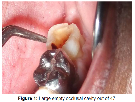
The tooth presented with an empty occlusal cavity 40%, an overflowing temporary or permanent dressing 50%, or decay 5%.
The teeth were pulped in 85% of the cases and the opposing tooth existed in all the cases and often alive. Adjacent teeth were present in 70% of cases and cases of teeth %
The fracture line is generally oblique in the buccal, palatal or lingual direction.
60% of oblique fractures were subgingival and 40% supragingival and the movable pan was most often vestibular 70 when the tooth was maxillary and in 60% lingual when the tooth was mandibular.
The maxillary teeth were the most represented in our sample 60%.
The teeth most affected by per masticatory fractures were, in order of frequency:
Upper 35%, upper premolars 30% and mandibular molars 25%.
Oblique radicular and vertical fractures represented 10% and concerned the incisor-canine group and the mandibular premolars. The living teeth 10% were all from the canine incisor group and accidental fractures (AVP, brawls, falls, etc.). A case of complete vertical fracture on a maxillary molar was noted. Regarding therapeutic management, simple conservative treatment was carried out when the pulp is not exposed 10%. cases lost to followup after removal of the fractured flap for the consultation represented approximately 20%. The tooth is preserved and the prosthetic crown is performed in 40% of cases and tooth extraction performed in 30% of cases.
Discussion
The diagnosis of these fractures is based on 8 elements according to the ADE (American Association of Endodontists) [2]:
A- The clinical interview emphasizes listening to the patient and researching the type of pain.
B- The bite test (bite on the cotton roll), the clinical reaction is the absence of pain on pressurization and sharp pain on release (Figure 1).
C- Trans-illumination using a light source focused by an optical fiber, which is applied to the buccal, lingual and palatal surfaces. The solution of continuity created by the fracture or crack, will prevent the correct diffusion of light through the crown.
D- Pulp sensitivity tests should be sought.
E- Periodontal percussion, palpation and probing tests should be sought.
F- The removal of the coronary restoration and direct visual examination of the desired pulp floor or ceiling.
G- Radiological examinations from the retro-alveolar to cone beam imaging.
H- Surgical exploration by avulsion of the most mobile fractured fragment epidemiologically, we noted a frequency of 9% of coronary radicular fractures diagnosed during our consultations, NDREASEN & ANDREASEN (2000) placed them at 7% for permanent teeth and 2 to 4% for teeth of milk [3].
In our study, the usual chewing of soft or slightly hard foods is the main circumstance of dental fractures in adults, unlike the literature which often incriminated trauma or impact on a hard object [4]. The accidentally bitten pebble is only found in 30% of cases. Pulpal or periodontal pain related to the mobility of the mobile fragment often motivates the consultation. Most fractured teeth are devitalized and have a temporary or permanent restoration. The loss of pulp vitality led to qualitative and quantitative changes in the residual dental structures. Qualitatively, it has long been assumed that the non-vital tooth becomes more fragile due to a decrease in its water content. The medical literature supports the concept of approximately 9% fluid loss in teeth.
Devitalized, but it is interesting to note that only the free water content, and not the water bound to the tissues, is changed [5].
During this study, we noted a high frequency of oblique subgingival coronary radicular fractures, whereas according to the literature [3] coronary fractures are generally oblique supragingival with or without pulp exposure.
The teeth most affected by per masticatory fractures were, in order of frequency:
The upper molars 35%, the upper premolars 30% and the mandibular molars 25%, the other 10% concern the single-rooted.
The maxillary molars, especially M1, are frequently fractured due to their age in the oral cavity and the occlusal pressure exerted by the mandibular antagonist during chewing. In addition, the second molar becomes fragile when the wisdom tooth is absent. However, the presence of a large occlusal cavity which may or may not be filled in a depulpated tooth seems to be very favorable conditions for the occurrence of fractures according to our study. Indeed, the extension of the coronary cavity and in particular the loss of the proximal ridges would lead to a loss of mechanical resistance of the order of 65%. The same authors concluded that the maximum weakening of the tooth was obtained after preparation of the tooth. 'an endodontic access cavity associated with a mesio-occluso-distal cavity (MOD) Reeh et al., [5].
In the premolars, oblique supragingival and subgingival fractures most often involve maxillary PM due to their larger occlusal anatomy and double or massive roots.
Oblique root fractures tend to occur in mandibular MPs with a more tapered root anatomy.
At the level of the incisors, the traumas and accidents of the public highway are responsible for oblique coronorradicular fracture above and subgingival oblique or radicular. The lower incisors were significantly less often involved than their upper antagonists (CALSIKAN & PEHLIVAN 1996) [6].
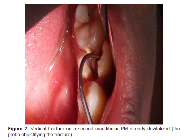
The observation made during this study shows that the fracture does not respect the root anatomy or the furcation of the multirooted cells. It never separates the tooth by the furcation the line is oblique vestibular subgingival or oblique lingual or palatine or even willingly vertical vestibulo palatine or vestibulo-lingual following the mesio-distal groove of the tooth, and being able to be complete as the 2 reported cases In this study (Figure 2).
Finally we note the almost permanent presence of an adjacent tooth which plays the role of vector agent of the fracture during chewing but the hardness of the food is not incriminated in this study because the patients all report that the fracture occurred when chewing soft foods. It is therefore more the fragility of the tooth that favors the occurrence of the fracture, contrary to certain data in the literature [4].
Coronoradicular fractures are the result of high mechanical stresses and subsequent shear forces determining the direction of the fracture line, so separation then divides the tooth into a coronal (peripheral) fragment and an apical (proximal) fragment [7].
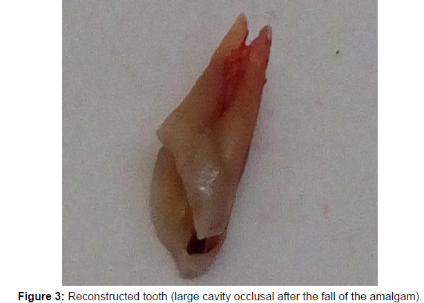
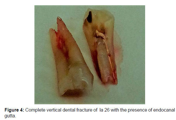
The molars are the most affected by caries disease and root canal treatments having resolved the issue of pain, most patients no longer continue care and the prosthetic restoration is not performed. Also, when the fracture occurs and the line is subgingival, the preservation of the tooth is difficult, which is why some of our colleagues in Africa do not hesitate to extract the fractured tooth. However, the discussion of treatment modalities should take into account the patient's age, the means of the patient and the fracture line. New therapeutic indications for fractures have been adopted according to AAE [2] and range from simple monitoring to tooth extraction. Removal of the fractured dental fragment and restoration with glued materials, periodontal surgery or root amputation can be performed as appropriate. In the case of combined lesions without dislocation of the coronary fragment, endodontic treatment must be instituted without delay, and in the event of dislocation of dental fragments, the clinical solutions currently proposed are conventionally divided into two categories: - The cast corono-radicular reconstruction (Figure 3). - Coronal-radicular reconstruction, with fiber anchoring [8] But it is usually not possible to keep teeth that have undergone vertical fracture or a very oblique fracture line (diagonal fracture), then the tooth must therefore be extracted (Figure 4).
Conclusion
Coronal radicular fractures are quite worrying for the practitioner by their frequency and their different types. The most affected teeth were, in order of frequency, the upper molars 35%, the upper premolars 30% and the mandibular molars 25%, the other 10% concerning the singlerooted. This is usually teeth already treated or at the end of treatment with a conservative restoration and the possibility of considering a prosthetic restoration or a prosthetic project.These corono-radicular fractures then occurring from usual chewing raise the question of whether or not the tooth is preserved. This will depend on the type of fracture and the height of the fracture, especially at the root level. The therapeutic possibilities range from simple restoration to the creation of an endoprosthetic complex or to tooth extraction.
References
- Rakotoarivony AE, Rakotoarison RA, Rakotoarimanana FV, ArijaonaAN, Rakoto-Alson S, et,al (2014) Epidemiology of Dento-Maxillofacial Trauma at CENHOSOA Antananarivo. Med Buccale Chir Buccale 20: 221-226.
- American Association of Endodontists (AAE) (2004) Recommended Guidelines of the AAE for the treatment of traumatic dental injuries.
- Andreasen JO, Bakland LK, Flores MT, Andreasen FM, Andersson L (2000) Traumatic Dental Injuries: A Manual. J Ortho 27: 361.
- Mahmoodi B, Rahimi-Nedjat R, Weusmann J, Azaripour A, Walter C, et.al (2015) Traumatic Dental Injuries in A University Hospital: A Four-Year Retrospective Study. BMC Oral Health 15: 139.
- Reeh ES, Messer HH, Douglas WH (1989) Reduction in Tooth Stiffness as A Result of Endodontic and Restorative Procedures. J Endod 15: 512-516.
- American Association of Endodontists (AAE) (2008) Cracking the Cracked Tooth Code: Detection and Treatment of Various Longitudinal Tooth Fractures.
- Brunner F, Krastl G, Filippi A (2009) Dental Trauma in Adults in Switzerland. Dent Traumatol 25: 181-184.
- Sharif MO, Tejani-Sharif A, Kenny K, Day FP (2015) A Systematic Review of Outcome Measures Used in Clinical Trials of Treatment Interventions Following Traumatic Dental Injuries. Dent Traumatol 31: 422-428.
Indexed at Google Scholar Crossref
Indexed at Google Scholar Crossref
Indexed at Google Scholar Crossref
Indexed at Google Scholar Crossref
Indexed at Google Scholar Crossref
Citation: Attogbain KP, Adou Akpe J, Amantchi D, Aka A (2022) Dental Corono- Radicular Fractures in Adult Subjects: Epidemiology, Etiopathogeny, Anatomic Pathology and Therapeutic Indications. J Diabetes Clin Prac 6: 114. DOI: 10.4172/jdpm.1000114
Copyright: © 2022 Attogbain KP, et al. This is an open-access article distributed under the terms of the Creative Commons Attribution License, which permits unrestricted use, distribution, and reproduction in any medium, provided the original author and source are credited.
Share This Article
Recommended Journals
Open Access Journals
Article Tools
Article Usage
- Total views: 1794
- [From(publication date): 0-2022 - Nov 24, 2024]
- Breakdown by view type
- HTML page views: 1415
- PDF downloads: 379
