Cystic Hygroma and COVID-19: Role of Vascular Endothelial Growth Factor C (VEGF-C)
Received: 23-Jan-2022 / Manuscript No. roa-22-55234 / Editor assigned: 25-Feb-2022 / PreQC No. roa-22-55234(PQ) / Reviewed: 01-Jan-1970 / QC No. roa-22- 55234 / Revised: 16-Mar-2022 / Manuscript No. roa-22-55234(R) / Published Date: 23-Mar-2022 DOI: 10.4172/2167-7964.1000367
Cystic hygromas are inherited defects of lymphatic origin. They are most commonly seen around the neck, clavicle, and axillary area [1]. Around half of the cases are usually present at birth, while the other half arises by age two [2]. These cysts may get infected and increase in size [3]. Manifestation of the disease depends on the site of anatomical obstruction [1]. Molecular research suggests that Vascular Endothelial Growth Factor C (VEGF-C) may be a key player in the development of these cysts [4].
Severe acute respiratory syndrome coronavirus-2 (Sars-CoV-2) is a new virus known to cause mild to moderate respiratory illness in healthy patients. However, patients with comorbidities such as obesity, diabetes, chronic respiratory disease and even cancer were found to suffer from a more severe form of the disease [5].
We describe a case of a previously healthy two years and a half old boy with a small neck mass that has evolved after Coronavirus disease (COVID-19 infection).
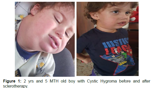
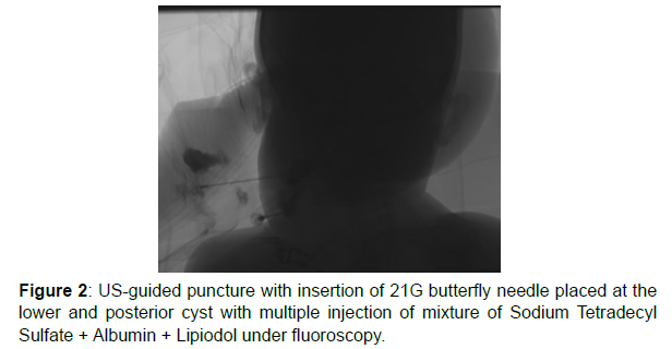
At 2 years of age, the patient presented to a private pediatric clinic with minimal right neck swelling (2x1.2 cm). It appeared as a small skin colored, transparent fluid-filled dot that was not painful and not inflamed. The patient was sent home on Clarithromycin for 1-week duration without any further evaluation. One week later, the mass remained unchanged despite antibiotics. The mass was observed since it was not painful and not evolving in size. It remained the same for 5 months (Figure 1 and 2).
Five Months later, the patient developed COVID-19 infection. During that time the parents reported that the neck mass was increasing dramatically in size (Figure 1). Upon presentation, the physician noticed that the mass became red to bluish in color and more firm in consistency causing discomfort upon neck deviation. Otherwise, the patient was clinically stable without any systemic symptoms. He also did not show any signs of respiratory distress.
Magnetic resonance imaging (MRI) of the neck showed a 7x4.8x9 cm cystic lesion spanning the right posterior and anterior cervical regions extending inferiorly to the upper mediastinum. These findings were compatible with the diagnosis of cystic hygroma with hemorrhagic content. The airway was patent and patient was cleared by otorhinolaryngology.
After contacting interventional radiology, the patient underwent right cystic mass sclerotherapy with Bleomycin under sedation with midazolam and fentanyl (Figure 2). Large quantities of blood and lymph were drained by a fine needle aspiration from the cystic mass, before the injection of the sclerosing agent. The patient recovered uneventfully and the mass shrunk in size over the next 3 months (Figure 1). He is now due for repeat procedure.
This case describes the quick growth of lymphatic cysts after COVID-19 infection. It illustrates the process of cystic enlargement after a cascade of inflammation induced by a COVID-19 infection.
Recent studies suggest a possible link between inflammation and the reactivation of dormant tumor cells. This phenomenon could be triggered by a change in their microenvironment [6]. Furthermore, COVID-19-induced inflammation may create this milieu that is harmful to cells. It promotes inflammation through the activation of immune cells that result in the release of pro-inflammatory cytokines such and markers [7]. Chronic inflammation and release of cytokines attract macrophages, which may be linked to epithelial cysts growth [8].
The literature about the role of COVID-19 infection in promoting tissue growth is limited. To our knowledge COVID-19 penetrates cells by attaching to Angiotensin-converting enzyme 2 (ACE2) and subsequently down regulating it. ACE2 is responsible for diminishing the release of cytokines most notably VEGF. As a result, VEGF levels are elevated in a patient with COVID-19 [7].
Although theoretical, this case highlights that COVID-19 is a potential powerful immune system instigator, we hypothesize that cystic enlargement may be related to a surge in many cytokines including VEGF in the context of Covid-19 acute infection course.
References
- Kumar N, Kohli M, Pandey S, Tulsi SPS (2010) Cystic hygroma. Natl J Maxillofac Surg 1: 81-85.
- Mirza B, Ijaz L, Saleem M, Sharif M, Sheikh A (2010) Cystic hygroma: an overview. J Cutan Aesthet Surg 3: 139–144.
- Vijay AP (2020) Lymphatic Malformation (Cystic Hygroma).
- Francescangeli F, Angelis MLD, Zeuner A (2020) COVID-19: a potential driver of immune-mediated breast cancer recurrence? Breast Cancer Res
- https://www.who.int/health-topics/coronavirus#tab=tab_1
- Francescangeli F, De Angelis ML, Baiocchi M, Rossi R, Biffoni M, et al. (2020) COVID-19-Induced modifications in the TUMOR Microenvironment: Do they affect Cancer Reawakening and Metastatic relapse?. Front Oncol 10: 592891
- Turkia M. (2020) COVID-19, Vascular Endothelial Growth Factor (VEGF) and Iodide. SSRN
- Karihaloo A (2015) Role of Inflammation in Polycystic Kidney Disease. In: Li X editor Polycystic Kidney Disease.
Indexed at, Google Scholar, Crossref
Indexed at, Google Scholar, Crossref
Indexed at, Google Scholar, Crossref
Indexed at, Google Scholar, Crossref
Citation: Hammoud M, Awad C, Sayad E (2022) Cystic Hygroma and COVID-19: Role of Vascular Endothelial Growth Factor C (VEGF-C). OMICS J Radiol 11: 366. DOI: 10.4172/2167-7964.1000367
Copyright: © 2022 Hammoud M, et al. This is an open-access article distributed under the terms of the Creative Commons Attribution License, which permits unrestricted use, distribution, and reproduction in any medium, provided the original author and source are credited.
Share This Article
Open Access Journals
Article Tools
Article Usage
- Total views: 4087
- [From(publication date): 0-2022 - Mar 29, 2025]
- Breakdown by view type
- HTML page views: 3561
- PDF downloads: 526


