Case Report Open Access
Cyst of the Canal of Nuck Mimicking Inguinal Hernia: A Case Report
Acu L1* and Acu B21Department of Radiology, Erbaa State Hospital, Erbaa/Tokat, Turkey
2Department of Radiology, Osmangazi University, Eskisehir, Turkey
- *Corresponding Author:
- Leyla Acu
Department of Radiology
Erbaa State Hospital
Erbaa/Tokat, Turkey
Tel: +90 (506) 5152679
Fax: +90 (356) 7151052
E-mail: leylaacu@gmail.com
Received Date: January 23, 2017; Accepted Date: January 25, 2017; Published Date: January 30, 2017
Citation: Acu L, Acu B (2017) Cyst of the Canal of Nuck Mimicking Inguinal Hernia: A Case Report. OMICS J Radiol 6:251. doi: 10.4172/2167-7964.1000251
Copyright: © 2017 Acu L, et al. This is an open-access article distributed under the terms of the Creative Commons Attribution License, which permits unrestricted use, distribution, and reproduction in any medium, provided the original author and source are credited.
Visit for more related articles at Journal of Radiology
Abstract
A 22-year-old female patient was referred to our department by general surgeon for a suspected left-sided inguinal hernia. She had a painful tender mass in her left groin. On physical examination, there was a palpable mass in the region of her left inguinal canal. US and pelvic CT examination were performed. Sonographic examination, revealed a well-defined cystic mass with internal septae and thin wall, located in the left inguinal canal extending along the course of the round ligament. On CT images, there was a thin-walled cystic mass in the left inguinal canal and intraabdominal extension of the canal of Nuck. Diagnosis of cyst of the canal of Nuck was confirmed by surgery and subsequent histopathologic evaluation. Hydrocele of the canal of Nuck is a rarely encountered entity. It should be on the differential diagnosis list of groin swellings in female patients. Radiologists must be aware of the imaging findings of hydrocele of the canal of Nuck to diagnose this entity before surgery.
Keywords
Hydrocele; Canal of Nuck; Nuck; Hydrocele; Female; Inguinal canal
Introduction
Hydrocele of the canal of Nuck, the female homologue of hydrocele of spermatic cord, is a rare cause of inguinal swelling in women. We report a case of a cyst of the canal of Nuck with Ultrasound (US) and Computerized Tomography (CT) findings that the female patient presenting swelling in the left inguinal region.
Case Report
A 22-year-old female patient was referred to our department by general surgeon for a suspected left-sided inguinal hernia. She had a painful tender mass in her left groin. In her history, she had noticed the palpable mass in her puberty. She had pain for 3 days and she had no problem before. She noticed that the size of the mass had grown in time.
On physical examination, there was a 3 × 2 cm palpable mass in the region of her left inguinal canal. The mass was non-reductable and tender. There was no discoloration of the overlying skin. There was no history of trauma. There was no sign of the intestinal obstruction.
US and pelvic CT examination were performed. The ultrasound examination of the left groin was performed with a high frequency (7.5 mHz) transducer.
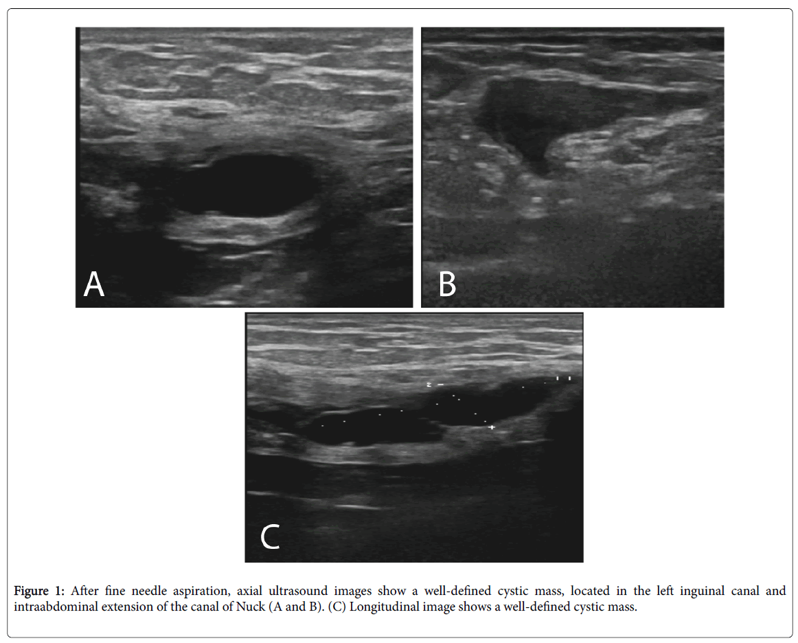
Sonographic examination revealed a well-defined cystic mass with internal septae and thin wall, measuring 3 × 2 cm, located in the left inguinal canal extending along the course of the round ligament.
The cyst was well marginated and had no internal echoes. Around the posterior margin of the cyst there was fluid accumulation and a proximal dilation at the inguinal canal leading to the internal inguinal ring connected to intraabdominal cavity (Figure 1).
There was no displacement during Valsalva maneuver on sonographic examination.
No internal flow was demonstrable in the cyst on a color Doppler examination.
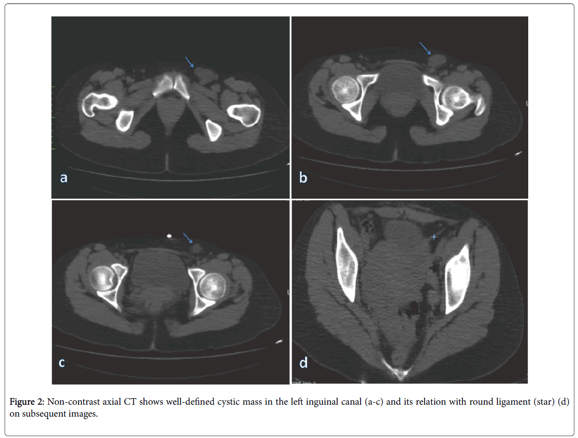
On CT images, there was a thin-walled cystic mass in the left inguinal canal and intraabdominal extension of the canal of Nuck (Figure 2).
The diagnosis of hydrocele of the canal of Nuck was suspected based on sonographic and CT findings.
Before surgical excision, the patient was punctured, followed by aspiration of 10 mL of clear fluid, which relieved her symptoms for a while. 2 months after referral to our department, she underwent surgery and the cyst was excised.
Diagnosis of cyst of the canal of Nuck was confirmed by surgery and subsequent histopathologic evaluation.
Discussion
Hydrocele of the canal of Nuck (cyst of the canal of Nuck) is the female homologue of hydrocele of the spermatic cord in males [1,2].
In the female, the round ligament is attached to the uterus near the origin of the fallopian tube and a small evagination of parietal peritoneum accompanies the round ligament through the inguinal ring into the inguinal canal. On occasion, the peritoneal evagination does not obliterate completely and is called the canal of Nuck. Normally, the canal of Nuck is completely closed in the first year of life. Incomplete closure of the canal results in an indirect inguinal hernia or a serous fluid-containing sac (a hydrocele or cyst of the canal of Nuck) [3,4].
The cyst formation is likely due to the imbalance of the secretion and absorption of the secretory membrane lining the processus vaginalis. Trauma or infection may cause disruption of lymphatic drainage, which can lead to the imbalance, but in most cases, it is idiopathic [2].
The patientis presented with an inguinal swelling and mostly underdiagnosed on physical examination.
Swelling in the inguinal region of a woman may result from several conditions, including adenopathy, inguinal hernia, cyst, abscess, tumor (lipoma, leiomyoma, sarcoma), or hydrocele of the canal of Nuck [3].
Sometimes history and clinical findings are nonspecific to distinguish these pathologies. At this point, imaging modalities are helpful in differential diagnosis.
Cyst of the canal of Nuck demonstrates varied appearances in sonography. In the literature sonographic appearance of hydrocele of the canal of Nuck shows thin walled, well defined, echo free, cystic structure varying from an anechoic, tubular, sausage, dumbbell or comma-shaped, ‘‘cyst within a cyst’’ to a multicystic appearance [2-6].
To our knowledge, reports of radiological findings of hydrocele of the canal of Nuck have been rare, and CT findings have not been previously described in the English literature. In our case, on CT images there was a thin-walled cystic mass in the left inguinal canal and intraabdominal extension of the canal of nuck. CT examination helps us to localize the lesion and its relationship with adjacent structures.
Conclusion
Hydrocele of the canal of Nuck is a rarely encountered entity. Its clinical presentation and history is usually subtle and not satisfactory. It should be on the differential diagnosis list of groin swellings in female patients. Radiologists must be aware of the imaging findings of hydrocele of the canal of Nuck to diagnose this entity before surgery.
References
- Jagdale R, Agrawal S, Chhabra S, Jewan SY (2012) Hydrocele of the canal of Nuck: value of radiological diagnosis. J Radiol Case Rep 6: 18-22.
- Stickel WH, Manner M (2004) Female Hydrocele (Cyst of the Canal of Nuck). J Ultrasound med 23: 429-432.
- Anderson CC, Broadie TA, Mackey JE, Kopecky KK (1995) Hydrocele of the canal of Nuck: ultrasound appearance. Am Surg 61: 959-961.
- Park SJ, Lee HK, Hong HS, Kim HC, Kim DH, et al. (2014) Hydrocele of the canal of Nuck in a girl: ultrasound and MR appearance. Br J Radiol 77: 243-244.
- Miklos JR, Karram MM, Silver E, Reid R (1995) Ultrasound and hookwire needle placement for localization of a hydrocele of the canal of Nuck. Obstet Gynecol 85: 884-886.
- Khanna PC, Ponsky T, Zagol B, Lukish JR, Markle BM (2007) Sonographic appearance of canal of Nuck hydrocele. Pediatr Radiol 37: 603-606.
Relevant Topics
- Abdominal Radiology
- AI in Radiology
- Breast Imaging
- Cardiovascular Radiology
- Chest Radiology
- Clinical Radiology
- CT Imaging
- Diagnostic Radiology
- Emergency Radiology
- Fluoroscopy Radiology
- General Radiology
- Genitourinary Radiology
- Interventional Radiology Techniques
- Mammography
- Minimal Invasive surgery
- Musculoskeletal Radiology
- Neuroradiology
- Neuroradiology Advances
- Oral and Maxillofacial Radiology
- Radiography
- Radiology Imaging
- Surgical Radiology
- Tele Radiology
- Therapeutic Radiology
Recommended Journals
Article Tools
Article Usage
- Total views: 12968
- [From(publication date):
February-2017 - Jul 05, 2025] - Breakdown by view type
- HTML page views : 11992
- PDF downloads : 976


