Customized Silicone Hydrogel Lens-A Case Study
Received: 03-Jun-2017 / Accepted Date: 31-Oct-2017 / Published Date: 03-Nov-2017 DOI: 10.4172/2476-2075.1000124
Abstract
We report a case in which customized contact lens were fitted in eyes of a high hypermetropic patient. Initially patient was using a readily available contact lens but complained about not compatible with those lens. In slit lamp examination over the old contact lens revealed those lenses were too loose fit and was folding when patient blinked. The symptoms resolved after custom made silicon hydrogel lens were given to the patient. These findings indicate that customized contact lens can help in certain high refractive error patient where ready stock contact lens may not be helpful.
Keywords: Contact lens; Silicone hydrogel; Customized contact lens
Introduction
Though not of many years old customized silicone hydrogel is still in its infancy in India. We convined patient for RGP lens use but she could not tolerate it even after 1 month period [1]. Then we tried with customized Silicone Hydrogel lens with gave a very positive feedback and created new hope.
When there is a high refractive error where few patients don’t readily agree to use RGPs and prefer using soft contact lens and as a practitioner you see there is no significant astigmatism to prescribe for RGP then you bound to use contact lens of limited power [2-4].
A young patient of age 21 came to our clinic for her regular eye checkup and had a enquiry if she could get rid of glasses. After her refraction we found she had High hypermetropia amounting +11.00/+1.00 × 180 in both eyes with best corrected visual acuity of 20/40 in both eyes as well. She had a history of using contact lens which she was very uncomfortable using [5]. The patient had no significant ocular history, such as trauma, amblyopia or strabismus and no family history of keratoconus [6]. Patient was earlier using contact lens which was very much uncomfortable and she wished if her thick glasses could be removed permanently. Owning to her request she was advised to undergo corneal topography, pachymetry and A-scan.
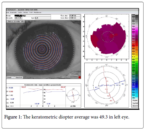
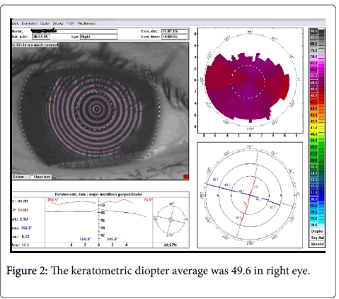
A complete ophthalmologic examination was unremarkable, Placido disc based corneal topography (Alcon) revealed steep corneal surface. This area appeared suspicious; especially the keratometric diopter average was 49.6 in right eye and 49.3 in left eye. Topography stated the patient has abnormal cornea curvature but no any thinning or keratoconus suspect [6]. Corneal pachymetry was 526 and 528 in right and left eye respectively. Her length showed just 17.16 mm in right eye and 17.12 mm in the left eye which explains her high hypermetropia. Her anterior chamber (A/C) depth was 3.53 mm in right eye and 3.54 mm in left eye (Figure 1).
After seeing her report doctor explained various options she could have to remove her glasses temporarily or permanently. She was explained about customized contact lens, Clear lens extraction with multifocal intraocular lens. When her IOL power was calculated in both eye we found it to be approximately +40 D in both eye under SRK-T with 118 A-constant [7]. Since there was no cataractous changes in crystalline lens she was asked to wait for CLE and explained about piggy back IOL can be useful given +40 D lens is not available in Indian market. Since she was very much keen on removing glasses she was explained how a CONTACARE made customized contact lens would her to get freedom from the glasses temporarily (Figure 2).
A custom made silicon hydrogel contact lens was advised to the patient in detail and she agreed to use it. A customize lens was ordered of following measurement (Table 1).
| Right eye | Left eye | |
|---|---|---|
| POWER | +13.50 | +13.50 |
| BASE CURVE | 7.7 mm | 7.7 mm |
| DIAMETER | 14 mm | 14 mm |
Table 1: The final measurement of the contact lens ordered.
After when her new contact lens fitted in her eyes it was very much well fitted and she was very happy with the fit and comfortability of the new lens. Her visual acuity with the contact lens was 20/40, N6 in both eyes. After 1 week of follow-up the patient was happy and satisfied with the new lens [8-14].
Discussion
In case presented the amount of hypermetropia is very high. Thus wearing such a thick glasses at any age would not be very comfortable to any patient [15-17]. Thus it is obvious that patient would be very much eager to remove their glasses either temporarily or permanently. Given her age is just 21 CLE with multifocal piggy back IOL could not be considered. That could have been done if patient was 10-15 years older [18].
In this case we found some new things related to hypermetropia. The 1st thing we notice was AC depth which is normal even the hypermetropia is high. The AC depth as mentioned earlier was in normal range [19]. The 2nd thing we noticed that given the cornea is so much steeper there was no keratoconic changes or thinning for cornea [20].
References
- Bennett ES, Henry VA (2013) Clinical Manual of Contact Lenses. Philadelphia: Lippincott 2nd ed.
- Milton H, Adrian B (2000) Manual of Contact Lens Prescribing and Fitting. Williams & Wilkins 2nd ed.
- Phillips AJ, Speedwell L (1997) Contact Lenses. Boston: Butterworth Heine- Mann 4th ed.
- Dingeldein, Klyce, Wilson (1989) Quantitative descriptors of corneal shape de- rived from computer-assisted analysis of photokeratographs. Refract Cor- neal Surg 5: 372–378.
- Reynolds (1959) Corneal topography as found by photo-electric keratoscopy. Con- tacto 3: 229–233.
- Bufidis, Konstas, Mamtziou (1998) The role of computerized corneal topography in rigid gas permeable contact lens fitting. CLAO J 24: 206–209.
- Jani, Szczotka. Efficiency and accuracy of two computerized topography software systems for fitting rigid gas permeable contact lenses. CLAO J 26: 91–96.
- Maeda, Klyce, Smolek, Thompson (1994) Automated keratoconus screening with corneal topography analysis. Invest Ophthalmol Vis Sci 35: 2749–2757
- Maguire, Bourne (1989) Corneal topography of early keratoconus. Am J Ophthal- mol 108: 107–112.
- McMahon, Robin, Scarpulla, Putz (1991) The spectrum of topography found in keratoconus. CLAO J 17: 198–204.
- Rabinowitz (1995) Videokeratographic indices to aid in screening for kerato- conus. J Refract Surg 11: 371–379.
- Rabinowitz, Rasheed (1999) KISA% index: a quantitative videokeratography al- gorithm embodying minimal topographic criteria for diagnosing kerato- conus J Cataract Refract Surg 25:1327–1335.
- Karabatsas, Cook, Sparrow (1999) Proposed classification for topographic pat- terns seen after penetrating keratoplasty Br J Ophthalmol 83:403–409.
- Rabinowitz, McDonnell (1989) Computer-assisted corneal topography in kera- toconus. Refract Corneal Surg 5: 400–408.
- Schwiegerling, Greivenkamp (1996) Keratoconus detection based on videokera- toscopic height data. Optom Vis Sci 73: 721–728.
- Lebow, Grohe (1999) Differentiating contact lens induced warpage from true ker- atoconus using corneal topography. CLAO J 25: 114–122.
- Novo, Pavlopoulos, Feldman (1995) Corneal topographic changes after refitting polymethylmethacrylate contact lens wearers into rigid gas permeable ma terials. CLAO J 21: 47–51.
- Maeda, Klyce, Hamano. Alteration of corneal asphericity in rigid gas per- meable contact lens induced warpage.
Citation: Pratyush D, Amrita S (2017) Customized Silicone Hydrogel Lens-A Case Study. Optom Open Access 2: 124. DOI: 10.4172/2476-2075.1000124
Copyright: © 2017 Pratyush D, et al. This is an open-access article distributed under the terms of the Creative Commons Attribution License, which permits unrestricted use, distribution, and reproduction in any medium, provided the original author and source are credited.
Share This Article
Recommended Journals
Open Access Journals
Article Tools
Article Usage
- Total views: 4886
- [From(publication date): 0-2017 - Apr 18, 2025]
- Breakdown by view type
- HTML page views: 4007
- PDF downloads: 879


