Research Article Open Access
CT scanning protocol in washin/washout method to reduce the effect of xenoninduced flow activation on xenon-enhanced computed tomography calculations: cerebral blood flow and partition coefficient
| Shigeru Sase1,2*, Hideharu Nakano3 and Mitsuru Honda4 | |
| 1Anzai Medical Co Ltd., Japan | |
| 2Department of Neurosurgery, Toho University Omori Medical Center, Japan | |
| 3Department of Radiology, Toho University Omori Medical Center, Japan | |
| 4Department of Critical Care Center, Toho University Omori Medical Center, Japan | |
| Corresponding Author : | Shigeru Sase Anzai Medical Co Ltd. 3-6-25 Nishi-Shinagawa Shinagawa-ku, Tokyo 141-0033,Japan Tel: +81-3-3779-1611 Fax: +81-3-3779-6606 E-mail: sase@anzai-med.co.jp |
| Received January 06, 2015; Accepted March 30, 2015; Published April 03, 2015 | |
| Citation: Sase S, Nakano H, Honda M (2015) CT scanning protocol in washin/washout method to reduce the effect of xenon-induced flow activation on xenon-enhanced computed tomography calculations: cerebral blood flow and partition coefficient. OMICS J Radiol 4:182. doi: 10.4172/2167-7964.1000182 | |
| Copyright: © 2015 Sase S, et al. This is an open-access article distributed under the terms of the Creative Commons Attribution License, which permits unrestricted use, distribution, and reproduction in any medium, provided the original author and source are credited. | |
Visit for more related articles at Journal of Radiology
Abstract
Background: It has been required to reduce the effect of xenon-induced flow activation (Xe activation) in xenonenhanced computed tomography (Xe-CT). The goal of this work was to propose a CT scanning protocol in the washin/washout method that could reduce the Xe-activation effect on calculated cerebral blood flow (CBF) and partition coefficient (lambda) in Xe-CT.
Methods: In the 4-min washin/4-min washout protocol with 1-min interval scans, the effect of Xe activation was calculated by theoretical simulations under the following scanning protocols based on the reported transcranial Doppler measurements: A: 1-min intervals, B: skip scans during the first 2 min in washout, and C: no scan in washout. For ten healthy subjects, CBF and lambda for the cortical regions of the right and left middle cerebral artery (MCA) territories were evaluated in protocols A and B.
Results: Due to Xe activation, simulated CBF for gray matter was changed by 13%, 7% and 6%; simulated lambda for gray matter was changed by 2%, 4% and 4%; simulated CBF for white matter was changed by 11%, 10% and -1%; and simulated lambda for white matter was changed by 14%, 14% and 133% in protocols A, B and C, respectively. For the ten subjects, the mean differences between protocols A and B in obtained CBF were 1.85 (P=0.0337) and 2.23 mL/100g/min (P=0.0077), and those in obtained lambda were -0.042 (P<0.0001) and -0.041 (P<0.0001) for the right and left MCA territories respectively. This demonstrated that gray matter CBF by protocol A was statistically larger than that by protocol B, and gray matter lambda by protocol A was statistically smaller than that by protocol B, which agreed with the theoretical simulation results.
| Keywords |
| Cerebral blood flow; Flow activation; Theoretical simulation; Xenon-enhanced computed tomography |
| Introduction |
| Xenon-enhanced computed tomography (Xe-CT) has long been developed as a noninvasive modality to obtain quantitative cerebral blood flow (CBF) maps [1,2]. In the Xe-CT washin/washout protocol, a constant concentration (about 30%) of non-radioactive xenon (Xe) gas is inhaled for several minutes (washin), then air is inhaled for another several minutes (washout), and the temporal changes in CT value [Hounsfield unit (HU)] produced by accumulated Xe in the tissue are measured by a series of CT scans. The sequential changes in CT value in the various tissue segments are computed in combination with Xe concentrations in the arterial blood to create CBF and tissueblood partition coefficient (lambda) maps. The washin/washout protocol can provide more accurate lambda values than the washinonly protocol under a short Xe-inhalation period like 4 minutes. Fat content in the human liver can be quantitatively evaluated using lambda in Xe-CT with the 4-min washin/4-min washout protocol [3]. |
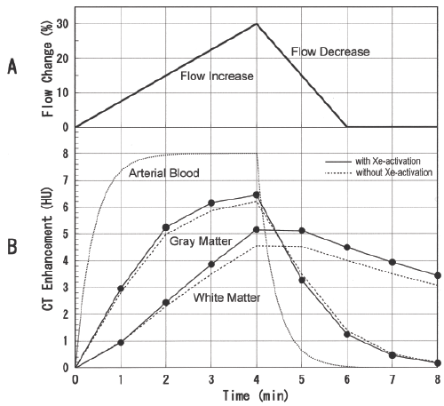
| In Xe-CT, CBF and lambda are calculated based on Fick’s law [4], and there is an assumption that CBF is stable during the examination: from the start of washin to the end of washout. However, Xe is known to be vasoactive like other inert gases, and there are reports that mean CBF increased by 16% to 30% during 5 to 6 min inhalation of 35% Xe in normal volunteers [5,6]. It was also reported that, in normal subjects, blood velocity in middle cerebral artery (MCA) gradually increased during 4.5-min inhalation of 33% Xe, reaching a peak value of 35 ± 13% (mean ± SD) above pre-inhalation baseline values (flow activation), followed by a rapid decline toward the baseline values in MCA blood velocity after terminating Xe inhalation [7]. This flow pattern can be simplified as shown in Figure 1A. There is an understanding of high correlation between the changes in CBF and those in MCA blood velocity [8]. |
| It is required to determine a CT scanning protocol that can reduce the effect of Xe-induced flow activation (Xe activation) on calculated CBF and lambda in Xe-CT. To reduce the Xe activation effect on CBF, the scanning protocol of 0.7-min scan intervals has been proposed in the 4-min washin-only protocol [7]. To our knowledge, there has been no report on how to reduce the effect of Xe activation in the washin/ washout protocol. The aim of this work was to propose an appropriate CT scanning protocol to reduce the Xe-activation effect on Xe-CT calculations (CBF and lambda), taking into account both the washinonly and the washin/washout protocols, by means of theoretical simulations based on the reported transcranial Doppler measurements by Obrist WD, et al. [7]. We analyzed Xe-CT data obtained from healthy volunteers in order to examine whether Xe activation could cause expected over- or under-estimation of CBF and lambda derived from the theoretical simulations. Since the 4-min washin/4-min washout protocol with 1-min CT-scan intervals has been commonly used for human measurements in Japan, this protocol was applied to the theoretical simulations and the assessment of the human results. |
| Materials and Methods |
| Theoretical simulations |
| When CBF is stable, tissue Xe concentration, C(t) (HU), can be described as |
 (1) (1) |
 (2) (2) |
| where, Ca(t) is arterial Xe concentration (HU), K is build-up rate (min-1) of tissue Xe concentration, λ is partition coefficient, f is CBF (mL/100 g tissue/min), and t is time (min) from the start of washin [9]. Ca (t) can be approximated by the following exponential equation [10]: |
 |
| τ= t when 0≤t≤W |
| = W when t>W (3) |
| where, Aa is saturation concentration (HU) of Xe in the arterial blood, Ra is rate constant (min-1) of arterial Xe concentration, and W is washin period (min). Rate constant of end-tidal Xe can be a substitute for Ra [11], and was reported to be 2.46 ± 0.54 (mean ± SD) min-1 for subjects without pulmonary function insufficiency [12]. To describe Xe activation, the following equations have been proposed [13,14]: |
 (5) (5) |
 (5) (5) |
| K( t) is proportional to CBF (f (t ) (equation 5) and reflects CBF increase and decrease. |
| In this work, we estimated the flow pattern in the 4-min washin/4- min washout protocol as follows based on the reported data of transcranial Doppler measurements by Obrist WD, et al. (1998) [7]: |
| 1. Linear flow increase beginning at the initiation of Xe inhalation, and reaching 30% at 4 min after the initiation of Xe inhalation. |
| 2. Linear flow decrease beginning at the termination of Xe inhalation, and returning to the baseline flow at 2 min after the termination of Xe inhalation. |
| This flow pattern is shown in Figure 1A. Then, K(t) is described as |
 (4 <t ≤ 6) (7) (4 <t ≤ 6) (7) |
 (6<t ≤8) (8) (6<t ≤8) (8) |
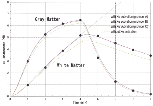
where, K(0) is equal to  . For the gray and white matter, baseline CBF values were assumed to be 80 and 20 mL/100 g/min, and lambda values were assumed to be 0.8 and 1.5, respectively [15]. Then, K(0) values for the gray and white matter were calculated to be 1 and 0.133 min-1 respectively. Time-course changes in CT enhancement [C( t) : t=1, 2, … , 8] for the gray and white matter in the 4-min washin/4-min washout protocol were calculated using equation 4 (Figure 1B). In this work, three CT scanning protocols were chosen for theoretical simulations: A: 1-min intervals, B: skip scans during the first 2 min in washout, and C: no scan in washout. . For the gray and white matter, baseline CBF values were assumed to be 80 and 20 mL/100 g/min, and lambda values were assumed to be 0.8 and 1.5, respectively [15]. Then, K(0) values for the gray and white matter were calculated to be 1 and 0.133 min-1 respectively. Time-course changes in CT enhancement [C( t) : t=1, 2, … , 8] for the gray and white matter in the 4-min washin/4-min washout protocol were calculated using equation 4 (Figure 1B). In this work, three CT scanning protocols were chosen for theoretical simulations: A: 1-min intervals, B: skip scans during the first 2 min in washout, and C: no scan in washout. |
| For each scanning protocol, CBF and lambda were calculated with the use of equations 1 and 2 using the data points as follows: |
| Protocol A: 8 data points of C (1) , C( 2) , … , C( 8) |
| Protocol B: 6 data points of C (1) , C( 2) , C( 3) , C( 4) , C( 7) , C( 8) |
| Protocol C: 4 data points of C (1) , C( 2) , C( 3) , C( 4) |
| Preliminarily, we evaluated by theoretical simulations the effect of Xe activation for every protocol in which one or two scans except for the baseline scan were skipped in the 4-min washin/4-min washout protocol with 1-min interval scans, and protocol B was found to be the most effective protocol to reduce the effect of Xe activation. |
| Subjects |
| The Ethics Committee of Toho University, Faculty of Medicine approved the study protocol and written informed consent was obtained from all subjects. Ten healthy volunteers (8 men and 2 women) were enrolled as the subjects in this work. Their ages ranged from 20 to 71 years [39.1 ± 17.4 (mean ± SD) years]. |
| Xe-CT studies |
| The CT scanner used was an X-vigor (Toshiba, Tokyo, Japan); 512 × 512 matrix and 10 mm slice thickness were used. Exposure factors were 120 kV (peak), 200 mA, and 2 s scans. The applied protocol was 4-min washin and 4-min washout. CT scanning was performed at 1- min intervals at the level of the basal ganglia (9 scans including the baseline scan). An AZ-725 (Anzai Medical Co., Ltd., Tokyo, Japan) was used as the Xe gas inhalation system, and the inhaled Xe concentration was 30%. CT images and respiratory xenon data were transferred to a workstation (AZ-7000Pro; Anzai Medical Co., Ltd.). On the workstation, unweighted filtering over a 9 × 9 neighborhood was performed on the CT images prior to computation in order to reduce noise contribution, and CBF and lambda maps were created using CT scanning protocols A and B for each subject. For statistical comparisons, paired Student’s t test was used and a P value of less than 0.05 was considered to indicate a statistically significant difference. Paired Student’s t test was applied because two values obtained in protocols A and B were compared for each of the 10 subjects. |
| Results |
| Simulated CBF, lambda, and build-up rate in CT scanning protocols A, B and C are listed in Table 1. In Figure 2, fitted curves using equation 1 for the gray and white matter in protocols A, B and C are shown with data points used for curve fitting. For the gray matter, the Xe-activation effect on simulated CBF was decreased from 12.5% to 7.4%, and that on simulated lambda was increased from 1.9% to 3.8%, by skipping scans during the first 2 min in washout. The effect of skipping the first 2 scans in washout was comparable to that of skipping all scans in washout (7.4% vs. 6.2% for CBF and 3.8% vs. 4.2% for lambda). For the white matter, the Xe-activation effect on simulated lambda was largely decreased from 133% to 14% by using washout scans. |
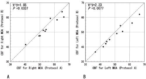
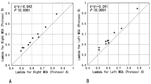
| CBF and lambda for the cortical regions of the right and left MCA territories were evaluated in protocols A and B for the 10 healthy subjects. As shown in Figures 3 and 4, the mean differences between protocols A and B in calculated CBF were 1.85 mL/100 g/min (P=0.0337) and 2.23 mL/100 g/min (P=0.0077), and those in calculated lambda were –0.042 (P < 0.0001) and –0.041 (P < 0.0001) for the right and left MCA territories respectively. |
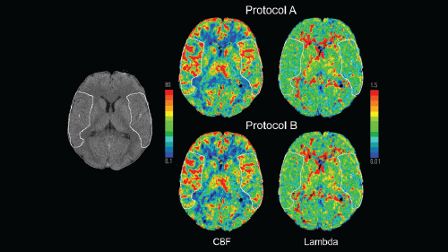
| This demonstrated that gray matter CBF by protocol A was statistically larger than that by protocol B, and gray matter lambda by protocol A was statistically smaller than that by protocol B. These differences between protocols A and B in gray matter CBF and lambda agreed with the theoretical simulation results in Table 1, and it was confirmed that Xe activation caused overestimation of CBF and underestimation of lambda. In Figure 5, CBF and lambda maps in protocols A and B are shown for a 27-year-old healthy man. It is visually recognized that the image quality of the maps in protocol B is close to that in protocol A. |
| Discussion |
| In Japan, Xe-CT for CBF measurement (3- or 4-min washin using 27 to 30% Xe gas and 4-min washout, and 1-min interval scans) has been a routine examination since the approval of this technology by the Japanese government in April 1992. As shown in Figure 1B reflecting a normal case, Xe concentration in the arterial blood almost reaches the saturation at 2 minutes after Xe initiation and becomes almost zero at 2 minutes after Xe termination; and Xe concentration in the gray matter is close to the saturation (>95% saturation) at 4 minutes after Xe initiation and is close to zero (<5% saturation) at 4 minutes after Xe termination. This means that the 4-min washin/4- min washout protocol can cover nearly the whole process of saturation and desaturation of Xe in the gray matter. Therefore, we consider the 4-min washin/4-min washout protocol would be the optimal one in the Xe-CT washin/washout method. The safety of Xe-CT has been well established, however, Xe-CT does expose the patient to some radiation (external exposure by X-ray), although there is no risk of internal exposure and no need of handling of radioactive isotopes. Decreasing the number of CT scans is an efficient way to reduce the radiation exposure in Xe-CT. It was reported that, in the 4-min washin/4-min washout protocol, 4 scans at 0 (baseline), 2, 4, and 8 min could provide CBF maps comparable to those by 1-min interval scans when head fixation was successful [16]. Skipping scans during the first 2 min in washout is beneficial to both reducing the effect of Xe activation and reducing the radiation exposure, and even if these scans are skipped, the image quality of obtained CBF and lambda maps would be close to that without skipping these scans in the 4-min washin/4-min washout protocol. |
| As shown in Figure 1A, the time-course flow change rate in the flow decrease is twice as large as that in the flow increase, and the duration of the flow decrease is 2 min. For the gray matter, the influence of Xe activation on CT enhancement is large at 1 and 2 min and is already small at 3 min after terminating Xe inhalation (Figure 1B). Therefore, in the case of 1-min interval scans, the rate constant (K ) of the fitted curve for the gray matter without using the data points during the first 2 min in washout (at 5 and 6 min after the start of washin) would be smaller than that with using these data points. Simulated K values were decreased from 1.10 to 1.03 min-1 for the gray matter by skipping the first 2 scans in washout (Table 1). This decrease in K contributed to the reduction in the Xe-activation effect on simulated CBF for the gray matter (from 12.5 to 7.4%). Simulated lambda for the white matter was largely affected (133% change) by Xe activation in protocol C (no scan in washout). This agrees with the reported simulation result for the white matter lambda (78% change) in the 4-min washin-only protocol with 0.7-min scan intervals [7]. Lambda is the ratio of the saturated Xe concentration in the tissue to that in the blood. The time-course CT enhancement for the white matter without Xe activation shows a rather linear change in washin between 1 and 4 min (Figure 2). Therefore, the extrapolated value of the saturated Xe concentration in the white matter using only washin data points is sensitive to Xe activation. In fact, a large saturated value is obtained due to Xe activation as shown in the white-matter graph of protocol C in Figure 2, and largely overestimated lambda is calculated for the white matter in protocol C (Table 1). Calculated white-matter lambda can be kept from being largely overestimated by using washout data points as well as washin data points because curve fitting is performed on the entire range which washin and washout data points cover. As demonstrated in Table 1, use of washout scans as well as washin scans (protocols A and B) can reduce the Xe-activation effect on simulated lambda for the white matter (from 133 to 14%). In this work, we did not perform the precision assessment; with less data point (6 for protocol B vs. 8 for protocol A), the precision of obtained CBF could deteriorate even though the accuracy of obtained CBF would improve. The degree of Xe activation varies individually. If any body-specific receptor responsible for Xe activation can be identified and this activation can be quenched effectively by working on the receptor, we can get rid of In conclusion, we propose to skip scans during the first 2 min in washout to reduce the Xe-activation effect on calculated CBF and lambda in the washin/washout protocol. Skipping these scans is beneficial to reducing the radiation exposure, and even if these scans are skipped, the image quality of obtained CBF and lambda maps would be close to that without skipping these scans in the 4-min washin/4-min washout protocol. |
References |
|
Figures at a glance
 |
 |
 |
 |
 |
| Figure 1 | Figure 2 | Figure 3 | Figure 4 | Figure 5 |
Relevant Topics
- Abdominal Radiology
- AI in Radiology
- Breast Imaging
- Cardiovascular Radiology
- Chest Radiology
- Clinical Radiology
- CT Imaging
- Diagnostic Radiology
- Emergency Radiology
- Fluoroscopy Radiology
- General Radiology
- Genitourinary Radiology
- Interventional Radiology Techniques
- Mammography
- Minimal Invasive surgery
- Musculoskeletal Radiology
- Neuroradiology
- Neuroradiology Advances
- Oral and Maxillofacial Radiology
- Radiography
- Radiology Imaging
- Surgical Radiology
- Tele Radiology
- Therapeutic Radiology
Recommended Journals
Article Tools
Article Usage
- Total views: 13519
- [From(publication date):
April-2015 - Apr 26, 2025] - Breakdown by view type
- HTML page views : 9136
- PDF downloads : 4383
