Research Article Open Access
CSF Biomarkers: Low Amyloid-β 1-42 and BDNF and High IFN γ Differentiate Children Exposed to Mexico City High Air Pollution V Controls. Alzheimer's Disease Uncertainties
Lilian Calderón-Garcidueñas1,2*, Chih-kai Chao1, Charles Thompson1, Joel Rodríguez-Díaz2, Maricela Franco-Lira3, Partha S. Mukherjee4 and George Perry5
1The Center for Structural and Functional Neurosciences, University of Montana, Missoula, MT, USA
2Escuela de Ciencias de la Salud, Universidad del Valle de México, Saltillo, Coahuila, 25204, México
3Hospital Central Militar, Mexico City, Mexico
4Department of Mathematics, Boise State University, Boise, Idaho, USA
5College of Sciences, University of Texas at San Antonio, Texas, USA
- Corresponding Author:
- Lilian Calderón-Garcidueñas
The Center for Structural and Functional Neurosciences
University of Montana,32 Campus Drive
287 Skaggs Building, Missoula, USA
Tel: 406-243-4785
Fax: 406-243-5228
E-mail: lilian.calderon-garciduenas@umontana.edu
Received date: April 17, 2015; Accepted date: May 13, 2015; Published date: May 20, 2015
Citation: Calderón-Garcidueñas L, Chao CK, Thompson C, Rodríguez-Díaz J, Franco-Lira M, et al. (2015) CSF Biomarkers: Low Amyloid-β1-42 and BDNF and High IFN γ Differentiate Children Exposed to Mexico City High Air Pollution V Controls. Alzheimer’sDisease Uncertainties. J Alzheimers Dis Parkinsonism 5:189. doi:10.4172/2161-0460.1000189
Copyright: © 2015 Calderón-Garcidueñas L, et al. This is an open-access article distributed under the terms of the Creative Commons Attribution License, which permits unrestricted use, distribution, and reproduction in any medium, provided the original author and source are credited.
Visit for more related articles at Journal of Alzheimers Disease & Parkinsonism
Abstract
Objective: Long-term exposure to fine particulate matter (PM2.5) and ozone above US EPA standards is associated with increased risk of Alzheimer’s disease (AD). Mexico City Metropolitan Area (MCMA) children have prenatal and lifelong exposures to high PM2.5 and O3. MCMA children and young adults exhibit frontal tau hyperphosphorylation (40%) and amyloid-β diffuse plaques (51%) and a brain imbalance in genes involved in oxidative stress, inflammation, innate and adaptive immune responses. Methods: We measured total tau, tau phosphorylated at threonine 181, amyloid-β1-42 (Fujirebio,US), brain derived neurotrophic factor (BDNF), inflammatory mediators, insulin, and leptin in normal CSF samples from 56 MCMA and 26 control children age 11.9 ± 4.7 years. Results: Aβ 1-42 concentrations were lower in MCMA children (p=0.001) and correlated with cumulative PM2.5 (R2= 0.70). MCMA children had low BDNF (p=0.02) and high IFN-γ (p=0.0003) versus controls. In MCMA children, leptin correlated with insulin and MCP-1 (rs 0.34 and 0.41). Conclusion: Low Aβ1-42 in normal CSF samples from megacity children is a major finding given the interpretation of Aβ 1-42 in the temporal evolution of AD biomarkers. Low CSF Aβ, high IFN γ -detected in early AD and a key neuroinflammatory mediator in AD models-, and low BDNF strongly suggest deleterious CSF changes are evolving in 12 y olds, historically showing deficits in attention and short-term memory, information processing speed and executive function, plus olfactory, auditory and metabolic brain changes. Consideration for a shift in the preclinical AD paradigm is put forward in the setting of severe air pollution exposures. CSF children’s derangements involving Aβ 1-42, BDNF and IFN-γ and the potential discontinuity in the leptin central signaling pathway could be signifying a vicious downward spiral towards AD. We need to aim our efforts to the identification and mitigation of environmental factors influencing Alzheimer’s disease.
Keywords
Air pollution; Alzheimer; Aβ1-42, BDNF; Children; Cerebrospinal fluid; Interferons; Mexico City
Introduction
Polluted environments have a negative central nervous system (CNS) impact on children ranging from delayed psychomotor development, increased risk of autism spectrum disorders, cognitive and olfaction deficits, brainstem auditory evoked potentials central delays, brain volumetric changes, systemic, intrathecal and brain inflammation, autoimmune responses and the hallmarks of Alzheimer disease (AD) [1-16]. Fossil-fuel combustion sources are consistently associated with both short- and long-term health adverse effects of fine particulate matter (PM2.5) exposures [17,18]. Neuroinflammation, altered brain-blood-barrier (BBB) permeability and preclinical markers of neurodegenerative disease are seen in experimental animals exposed to traffic-generated air pollutants and diesel exhaust particles (DEP) [19-21]. Recent work strongly suggests that long-term exposure to O3 and PM2.5 above the current US EPA standards is associated with increased risk of AD [22].
Mexico City Metropolitan Area (MCMA) children with no known risk factors for neurological or cognitive disorders and intrauterine and postnatal lifetime exposures to PM2.5 and ozone above current EPA standards exhibit impaired attention, short-term memory and learning abilities that are commonly seen in neurodegenerative disorders [6-8]. We previously showed that cerebrospinal fluid (CSF) concentrations of macrophage inhibitory factor (MIF), interleukin 6 (IL6), interleukin 1 receptor antagonist (IL1Ra), IL-2, and cellular prion protein PrP (C) can differentiate air pollution exposures in children [16]. Further, CSF myelin basic protein autoantibodies and nickel concentrations are higher in MCMA v low pollution controls [14]. Breakdown of epithelial and endothelial barriers, including the BBB and the GI barrier are common in Mexico City residents and constitute a direct pathway of entrance of air pollutant components to the systemic circulation and the brain [23-25]. In association to cognitive deficits, systemic, neural inflammation and autoimmunity, and structural and metabolic brain abnormalities, young MCMA residents exhibit the same neuropathological hallmarks of AD patients i.e., amyloid beta 42 (Aβ42) plaques, and tau hyperphosphorylation with pre-tangles [10,11,23,24].
Thus, concerning current challenges for mechanisms involved in the development of neuroinflammation and neurodegeneration and the early identification of individuals at risk for AD, we expected the measurement of key CSF Alzheimer biomarkers in highly exposed children, to be relevant for the critical debate on: i. the earliest age CSF key AD-associated proteins show significant differences in contrasting polluted environments, ii. The CSF temporal evolution of inflammatory biomarkers, and iii. The contribution of air pollutants to changes in biomarkers classically associated with Alzheimer’s disease development [22,26-32].
The purpose of the present study was to assess, in MCMA versus low pollution matched children, the impact of their residency on key CSF Alzheimer’s disease biomarkers, neurotrophic factors, inflammatory mediators, glucose metabolism players and food reward hormones, all integral to the air pollution associated pathophysiology of neuroinflammation and neurodegeneration [19,20,23-25,33-38]. Our special interest in the relationship between adipokines, food reward hormones with insulin resistance, metabolic syndrome and Alzheimer risk is based on our data on MCMA lean children showing higher serum leptin and decreases in glucagon-like peptide-1 (GLP1), ghrelin, and glucagon v controls [39].
Our results identify lower concentrations of amyloid β1-42 in normal CSF samples of MCMA children, significantly correlating with PM2.5 cumulative exposures. Low BDNF, high IFN-γ, and strong correlations between leptin/ insulin/ monocyte chemo attractant protein-1/ chemokine (C-C motif) ligand 2 (MCP-1/CCL2), characterized megacity children’s CSF samples. If indeed low concentrations of amyloid β1-42 in CSF should be a key preclinical biomarker of AD and if the current accepted mantra is that there is a trend for a decrease in Aβ1-42 15-20 years before expected onset of clinical symptoms, we need to reconsider the earliest age CSF key AD-associated proteins show significant changes in the setting of severe air pollution. We can’t ignore these children are on average 12 years old and that 5 decades of evolving AD changes will be in place before they come to the neurologist attention. These CSF findings have to be evaluated in light of the wide spectrum of clinical, cognitive, imaging, molecular and neuropathological data already published in similar cohorts. Paediatric CSF data and their proper interpretation could provide new paths towards the unprecedented opportunity for early neuroprotection and Alzheimer disease prevention.
Procedure
This prospective pilot study was approved by the review boards and ethics committees at the Hospital Central Militar. Children had been admitted to the hospital with a work up diagnosis of acute lymphoblastic leukemia (ALL) entering a clinical protocol, which included a spinal tap. The normal CSF samples used for this study were destined to be destroyed after the diagnosis of normal CSF was done.
Study Cities and Air Quality
Children’s cohorts were selected from the Mexico City Metropolitan Area (MCMA) and small cities in Mexico (Zacatlán and Huachinango, Puebla;Zitácuaro, Michoacán; Puerto Escondido, Oaxaca; Chalma, Veracruz;Tlaxcala, Tlaxcala).The control cities have <75,000 inhabitants and are characterized by concentrations of the six criteria air pollutants (ozone, particulate matter, sulfur dioxide, nitrogen oxides, carbon monoxide and lead) below the current US EPA standards [40]. Mexico City Metropolitan Area is an example of extreme urban growth and accompanying environmental pollution [41-45]. The metropolitan area of over 2,000 km2 lies in an elevated basin 7400 feet above sea level surrounded on three sides by mountain ridges. MCMA has nearly 24 million inhabitants, over 50,000 industries, and >5 million vehicles consume more than 50 million litres of petroleum fuels per day [46]. MCMA motor vehicles produce abundant amounts of primary PM2.5, elemental carbon, particle-bound polycyclic aromatic hydrocarbons, carbon monoxide, and a wide range of air toxins, including lipopolysaccharides, formaldehyde, acetaldehyde, benzene, toluene, and xylenes [47-48]. The high altitude and tropical climate facilitate ozone production all year and contribute to the formation of fine secondary particulate matter. Air quality is worse in the winter, when rain is scanty and thermal inversions are frequent. Children from MCMA were residents in the northern-industrialized and southernresidential zones. Southern Mexico City children have been exposed to significant concentrations of ozone, secondary tracers (NO3) and PM-LPS, while northern children have been exposed to higher concentrations of volatile organic compounds (VOCs), PM2.5, and its constituents: organic and elemental carbon including polycyclic aromatic hydrocarbons, secondary inorganic aerosols (SO42-, NO3-,NH4+), and metals (Zn, Cu, Pb, Ti, Mn, Sn, V, Ba) [42,44,48]. Recent studies on the composition of PM2.5 with regards to sites and samples collected in 1997 show that composition has not changed during the last decade [42]. In general, the PM2.5 concentrations coincide with the times children are outdoors during the school recess and physical education periods and the higher O3 concentrations when they play outdoors at home [49].
Participant children
This work includes data from 56 children from Mexico City (22F/34M, Mean age = 11.09 years, SD = 5.5) and 26 control children (12F/14M, Mean age = 12.8 years, SD = 4.0). Children entering a haematology protocol, which included a spinal tap, had been admitted to the hospital from either MCMA or a low polluted city.These selected children had no previous oncologic and/or hematologic treatments, their CSF samples were read as normal and CNS involvement was ruled out at the time of their hospitalization. Children’s clinical inclusion criteria were:negative smoking history and environmental tobacco exposure, lifelong residency in MCMA or the control city, residency within 5 miles of the city monitoring stations, full term birth, and unremarkable clinical histories prior to their admission to the hospital. We specifically excluded children with a history of active participation in team sports with high incidence of head trauma, including soccer. Mothers had unremarkable, full term pregnancies with uncomplicated vaginal deliveries and took no drugs, including alcohol and tobacco. These children had a history of breast feeding for a minimum of 6 months and were introduced to solid foods after age 4 months. Participants were from middle class families, living in single-family homes with no indoor pets, used natural gas for cooking and kitchens were separated from the living and sleeping areas. Low and high pollution exposed children were matched by age, gender and socioeconomic status.
Cerebrospinal fluid (CSF) samples
Spinal tap was performed in the lateral recumbent position from lumbar levels using a standard 22 spinal needle. Spinal taps were performed between 8 and 10 am. CSF was collected dripping in free air in1 ml aliquot into Nalge Nunc polypropylene CryoTubes. Lumbar puncture samples were collected during non-traumatic, noncomplicated procedures. CSF were stored at −80°C immediately after examination to determine haematological involvement (blasts present in a cytospin) [50], and kept frozen until the current analysis. CSF pleocytosis was defined as CSF white blood cell (WBC) counts of >7 cells per mm3. We performed the T-tau, P-tau181P and beta amyloid 1-42HS from Fujirebio-US, Seguin,TX. Human Metabolic Hormone 5 plex Discovery Assay (Ghrelin (active),Gastric Inhibitory peptide GIP (total), Insulin, Leptin, MCP-1) and the Human Cytokine 6 plex assay (TNF-α, IL-1β, IL-2,IL-6, IL-10, IFN-γ) were custom made human Multiplexing Laser Bead Technology, Bio-Rad Human Diabetes (Eve Technologies Corporation, Calgary, Alberta, Canada). BDNF was done using the Boster Biology Tech ELISA kit EK0307, Pleasanton, CA.
Data analysis
We performed three types of analyses: [1] Calculated mean and standard deviation of the variables of interest in low and high pollution exposed children, adjusted for age and gender [2].We calculated Spearman’s partial rank correlations between variables of interest after adjusting age within each group of low and high pollution exposed children [3]. We also calculated the p-values of those Spearman’s partial rank correlations. We modeled Aβ1-42 concentrations as a function of age and the annual mean accumulated PM2.5 concentrations in Mexico City using a non-linear correlation surface map resulting in a 3D Gaussian graphic. The strength of simple linear relationship was summarized as r-squares. All tests were two-sided and significance was assumed when a p-value is less than 0.05. The statistical analyses were performed using the statistical software `R’.
Results
Air pollution levels
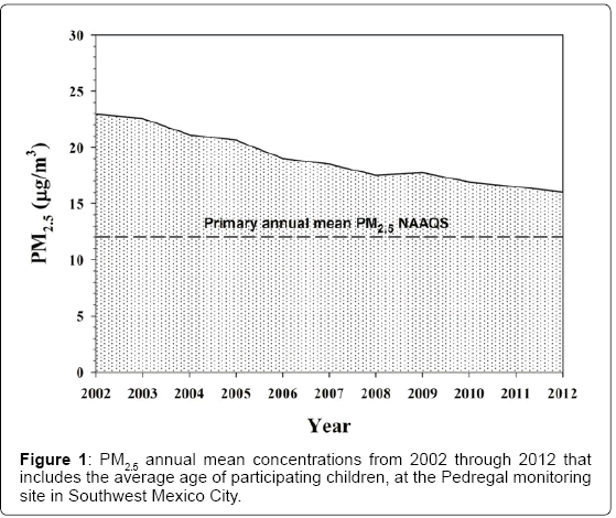
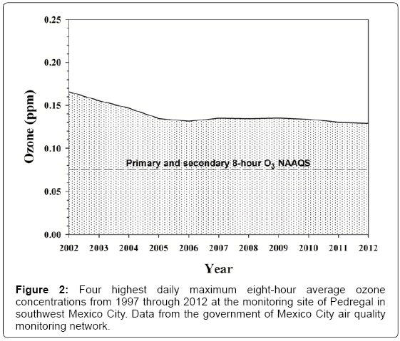
MCMA children in this study have been exposed to significant concentrations of fine particulate matter (PM2.5) and O3 for their entire life [41,42,44,45,48,49]. The climatic conditions in MCMA are relatively stable through the year, thus pollutant concentrations are relatively uniform without significant variations. According to data from the government air quality monitoring network , during the 1997- 2012 period that includes the period the children have lived in MCMA, the PM2.5 three-year averages of annual average concentrations in the representative southwest (Pedregal) monitoring station were above the respective primary PM2.5 US EPA annual standard of 12 μg/m3 (Figure 1). This standard is attained when the 3-year average of annual means is less than or equal to the above mentioned concentration. In addition, the four highest daily maximum eight-hour average concentrations for each of 3 consecutive years for the ozone monitor in the same monitoring site for the 1997-2012 period (the current 8-hr average ozone NAAQS is of 75 ppb) (Figure 2), shows that after a clear decrease from 2002 through 2005 (from 0.16 to 0.13 ppm) the average levels until 2012 have not significantly changed. Fine particulate matter and ozone data clearly show MCMA children in this study have been exposed their entire prenatal and postnatal life to concentrations of PM2.5 and ozone above current US standards. Criteria pollutants in control cities have been below the USA EPA air quality standards [40].
Cerebrospinal fluid results
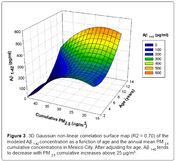
CSF samples were colorless, with a normal opening pressure, a mean WBC count of 2.4± 1 cells per mm3. Table 1 shows the Mean ±SD results in pg/ml and p values adjusted for age and gender, in control versus MCMA children. Aβ1-42 concentrations were significantly lower in MCMA children (p=0.0016) v controls. BDNF was also significantly lower (p=0.02), while IFN-γ was higher (p=0.0003). In MCMA children, Aβ1-42 positively correlated with MCP-1 and IL-6 (rs 0.4), while leptin correlated with insulin and MCP-1 (rs 0.34 and 0.41) (Table 2). The correlations in control children were significantly different from MCMA children. Strong negative correlations were seen between Aβ1-42 , IL1β (rs -0.65) and ghrelin (rs -0.6), while ghrelin positively correlated with IL1β (rs 0.85) and IFN γ(rs 0.71). Interestingly, leptin in control children show no significant correlations with any of the variables. Strong correlations between cumulative PM2.5 and Aβ1-42 (R2 =0.70) were seen in MCMA children (Figure 3).
| CSF variables pg/ml | Controls n:26 | MCMA children n:56 | p value adjusted for age and gender |
|---|---|---|---|
| Amyloid β 1-42 | 312.5±106.2 | 224.8±99.38 | 0.0016 |
| Total tau | 9.89±6.9 | 10.49±8.25 | 0.7245 |
| h tau | 13.88±9.5 | 14.11±8.9 | 0.827 |
| BDNF | 49.51±65.61 | 24.72±22.14 | 0.0236 |
| TNF α | 0.60±0.47 | 1.69±8.9 | 0.122 |
| IL1β | 0.43±0.10 | 0.41±0.1 | 0.343 |
| IL2 | 0.43±0.13 | 0.39±0.09 | 0.131 |
| IL6 | 5.5±14.62 | 1.35±4.16 | 0.0457 |
| IL10 | 0.70±0.81 | 0.57±0.23 | 0.217 |
| IFN γ | 0.28±0.09 | 0.40±1.34 | 0.0003 |
| MCP-1 | 122.2±76 | 162.6±119 | 0.121 |
| Leptin | 54.46±38.8 | 50.34±25.02 | 0.740 |
| Insulin | 60.1±24 | 62.0±31.9 | 0.955 |
| Ghrelin | 5.18±1.2 | 5.2±0.88 | 0.747 |
| GIP | 2.12±2.5 | 2.60±4.09 | 0.961 |
Table 1: Alzheimer, BDNF, cytokines and metabolic CSF variables values and p values in Control v Metropolitan Mexico City area children.
| Control | |||||||||
| Aβ 1-42 | BDNF | Insulin | MCP1 | IFNγ | IL6 | IL1β | Ghrelin | Leptin | |
| Aβ 1-42 | 1 | -0.28 | -0.23 | -0.32 | -0.46 | -0.23 | -0.65 | -0.6 | -0.08 |
| BDNF | -0.28 | 1 | 0.14 | -0.01 | 0.51 | 0.26 | 0.57 | 0.49 | 0.06 |
| Insulin | -0.23 | 0.14 | 1 | -0.1 | 0.05 | -0.3 | -0.11 | 0.11 | 0.24 |
| MCP1 | -0.32 | -0.01 | -0.1 | 1 | -0.32 | 0.15 | 0.35 | 0.28 | 0.36 |
| IFN γ | -0.46 | 0.51 | 0.05 | -0.32 | 1 | 0 | 0.6 | 0.71 | 0.01 |
| IL6 | -0.23 | 0.26 | -0.3 | 0.15 | 0 | 1 | 0.18 | -0.01 | -0.43 |
| IL1β | -0.65 | 0.57 | -0.11 | 0.35 | 0.6 | 0.18 | 1 | 0.85 | 0.17 |
| Ghrelin | -0.6 | 0.49 | 0.11 | 0.28 | 0.71 | -0.01 | 0.85 | 1 | 0.22 |
| Leptin | -0.08 | 0.06 | 0.24 | 0.36 | 0.01 | -0.43 | 0.17 | 0.22 | 1 |
| MexCity | |||||||||
| Aβ 1-42 | 1 | 0.01 | 0.2 | 0.4 | -0.3 | 0.4 | -0.08 | 0.15 | 0.13 |
| BDNF | 0.01 | 1 | -0.03 | -0.09 | 0.04 | -0.19 | -0.12 | 0.1 | -0.17 |
| Insulin | 0.2 | -0.03 | 1 | 0.3 | -0.11 | -0.02 | -0.09 | 0.26 | 0.34 |
| MCP1 | 0.4 | -0.09 | 0.3 | 1 | -0.04 | 0.49 | -0.1 | 0.05 | 0.41 |
| IFN γ | -0.03 | 0.04 | -0.11 | -0.04 | 1 | 0.08 | 0.09 | -0.19 | -0.17 |
| IL6 | 0.4 | -0.19 | -0.02 | 0.49 | 0.08 | 1 | 0.03 | 0.08 | 0.15 |
| IL1β | -0.08 | -0.12 | -0.09 | -0.1 | 0.09 | 0.03 | 1 | 0 | 0.19 |
| Ghrelin | 0.15 | 0.1 | 0.26 | 0.05 | -0.19 | 0.08 | 0 | 1 | 0.03 |
| Leptin | 0.13 | -0.17 | 0.34 | 0.41 | -0.17 | 0.15 | 0.19 | 0.03 | 1 |
Table 2: Spearman partial rank correlations values of key CSF variables after adjusting for age in Control and Metropolitan Mexico City area children.
Discussion
Metropolitan Mexico City children with lifelong exposures to fine particulate matter and ozone above current standards versus low pollution controls, show statistically significant changes in key CSF biomarkers associated with Alzheimer disease: low Aβ1-42 and BDNF, concurrently with an inflammatory profile characterized by high IFN-γ. Strikingly, after adjusting for age, Aβ1-42 tends to decrease with PM2.5 cumulative increases >25 μg/m3. MCMA children showed positive leptin correlations with insulin and monocyte chemoattractant protein-1/chemokine (C-C motif) ligand 2 (MCP-1/CCL2), suggesting a discontinuity in the leptin central signaling pathway and its critical relationship with neuroinflammation [16,33,39,51].
Low CSF Aβ1-42 concentrations in MCMA children is an important finding taking into consideration long-term exposure to O3 and PM2.5 above current US EPA standards is associated with increased risk of AD [22]. Levels of Aβ1-42 reach maximum abnormality level in the asymptomatic stage of Alzheimer [26] and there is a known consensus in AD familial cases of a trend for the decrease in CSF Aβ1-42 15- 20 years before expected onset of clinical symptoms [28]. There is a current agreement that CSF changes in Aβ1-42, T-tau and P-tau181P are diagnostic of AD in its prodromal stage and have proven diagnostic accuracy for mild cognitive impairment and Alzheimer’s disease [27,52-55].Conversely, having all three biomarkers in the normal range rules out AD [27]. There is also agreement in that levels of CSF Aβ1- 42 are regulated age-dependently [56], genetic variations modify the association between AD biomarkers and neurodegeneration [57-59] and AD risk loci polygenically contribute to Aβ pathology in the CSF [60]. Development of low CSF Aβ1-42 values in cognitively healthy individuals in the lower tertile of the reference range, in a 3 year longitudinal follow-up predicted future Aβ positivity [61]. Low CSF Aβ1-42 however, does not always translates in brain florbetapir imaging Aβ accumulation as shown in Mattsson et al., in cognitively healthy people [62]. Thus, if indeed molecular changes in the brain extracellular and interstitial environments are reflected in CSF, if reduced CSF amyloid-β may be more strongly related to early stage AD, [62,63], and if the presence of frontal diffuse amyloid plaques in highly exposed children is taken into account, these CSF results could also be interpreted as reflecting brain Aβ alterations paralleling those of AD. There is one more important consideration in evaluating CSF Aβ1-42 in severely exposed MCMA children: they are not asymptomatic. They have already significant cognitive deficits compared to matched low air pollution children and display white matter volumetric and structural changes concordant with specific inflammatory profiles [6-8].Their central delay in brainstem auditory evoked potentials (BAEPS) relate to the accumulation of α synuclein and/or Aβ1-42 in key brainstem nuclei [9], and their olfactory deficits are important and progressive [10,64]. Equally critical is the role APOE plays in the air pollution scenario both in clinical and neuropathology grounds [23,24,64]. We have reported that MCMA APOE4 teens had greater hyperphosphorylated tau and diffuse Aβ plaques versus E3 carriers (Q = 7.82, p = 0.005) [24] and we can’t dismissed Mexico City APOE 4 v 3 children have reduced NAA/ Cr ratios in the right frontal white matter and decrements on attention, and short-term memory, including >10 point deficit in Verbal and Full Scale IQ [64]. CSF Aβ1-42 low concentrations have been reported in APOE 4 carriers with a diagnosis of AD, prodromal AD, and stable mild cognitive impairment [29]. In contrast, in 105 non-demented controls ages 20-34 years, APOE4 status did not influence CSF Aβ1-42 levels [29]. In this regard, consensus has been reached there is no need to use different cutoffs for the AD CSF biomarkers for different age groups depending on APOE status [27]. Thus, although we do not have APOE genotyping in our cohorts, the CSF Aβ1-42 low concentrations carry strong significance.
We argue consideration for a shift in the preclinical AD paradigm should be entertained in the setting of severe air pollution exposures and although we do not foresee and we do not support taking CSF samples to seemingly healthy children, we should not ignore the weight of clinical, cognitive, laboratory and brain imaging evidence in highly exposed pediatric populations. Certainly the idea that Alzheimer’s changes start in childhood in a stressed environment, is biologically plausible [65,66].
A growing body of evidence demonstrates that abnormalities in the BDNF system are altered in CSF and peripheral blood from AD patients, animal models of AD and in late-life major depression (LLD) and bipolar disorder [67-71]. Of key relevance for this work, BDNF has widespread roles in regulating energy homeostasis by controlling patterns of feeding and physical activity, and by modulating glucose metabolism in peripheral tissues [72]. BDNF mediates the beneficial effects of energetic challenges such as vigorous exercise and fasting on cognition, mood, cardiovascular function, and on peripheral metabolism. Glucose transport and mitochondrial biogenesis are stimulated and BDNF bolsters cellular bioenergetics, protects neurons against injury and disease and increases insulin sensitivity and parasympathetic tone [72].Thus, significantly low CSF concentrations of BDNF cannot be dismissed in urban children, as the reduction of the availability of BDNF in the CNS may indicate loss of neuroprotection and increased vulnerability to the development of several neuropsychiatric disorders as well as to adverse cognitive outcomes [73-77]. Naert and Rivest’s paper [78] is of utmost relevance to us. Their work associates progressive cognitive decline with the accumulation of soluble Aβ, disruption of synaptic activity, alteration in the BDNF system, and a defective production in the subset of CX(3)CR1(low)Ly6- C(high)CCR2(+)Gr1(+) monocytes in APP(Swe)/PS1 mice and their age-matched wild-type (WT) littermates. Its relevance is centred in the BDNF system alteration given that MCMA children exhibit altered monocyte and lymphocyte populations and an endotoxin tolerance-like state that could play a role in the peripheral BDNF production [79-82]. The issue is critical because exercise has been linked to neuroprotection with BDNF playing a key role [73,83-86].Notably, aerobic training in an urban environment with high traffic-related air pollution increases inflammatory biomarkers, and obliterates the BDNF positive responses [87,88]. Equally relevant, ultrafine particulate matter exposures during forced exercise in rats decrease their hippocampal BDNF expression [89]. Bos and colleagues concluded that traffic-related air pollution exposure during exercise may inhibit the positive effect of exercise on cognition [90]. We fully agreed with them.
IFN-γ produced by CD4 Th1, CD8, gamma delta T, and natural killer (NK) cells, is an interesting player in the high PM2.5 scenario. Interferons, a super-family of cytokines with major roles in host immune responses to pathogens, antiviral defense, and tumor surveillance [91-93] can induce pro-inflammatory gene transcription leading to the secretion of powerful inflammatory cytokines including IL1β, IL-6 and TNF-α, all of them up-regulated both peripherally and in the brain of MCMA children [23]. Also relevant to MCMA cohorts is the role of IFN-γ as a central mediator of Th1-mediated autoimmune disorders by deflecting the immune response toward a Th1 phenotype and inhibiting the development of Th2 cells in autoimmune disorders [94], a major issue given the CSF production of autoantibodies to myelin basic protein in similar children cohorts [14].The work of Taylor et al support the strong involvement of IFN signaling in the regulation of the neuroinflammation and neuronal cell death in animal models of AD and increased expression of IFNα and β in human prefrontal cortex of AD patients [92]. Given that IFN type 1 responses are considered the master regulators of cytokine production within the innate immune response [95], Taylor et al suggest IFN responses are critical to the development of neuroinflammation [92]. The issue is more relevant in PM-LPS exposed children (related to the thousands of tons of fecal dog and human material deposited daily in Mexico City streets) because type-1 IFNs are shown to be critically involved in the priming and activation of the NALP3 inflammasome (brain activated in similar cohorts [24] by inducing caspase-11 to cleave pro-caspase-1 to its active form [96].This is an interesting concept, because indeed MCMA 12y old children have a robust CSF IFN-γ response contrasting with the blood lower concentrations of IFN-γ and low numbers of NK cells present in MCMA younger children [79].The peripheral blood and CSF IFN γ signaling is likely key in the air pollution responses and needs to be defined in PM2.5 exposed children.
An increasing number of studies provide support to the relationships between leptin dysregulation and Alzheimer’s disease, insulin resistance, cerebral volumes, learning and memory performance [34-36,97]. Leptin associations with AD are discordant, while some works show significant leptin elevations in CSF and hippocampal tissue of AD patients v controls, along with level of leptin receptor mRNA decreased in AD brain [39], others show CSF leptin levels unchanged as subjects progress to AD [33]. Dysregulated leptin-signaling circuitry, however appears to be a common AD denominator [33,34,36,39,97]. We found no differences in CSF leptin in high versus low pollution 12 year old children, but the relationship between leptin and insulin is significantly different among cohorts. In MCMA children CSF leptin correlates positively with insulin while control children exhibit no correlations with insulin at all. This is key, because in similar age cohorts, lean MCMA children v controls exhibit significantly higher serum leptin that correlated positively with cumulative values of PM2.5 (Calderón-Garcidueñas and Perry personal communication). These CSF leptin results and our serum previous data, support the hypothesis of a significant leptin dysregulation in highly exposed PM2.5 children and an extension of this dysregulatory process to their brain. If indeed the provocative emerging data of brain insulin resistance leading to AD neurodegeneration [36] is at play here, we have the unfortunate perfect scenario in the developing brain of urban children. In this regard, the positive significant association between CSF leptin and MCP-1 in highly exposed children is also very relevant in the setting of neuroinflammation. A potent monocyte attractant with key roles in neuroinflammatory diseases, including AD and with a striking role in BBB integrity has to be contemplated as a chemokine with pathophysiological significance in the air pollution scenario where the BBB is damaged [24,98-102]. Although there is significant controversy in the interpretation of CSF MCP-1 in AD, i.e., increased levels linked to the transition from MCI to AD, high persistent levels in AD patients [103], overlaps between controls, MCI and AD patients have also been reported [102] and age seem also to relate to high MCP-1 [104]. The important issue here is: these are children and since MCP-1 is a biomarker of microglial activity, the intriguing associations with leptin (in a clear evolving chronic neuroinflammatory process with strong microglia activation) obligates us to carefully monitor this biomarker in exposed populations.
Looking forward, limitations and summary
Strong evidence supports a link between oxidative stress, abnormal lipid, glucose and insulin metabolism, inflammasomes, neuroinflammation and Alzheimer’s disease [33-39,105-115]. The work of Xia et al., [110] is of key relevance to our findings in highly exposed urban children: increased oxidative stress, along with alterations in lipid metabolism in neurons, may be some of the very early events occurring in AD pathology. Brain vascular and mitochondrial abnormalities are also emerging as a common feature in AD cases [111], an important issue in urban children with extensive small arterioles and postcapillary venule damage, breakdown of the BBB, and mitochondrial abnormalities directly associated with the presence of nano size particles [14,23,24].
In the natural setting of severe PM2.5 exposures [6-16,22-25] or in the results of animal models exposed to air pollution components [19-21], the notion that neurodegeneration results from an active host response or environmental adaptation [106], is biologically plausible. A new brain health paradigm should be entertained [113], the Alzheimer research community must acknowledge all aspects of disease pathogenesis [109] and support should be allocated to explore air pollution as a key factor in the development of Alzheimer’s disease in pediatric and young adult cohorts.
We acknowledge our main limitation, the number of CSF samples, based on ethical considerations: under no circumstances as physicians we will jeopardize children’s health by using an invasive spinal tap, thus our strategy of using CSF normal samples in children undergoing a spinal tap within a hemato-oncological protocol and no CNS involvement [50].
CSF derangements involving key proteins, inflammatory cytokines, chemokines and adipokines in urban children are likely representing a vicious downward spiral towards Alzheimer’s disease. We could ignore current evidence, pretend air pollution is not affecting our children’s brain and disregard we have a 50 year window of opportunity between the time urban 12y olds have the CSF Alzheimer-related changes and the wide spectrum of clinical, cognitive, olfactory and imaging alterations we have been describing and need a neurologist. Facing the current clinical, cognitive, laboratory and imaging evidence impacting the health of millions of urban children is imperative if we are aiming our efforts to identify and mitigate environmental factors influencing Alzheimer’s disease [106]. If indeed, reduced CSF amyloid-β is strongly related to early stage AD, consideration for a shift in the preclinical AD paradigm is put forward in the setting of severe air pollution exposures. Defining the linkage and the health consequences of chronic exposures to air pollutants in the developing brain and keeping in mind the relentless path towards Alzheimer’s disease ought to be of pressing importance for public health.
Acknowledgements
We are very grateful to Dr. Ricardo Torres-Jardón from the Centro de Ciencias de la Atmósfera, Universidad Nacional Autónoma de México, Mexico City, who generously elaborated Figures 1-3 and reviewed the manuscript.
References
- Suglia SF, Wright RO, Schwartz J, Wright RJ (2008) Association between lung function and cognition among children in a prospective birth cohort study. Psychosom Med 70: 356-362
- Lucchini RG, Dorman DC, Elder A, Veronesi B (2012) Neurological impacts from inhalation of pollutants and the nose-brain connection. Neurotoxicology 33: 838-841.
- Becerra TA, Wilhelm M, Olsen J, Cockburn M, Ritz B (2013) Ambient air pollution and autism in Los Angeles county, California. Environ Health Perspect 121: 380-386.
- Kalkbrenner AE, Windham GC, Serre ML, Akita Y, Wang X, et al. (2015) Particulate matter exposure, prenatal and postnatal windows of susceptibility, and autism spectrum disorders. Epidemiology 26: 30-42.
- Jedrychowski WA, Perera FP, Camann D, Spengler J, Butscher M, et al. (2015) Prenatal exposure to polycyclic aromatic hydrocarbons and cognitive dysfunction in children. Environ Sci Pollut Res Int 22: 3631-3639.
- Calderón-Garcidueñas L, Mora-Tiscareño A, Ontiveros E, Gómez-Garza G, Barragán-MejÃa G, et al. (2008) Air pollution, cognitive deficits and brain abnormalities: a pilot study with children and dogs. Brain Cogn 68: 117-127.
- Calderón-Garcidueñas L, Engle R, Mora-Tiscareño A, Styner M, Gómez-Garza G, et al. (2011) Exposure to severe urban air pollution influences cognitive outcomes, brain volume and systemic inflammation in clinically healthy children. Brain Cogn 77: 345-355.
- Calderón-Garcidueñas L, Mora-Tiscareño A, Styner M, Gómez-Garza G, Zhu H, et al. (2012) White matter hyperintensities, systemic inflammation, brain growth, and cognitive functions in children exposed to air pollution. J Alzheimers Dis 31: 183-191.
- Calderón-Garcidueñas L, D'Angiulli A, Kulesza RJ, Torres-Jardón R, Osnaya N, et al. (2011) Air pollution is associated with brainstem auditory nuclei pathology and delayed brainstem auditory evoked potentials. Int J Dev Neurosci 29: 365-375.
- Calderón-Garcidueñas L, Franco-Lira M, HenrÃquez-Roldán C, González-Maciel A, Reynoso-Robles R, et al. (2010) Urban air pollution: influences on olfactory function and pathology in exposed children and young adults. Exp Toxicol Pathol 62: 91-102.
- Calderón-Garcidueñas L, Franco-Lira M, Mora-Tiscareño A, Medina-Cortina H, Torres-Jardón R, et al. (2013) Early Alzheimer’s and Parkinson’s disease pathology in urban children: Friend versus Foe responses-it is time to face the evidence. Biomed Res Int doi: 10.1155/2013/161687.
- Calderón-Garcidueñas L, Torres-Jardón R (2012) Air Pollution, Socioeconomic Status, and Children's Cognition in Megacities: The Mexico City Scenario. Front Psychol 3: 217.
- Calderón-Garcidueñas L, MacÃas-Parra M, Hoffmann HJ, Valencia-Salazar G, HenrÃquez-Roldán C, et al. (2009) Immunotoxicity and environment: immunodysregulation and systemic inflammation in children. Toxicol Pathol 37: 161-169.
- Calderón-Garcidueñas L, Vojdani A, Blaurock-Busch E, Busch Y, Friedle A, et al. (2015) Air pollution and children: neural and tight junction antibodies and combustion metals, the role of barrier breakdown and brain immunity in neurodegeneration. J Alzheimers Dis 43: 1039-1058.
- Calderón-Garcidueñas L, Serrano-Sierra A, Torres-Jardón R, Zhu H, Yuan Y, et al. (2013) The impact of environmental metals in young urbanites' brains. Exp Toxicol Pathol 65: 503-511.
- Calderón-Garcidueñas L, Cross JV, Franco-Lira M, Aragón-Flores M, Kavanaugh M, et al. (2013) Brain immune interactions and air pollution: macrophage inhibitory factor (MIF), prion cellular protein (PrPC),interleukin-6 (IL6), interleukin 1 receptor antagonist (IL-1Ra), and interleukin-2 (IL-2) in cerebrospinal fluid and MIF in serum differentiate urban children exposed to severe vs low air pollution. Front Neurosci doi: 10.3389/fnins.2013.00183.
- Lippmann M, Chen LC, Gordon T, Ito K, Thurston GD (2013) National Particle Component Toxicity (NPACT) Initiative: integrated epidemiologic and toxicologic studies of the health effects of particulate matter components. Res Rep Health Eff Inst : 5-13.
- Ailshire JA, Crimmins EM (2014) Fine particulate matter air pollution and cognitive function among older US adults. Am J Epidemiol 180: 359-366.
- Levesque S, Surace MJ, McDonald J, Block ML (2011) Air pollution and the brain: Subchronic diesel exhaust exposure causes neuroinflammation and elevates early markers of neurodegenerative disease. J Neuroinflammation doi: 10.1186/1742-2094-8-105
- Levesque S, Taetzsch T, Lull ME, Kodavanti U, Stadler K, et al. (2011) Diesel exhaust activates and primes microglia: air pollution, neuroinflammation, and regulation of dopaminergic neurotoxicity. Environ Health Perspect 119: 1149-1155.
- Oppenheim HA, Lucero J, Guyot AC, Herbert LM, McDonald JD, et al. (2013) Exposure to vehicle emissions results in altered blood brain barrier permeability and expression of matrix metalloproteinases and tight junction proteins in mice. Part Fibre Toxicol doi: 10.1186/1743-8977-10-62
- Jung CR, Lin YT, Hwang BF (2015) Ozone, particulate matter, and newly diagnosed Alzheimer's disease: a population-based cohort study in Taiwan. J Alzheimers Dis 44: 573-584.
- Calderón-Garcidueñas L, Solt AC, HenrÃquez-Roldán C, Torres-Jardón R, Nuse B, et al. (2008) Long-term air pollution exposure is associated with neuroinflammation, an altered innate immune response, disruption of the blood-brain-barrier, ultrafine particulate deposition, and accumulation of amyloid beta-42 and alpha-synuclein in children and young adults. Toxicol Pathol 36: 289-310.
- Calderón-Garcidueñas L, Kavanaugh M, Block ML, D'Angiulli A, Delgado-Chávez R, Torres-Jardón R, et al. (2012) Neuroinflammation, hyperphosphorilated tau, diffuse amyloid plaques and down- regulation of the cellular prion protein in air pollution exposed children and adults. Journal Alzheimer Disease 28: 93-107
- Calderón-Garcidueñas L, Gónzalez-Maciel A, Vojdani A, Franco-Lira M, Reynoso-Robles R, et al. (2015) The intestinal barrier in air pollution-associated neural involvement in Mexico City residents: Mind the gut, the evolution of a changing paradigm relevant to Parkinson disease risk. J Alz Dis Parkinsonism
- Susanto TA, Pua EP, Zhou J,Alzheimer’s Disease Neuroimaging Initiative (2015) Cognition, brain atrophy, and cerebrospinal fluid biomarkers changes from preclinical to dementia stage of Alzheimer's disease and the influence of apolipoprotein e. J Alzheimers Dis 45: 253-268.
- Bertens D, Knol DL, Scheltens P, Visser PJ (2014) Alzheimer’s Disease Neuroimaging Initiative. Temporal evolution of biomarkers and cognitive markers in the asymptomatic, MCI and dementia stage of Alzheimer’s disease. Alzheimers Dement doi:10.1016/j.alz.2014.05.1754
- Molinuevo JL, Sánchez-Valle R, Lladó A, Fortea J, Bartrés-Faz D, et al. (2012) Identifying earlier Alzheimer's disease: insights from the preclinical and prodromal phases. Neurodegener Dis 10: 158-160.
- Thordardottir S, Ståhlbom AK, Ferreira D, Almkvist O, Westman E, et al. (2015) Preclinical cerebrospinal fluid and volumetric magnetic resonance imaging biomarkers in Swedish familial Alzheimer's disease. J Alzheimers Dis 43: 1393-1402.
- Lautner R, Palmqvist S, Mattsson N, Andreasson U, Wallin A, et al. (2014) Apolipoprotein E genotype and the diagnostic accuracy of cerebrospinal fluid biomarkers for Alzheimer disease. JAMA Psychiatry 71: 1183-1191.
- Rosén C, Hansson O, Blennow K, Zetterberg H (2013) Fluid biomarkers in Alzheimer's disease - current concepts. Mol Neurodegener 8: 20.
- Weuve J (2014) Invited commentary: how exposure to air pollution may shape dementia risk, and what epidemiology can say about it. Am J Epidemiol 180: 367-371.
- Maioli S, Lodeiro M, Merino-Serrais P, Falahati F, Khan W, et al. (2015) Alterations in brain leptin signalling in spite of unchanged CSF leptin levels in Alzheimer's disease. Aging Cell 14: 122-129.
- Arnoldussen IA, Kiliaan AJ, Gustafson DR (2014) Obesity and dementia: adipokines interact with the brain. Eur Neuropsychopharmacol 24: 1982-1999.
- de la Monte SM (2014) Type 3 diabetes is sporadic Alzheimer׳s disease: mini-review. Eur Neuropsychopharmacol 24: 1954-1960.
- Farr OM1, Tsoukas MA2, Mantzoros CS2 (2015) Leptin and the brain: influences on brain development, cognitive functioning and psychiatric disorders. Metabolism 64: 114-130.
- Kinawy AA, Ezzat AR, Al-Suwaigh BR(2014) Inhalation of air polluted with gasoline vapours alters the levels of amino acid neurotransmitters in the cerebral cortex, hippocampus, and hypothalamus of the rat. Exp Toxicol Pathol 66: 219-224.
- Allen JL, Liu X, Weston D, Prince L, Oberdörster G, et al. (2014) Developmental exposure to concentrated ambient ultrafine particulate matter air pollution in mice results in persistent and sex-dependent behavioural neurotoxicity and glial activation. Toxicol Sci 140:160-178.
- Bonda DJ1, Stone JG, Torres SL, Siedlak SL, Perry G, et al. (2014) Dysregulation of leptin signaling in Alzheimer disease: evidence for neuronal leptin resistance. J Neurochem 128: 162-172.
- US EPA (2014) National Ambient Air Quality Standards. (NAAQS). Air and Radiation. U. S. Environmental Protection Agency. http://www.epa.gov/air/criteria.html.
- Bravo-Alvarez HR, Torres-Jardón RJ (2002) Air pollution levels and trends in the Mexico City metropolitan area. In: Urban air pollution and forests: resources at risk in the Mexico City Air Basin Ecological Studies, Fenn M, Bauer L, Hernández T, eds. Springer-Verlag, New York: 121-159.
- Molina LT, Madronich S, Gaffney JS, Apel E, de Foy B,et al. (2010) An overview of the MILAGRO 2006 Campaign: Mexico City emissions and their transport and transformation. Atmos Chem Phys 10: 8697–8760.
- Rosas Pérez I, Serrano J, Alfaro-Moreno E, Baumgardner D, GarcÃa-Cuellar C, et al. (2007) Relations between PM10 composition and cell toxicity: a multivariate and graphical approach. Chemosphere 67: 1218-1228.
- Querol X, Pey J, Minguillón MC, Pérez N, Alastuey A, et al. (2008) PM speciation and sources in Mexico during the MILAGRO-2006 Campaign. Atmos Chem Phys 8: 111-121.
- Valle-Hernández BL, Mugica-Alvarez V, Salinas-Talavera E, Amador-Muñoz O, Murillo-Tovar MA, et al. (2010) Temporal variation of nitro-polycyclic aromatic hydrocarbons in PM10 and PM2.5 collected in Northern Mexico City. Sci Total Environ 408: 5429-5438.
- SMA SecretarÃa del Medio Ambiente del Gobierno del Distrito Federal. (2014) Dirección General de Gestión de la Calidad del Aire. Sistema de monitoreo Atmosférico de la ciudad de México. Dirección de Monitoreo Atmosférico.2014. http://www.sma.df.gob.mx/simat2/.
- Estrada-Garcia T, Cerna JF, Thompson MR, Lopez-Saucedo C (2002) Faecal contamination and enterotoxigenic Escherichia coli in street-vended chili sauces in Mexico and its public health relevance. Epidemiol Infect 129: 223-226.
- Dzepina K, Arey J, Marr L, Worsnop DR, Salcedo D, et al. (2007) Detection of particle-phase polycyclic aromatic hydrocarbons in Mexico City using an aerosol mass spectrometer. International Journal of Mass Spectrometry 263: 152–170.
- Villarreal-Calderón A, Acuña H, Villarreal-Calderón J, Garduño M, HenrÃquez-Roldán CF, et al. (2002) Assessment of physical education time and after-school outdoor time in elementary and middle school students in south Mexico City: the dilemma between physical fitness and the adverse health effects of outdoor pollutant exposure. Arch Environ Health 57: 450-460.
- Gassas A1, Krueger J, Alvi S, Sung L, Hitzler J, et al. (2014) Diagnosis of central nervous system relapse of pediatric acute lymphoblastic leukemia: Impact of routine cytological CSF analysis at the time of intrathecal chemotherapy. Pediatr Blood Cancer 61: 2215-2217.
- Anderson AM, Harezlak J, Bharti A, Mi D, Taylor MJ, et al. (2015) Plasma and cerebrospinal fluid biomarkers predict cerebral injury in HIV-infected individuals on stable combination antiretroviral therapy. J Acquir Immune Defic Syndr Jan 23
- Blennow K, Zetterberg H (2009) Cerebrospinal fluid biomarkers for Alzheimer's disease. J Alzheimers Dis 18: 413-417.
- Bocchetta M, Galluzzi S, Kehoe PG, Aguera E, Bernabei R, et al. (2015) The use of biomarkers for the etiologic diagnosis of MCI in Europe: an EADC survey. Alzheimers Dement 11: 195-206.
- Seeburger JL, Holder DJ, Combrinck M, Joachim C, Laterza O, et al. (2015) Cerebrospinal fluid biomarkers distinguish post-mortem confirmed Alzheimer’s disease from other dementias and healthy controls in the OPTIMA cohort. J Alzheimers Dis 44:525-539
- Ritchie C, Smailagic N, Noel-Storr AH, Takwoingi Y, Flicker L, et al. (2014) Plasma and cerebrospinal fluid amyloid beta for the diagnosis of Alzheimer's disease dementia and other dementias in people with mild cognitive impairment (MCI). Cochrane Database Syst Rev 6: CD008782.
- Shoji M, Kanai M, Matsubara E, Tomidokoro Y, Shizuka M, et al. (2001) The levels of cerebrospinal fluid Abeta40 and Abeta42(43) are regulated age-dependently. Neurobiol Aging 22: 209-215.
- Hohman TJ, Koran ME, Thornton-Wells TA (2014) for the Alzheimer’s Neuroimaging Initiative. Genetic variation modifies risk for neurodegeneration based on biomarker status. Frontiers Aging Neuroscience doi: 10.3389/fnagi.2014.00183
- Kettunen P1, Larsson S, Holmgren S, Olsson S, Minthon L, et al. (2015) Genetic variants of GSK3B are associated with biomarkers for Alzheimer's disease and cognitive function. J Alzheimers Dis 44: 1313-1322.
- Ramirez A, van der Flier WM, Herold C, Ramonet D, Heilmann S, et al. (2014) SUCLG2 identified as both a determinator of CSF Aβ1-42 levels and an attenuator of cognitive decline in Alzheimer's disease. Hum Mol Genet 23: 6644-6658.
- Martiskainen H, Helisalmi S, Viswanathan J, Kurki M, Hall A, et al. (2015) Effects of Alzheimer's disease-associated risk loci on cerebrospinal fluid biomarkers and disease progression: a polygenic risk score approach. J Alzheimers Dis 43: 565-573.
- Mattsson N1, Insel PS2, Donohue M3, Jagust W4, Sperling R5, et al. (2015) Predicting Reduction of Cerebrospinal Fluid β-Amyloid 42 in Cognitively Healthy Controls. JAMA Neurol 72: 554-560.
- Mattsson N, Insel PS, Donohue M, Landau S, Jagust WJ, et al. (2015) Independent information from cerebrospinal fluid amyloid-β and florbetapir imaging in Alzheimer's disease. Brain 138: 772-783.
- Mattsson N1, Blennow K, Zetterberg H (2009) CSF biomarkers: pinpointing Alzheimer pathogenesis. Ann N Y Acad Sci 1180: 28-35.
- Calderón-Garcidueñas L, Mora-Tiscareño A, Franco-Lira M, Zhu H, Lu Z, et al. (2015) Decreases in short term memory, IQ and altered brain metabolic ratios in urban apolipoprotein e4 children exposed to air pollution. APOE modulates children’s brain air pollution responses. J Alzheimer Dis 45:757-770
- Kristiansson M, Sörman K, Tekwe C, Calderón-Garcidueñas L (2015) Urban air pollution, poverty, violence and health-Neurological and immunological aspects as mediating factors. Environmental Res 140:1-3.
- Moffitt TE1; Klaus-Grawe 2012 Think Tank (2013) Childhood exposure to violence and lifelong health: clinical intervention science and stress-biology research join forces. Dev Psychopathol 25: 1619-1634.
- Diniz BS, Teixeira AL, Machado-Vieira R, Talib LL, Radanovic M, et al. (2014) Reduced cerebrospinal fluid levels of brain-derived neurotrophic factor is associated with cognitive impairment in late-life major depression. J Gerontol B Psychol Sci Soc Sci 68:845-851.
- Hashimoto K (2010) Brain-derived neurotrophic factor as a biomarker for mood disorders: an historical overview and future directions. Psychiatry Clin Neurosci 64: 341-357.
- Sopova K, Gatsiou K, Stellos K, Laske C (2014) Dysregulation of neurotrophic and haematopoietic growth factors in Alzheimer's disease: from pathophysiology to novel treatment strategies. Curr Alzheimer Res 11: 27-39.
- Rodrigues R, Petersen RB, Perry G (2014) Parallels between major depressive disorder and Alzheimer's disease: role of oxidative stress and genetic vulnerability. Cell Mol Neurobiol 34: 925-949.
- Molendijk ML, Spinhoven P, Polak M, Bus BA, Penninx BW, et al. (2014) Serum BDNF concentrations as peripheral manifestations of depression: evidence from a systematic review and meta-analyses on 179 associations (N=9484). Mol Psychiatry 19: 791-800.
- Marosi K, Mattson MP (2014) BDNF mediates adaptive brain and body responses to energetic challenges. Trends Endocrinol Metab 25: 89-98.
- Bechara RG, Lyne R, Kelly AM (2014) BDNF-stimulated intracellular signalling mechanisms underlie exercise-induced improvement in spatial memory in the male Wister rat. Behav Brain Res 275: 297-306.
- Diniz BS, Teixeira AL (2011) Brain-derived neurotrophic factor and Alzheimer's disease: physiopathology and beyond. Neuromolecular Med 13: 217-222.
- Zhang J, Sokal I, Peskind ER, Quinn JF, Jankovic J, et al. (2008) CSF multianalyte profile distinguishes Alzheimer and Parkinson diseases. Am J Clin Pathol 129: 526-529.
- Meeker RB, Poulton W, Markovic-Plese S, Hall C, Robertson K (2011) Protein changes in CSF of HIV-infected patients: evidence for loss of neuroprotection. J Neurovirol 17: 258-273.
- .Li G, Peskind ER, Millard SP, Chi P, Sokai I, et al.(2009) Cerebrospinal fluid concentration of brain-derived neurotrophic factor and cognitive function in non-demented subjects. PLoS One doi: 10.1371/journal.pone.0005424
- Naert G, Rivest S (2012) Age-related changes in synaptic markers and monocyte subsets link the cognitive decline of APP(Swe)/PS1 mice. Front Cell Neurosci 6: 51.
- Calderón-Garcidueñas L, MacÃas-Parra M, Hoffmann HJ, Valencia-Salazar G, HenrÃquez-Roldán C, et al. (2009) Immunotoxicity and environment: immunodysregulation and systemic inflammation in children. Toxicol Pathol 37: 161-169.
- Tong J, Buch S, Yao H, Wu C, Tong H, et al. (2014) Monocytes-derived macrophages mediated stable expression of human brain-derived neurotrophic factor, a novel therapeutic strategy for neuroAIDS. PLoS One 9(2):e82030. doi: 10.1371/journal.pone.0082030.
- Linker RA, Lee DH, Demir S, Wiese S, Kruse N, et al. (2010) Functional role of brain-derived neurotrophic factor in neuroprotective autoimmunity: therapeutic implications in a model of multiple sclerosis. Brain 133:2248-2263.
- Guidotti A, Auta J, Davis JM, Dong E, Gavin DP, et al. (2014) Toward the identification of peripheral epigenetic biomarkers of schizophrenia. J Neurogenet 28: 41-52.
- Huang T, Larsen KT, Ried-Larsen M, Møller NC, Andersen LB (2014) The effects of physical activity and exercise on brain-derived neurotrophic factor in healthy humans: A review. Scand J Med Sci Sports 24: 1-10.
- Brunelli A, Dimauro I, Sgrò P, Emerenziani GP, Magi F, et al. (2012) Acute exercise modulates BDNF and pro-BDNF protein content in immune cells. Med Sci Sports Exerc 44: 1871-1880.
- Szuhany KL, Bugatti M, Otto MW (2015) A meta-analytic review of the effects of exercise on brain-derived neurotrophic factor. J Psychiatr Res 60: 56-64.
- Phillips C, Baktir MA, Srivatsan M, Salehi A (2014) Neuroprotective effects of physical activity on the brain: a closer look at trophic factor signaling. Front Cell Neurosci 8: 170.
- Bos I, Jacobs L, Nawrot TS, de Geus B, Torfs R, et al. (2011) No exercise-induced increase in serum BDNF after cycling near a major traffic road. Neurosci Lett 500: 129-132.
- Bos I, De Boever P, Vanparijs J, Pattyn N, Panis L, et al. (2013) Subclinical effects of aerobic training in urban environment. Med Sci Sports Exerc 45: 439-447.
- Bos I, De Boever P, Panis L, Sarre S, Meeusen R (2012) Negative effects of ultrafine particle exposure during forced exercise on the expression of Brain-Derived Neurotrophic Factor in the hippocampus of rats. Neuroscience 223:131-139.
- Bos I, De Boever P, Int Panis L, Meeusen R (2014) Physical activity, air pollution and the brain. Sports Med 44: 1505-1518.
- de Weerd NA, Nguyen T (2012) The interferons and their receptors--distribution and regulation. Immunol Cell Biol 90: 483-491.
- Taylor JM, Minter MR, Newman AG, Zhang M, Adlard PA, et al. (2014) Type-1 interferon signaling mediates neuro-inflammatory events in models of Alzheimer's disease. Neurobiol Aging 35: 1012-1023.
- Paolini R, Bernardini G, Molfetta R, Santoni A (2015) NK cells and interferons. Cytokine Growth Factor Rev 26: 113-120.
- Zhang HL, Wu L, Wu X, Zhu J (2014) Can IFN-γ be a therapeutic target in Guillain-Barré syndrome? Expert Opin Ther Targets 18: 355-363.
- Kawai T, Akira S (2006) Innate immune recognition of viral infection. Nat Immunol 7: 131-137.
- Rathinam VA, Vanaja SK, Waggoner L, Sokolovska A, Becker C, et al. (2012) TRIF licenses caspase-11-dependent NLRP3 inflammasome activation by gram-negative bacteria. Cell 150: 606-619.
- Lee S, Tong M, Hang S, Deochand C, de la Monte S4(2013) CSF and Brain Indices of Insulin Resistance, Oxidative Stress and Neuro-Inflammation in Early versus Late Alzheimer's Disease. J Alzheimers Dis Parkinsonism 3: 128.
- Yadav A, Saini V, Arora S (2010) MCP-1: chemoattractant with a role beyond immunity: a review. Clin Chim Acta 411: 1570-1579.
- Conductier G, Blondeau N, Guyon A, Nahon JL, Rovère C (2010) The role of monocyte chemoattractant protein MCP1/CCL2 in neuroinflammatory diseases. J Neuroimmunol 224: 93-100.
- Yao Y, Tsirka SE (2014) Monocyte chemoattractant protein-1 and the blood-brain barrier. Cell Mol Life Sci 71: 683-697.
- Yao Y, Tsirka SE (2011) Mouse MCP1 C-terminus inhibits human MCP1-induced chemotaxis and BBB compromise. J Neurochem 118: 215-223.
- Mattsson N, Tabatabaei S, Johansson P, Hansson O, Andreasson U, et al. (2011) Cerebrospinal fluid microglial markers in Alzheimer’s disease: elevated chitotriosidase activity but lack of diagnostic utility. Neuromol Med 13:151-159.
- Galimberti D, Schoonenboom N, Scheltens P, Fenoglio C, Bouwman F, et al. (2006) Intrathecal chemokine synthesis in mild cognitive impairment and Alzheimer disease. Arch Neurol 63: 538-543.
- Blasko I, Lederer W, Oberbauer H, Walch T, Kemmler G, et al. (2006) Measurement of thirteen biological markers in CSF of patients with Alzheimer's disease and other dementias. Dement Geriatr Cogn Disord 21: 9-15.
- Nunomura A, Moreira P, Castellani RJ, Lee HG, Zhu X, et al. (2012) Oxidative damage to RNA in aging and neurodegenerative disorders. Neurotox Res 22: 231-248.
- Castellani RJ, Perry G (2014) The complexities of the pathology-pathogenesis relationship in Alzheimer disease. Biochem Pharmacol 88: 671-676.
- Butterfield DA, Di Domenico F, Barone E (2014) Elevated risk of type 2 diabetes for development of Alzheimer disease: a key role for oxidative stress in brain. Biochim Biophys Acta 1842: 1693-1706.
- Wang X, Wang W, Li L, Perry G, Lee HG, et al. (2014) Oxidative stress and mitochondrial dysfunction in Alzheimer's disease. Biochim Biophys Acta 1842: 1240-1247.
- Bonda DJ, Wang X, Lee HG, Smith MA, Perry G, et al. (2014) Neuronal failure in Alzheimer's disease: a view through the oxidative stress looking-glass. Neurosci Bull 30: 243-252.
- Xia J, Rocke DM, Perry G, Ray M (2014) Differential network analyses of Alzheimer's disease identify early events in Alzheimer's disease pathology. Int J Alzheimers Dis 2014: 721453.
- Carvalho C, Correia SC, Perry G, Castellani RJ, Moreira PI (2015) Cerebrovascular and mitochondrial abnormalities in Alzheimer's disease: a brief overview. J Neural Transm.
- Fornicola W, Pelcovits A, Li BX, Heath J, Perry G, et al. (2014) Alzheimer Disease Pathology in Middle Age Reveals a Spatial-Temporal Disconnect Between Amyloid-β and Phosphorylated Tau. Open Neurol J 8: 22-26.
- Petersen RB, Lissemore FM, Appleby B, Aggarwal N, Boyatzis R, et al. (2015) From Neurodegeneration to Brain Health: An Integrated Approach. J Alzheimers Dis.
- Singhal G, Jaehne EJ, Corrigan F, Toben C, Baune BT (2014) Inflammasomes in neuroinflammation and changes in brain function: a focused review. Front Neurosci 8: 315.
- Belkhelfa M, Rafa H, Medjerber O, Arroul-Lammali A, Behairi N, et al. (2014) IFN-? and TNF-? are involved during Alzheimer disease progression and correlate with nitric oxide production: a study in Algerian patients. J Interferon Cytokine Res 34:839-847
Relevant Topics
- Advanced Parkinson Treatment
- Advances in Alzheimers Therapy
- Alzheimers Medicine
- Alzheimers Products & Market Analysis
- Alzheimers Symptoms
- Degenerative Disorders
- Diagnostic Alzheimer
- Parkinson
- Parkinsonism Diagnosis
- Parkinsonism Gene Therapy
- Parkinsonism Stages and Treatment
- Stem cell Treatment Parkinson
Recommended Journals
Article Tools
Article Usage
- Total views: 15096
- [From(publication date):
September-2015 - Nov 21, 2024] - Breakdown by view type
- HTML page views : 10689
- PDF downloads : 4407


