Research Article Open Access
Correlation between Clinical and Histopathologic Chorioamnionitis in HIV-positive Women
| Hannah Anastasio1*, Meredith Birsner1, Cynthia Argani1, Jean Anderson1, Frederic Askin2 and Zahra Maleki2 | |
| 1Department of Gynaecology and Obstetrics, Johns Hopkins University, Baltimore, Maryland, USA | |
| 2Department of Pathology, Johns Hopkins University, Baltimore, Maryland, USA | |
| Corresponding Author : | Hannah Anastasio Department of Gynaecology and Obstetrics Johns Hopkins University Baltimore, Maryland, USA Tel: 410-955-6710 E-mail: hbrotzm1@jhmi.edu |
| Received: January 13, 2016; Accepted: February 04, 2016; Published: February 11, 2016 | |
| Citation: Anastasio H, Birsner M, Argani C, Anderson J, Askin F, et al. (2016) Correlation between Clinical and Histopathologic Chorioamnionitis in HIV-positive Women. J Preg Child Health 2:217. doi:10.4172/2376-127X.1000217 | |
| Copyright: © 2016 Anastasio H, et al., This is an open-access article distributed under the terms of the Creative Commons Attribution License, which permits unrestricted use, distribution, and reproduction in any medium, provided the original author and source are credited. | |
Visit for more related articles at Journal of Pregnancy and Child Health
Abstract
Objective Histopathologic findings of intraamniotic infection include chorioamnionitis and funisitis. Chorioamnionitis is a recognized risk factor for intrapartum HIV transmission. We sought to determine whether histologic placental findings in patients with clinical chorioamnionitis were different between HIV-positive and HIV-negative patients. Study design Retrospective case control study of all cases of clinical chorioamnionitis at The Johns Hopkins Hospital between 1996 and 2010, diagnosed by presence of maternal fever and one or more of the following criteria: maternal or fetal tachycardia, fundal tenderness, malodorous amniotic fluid, or elevated WBC. Placentas of HIV-positive women were matched 1:2 to HIV-negative controls. Placentas were reviewed by two independent pathologists, blinded to HIV status, using 2003 Society for Pediatric Pathology criteria. Primary outcome was histopathologic finding of chorioamnionitis by maternal grade/stage and fetal grade/stage. Results Of 28,915 deliveries, 2,228 met criteria for chorioamnionitis (7.7%), and 11 were HIV-positive (0.5%); one had dichorionic-diamnionic twins with placentas evaluated separately. Parity, gestational age, mode of delivery, Apgar scores, and infant birth weight were similar between groups. WBC at delivery was significantly lower in HIVpositive patients (9 vs. 13 p<0.01), and women with HIV were significantly older (34.4 vs 28.9 years, p=0.05). HIVpositive patients were no more likely to have histologic chorioamnionitis than their HIV-negative controls. Maternal and fetal stage/grade did not vary significantly between groups. Conclusion Among patients with clinical chorioamnionitis, histopathologic confirmation does not differ significantly between HIV-positive and -negative patients. Additionally, histologic chorioamnionitis in the HIV-positive population does not appear to differ in severity from that in the HIV-negative population. White blood cell count at delivery is significantly lower in HIV-positive women with chorioamnionitis compared to HIV-negative controls.
| Keywords |
| Chorioamnionitis; Human Immunodeficiency Virus (HIV); Pregnancy |
| Introduction |
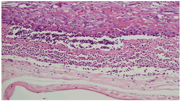
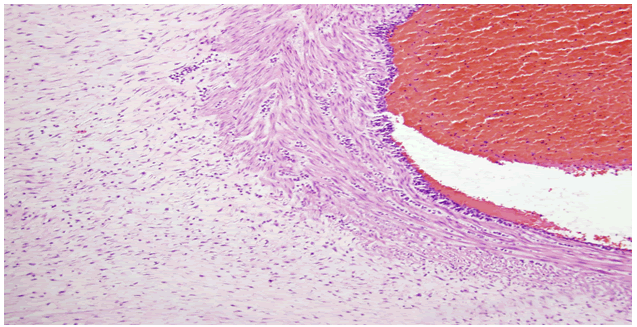
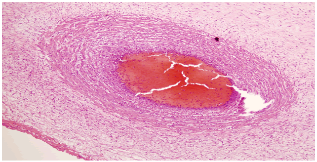
| Chorioamnionitis is infection of the placental membranes surrounding the fetus. Diagnosis can be made either clinically or histologically. Clinical chorioamnionitis is diagnosed by presence of maternal fever in conjunction with one or more of the following: maternal tachycardia (>100 beats per minute), fetal tachycardia (>160 beats per minute), fundal tenderness, or malodorous discharge or leukocytosis (>15,000 cells/mm3). Histologic diagnosis of chorioamnionitis is made by presence of neutrophilic infiltrate of the chorionic plate and membranes, possibly involving the walls of the umbilical blood vasculature (vasculitis) or progressing to infiltrate the stroma of the cord surrounding the cord vessels (funisitis), as demonstrated by Figures 1-3. |
| At our institution, department protocol dictates that placentas of all mothers with clinical chorioamnionitis be sent for evaluation by a pathologist for histopathologic confirmation of diagnosis. |
| Histopathologic evaluation is considered the gold standard for making the pathologic diagnosis of chorioamnionitis [1]. A number of studies have evaluated the degree to which clinical and histologic diagnosis of chorioamnionitis overlap, and have found clinical diagnosis to be less sensitive than histopathologic diagnosis [2]. Published estimates show that histologic chorioamnionitis is observed in 10-38% of term placentas, whereas clinical chorioamnionitis is recognized in 5-12% of term pregnancies [3]. These ranges of values are likely due to variable definitions both of clinical and histopathologic definitions of chorioamnionitis, which can differ by institution. |
| Chorioamnionitis has important clinical implications for mother and fetus. Mothers with chorioamnionitis are at increased risk of postpartum hemorrhage and endomyometritis, and those that deliver by cesarean have an increased risk of postoperative surgical site infection [4,5]. Infants born to mothers with chorioamnionitis are at risk for sepsis and often undergo prolonged observation in the neonatal intensive care unit, and may undergo invasive procedures such as blood cultures and lumbar puncture to rule out sepsis. Additionally, chorioamnionitis has been associated with cerebral palsy in both preterm and term infants [6,7]. Diagnosis of chorioamnionitis is of particular importance among HIV-positive women, because it is a known risk factor for vertical HIV transmission [8]. In the literature, the histologic features of the placentas of HIV-positive women have been characterized [9]. However, whether a difference exists in the histologic features of HIV-positive and HIV-negative women with chorioamnioinitis has not yet been evaluated. We sought to determine whether a difference exists histologically between these two patient populations. We hypothesized that given the immunocompromised characteristic of HIV, that a difference would be observed in the histologic features of these two populations. |
| Materials and Methods |
| Study permission was granted from the Johns Hopkins Institutional Review Board. The study was performed as a retrospective case-control study. All cases of clinical chorioamnionitis between 1996 and 2010 were identified. Patients with HIV were identified, and were contemporaneously matched 1:2 with HIV-negative patients. Maternal demographic and medical information including age, parity, gestational age, presence/absence of spontaneous labor, CD4 count, viral load, and white blood cell (WBC) count on admission, and presence/absence of intrapartum fever were obtained from the medical record. Delivery information including mode of delivery, neonatal Apgar scores, NICU admission, use of antiretroviral medications and antibiotics in labor, and neonatal arterial cord gas information were also abstracted from the medical record. Placentas for study patients were reviewed by two independent pathologists (ZM, FA) both blinded to HIV status, using 2003 Society for Pediatric Pathology criteria [9], and were analyzed for presence of histologic chorioamnionitis as well as maternal and fetal grade and stage of the placenta (Table 1). |
| The primary study outcome was histopathologic findings of chorioamnionitis (including maternal grade and stage and fetal grade and stage). Secondary outcomes included mode of delivery, APGAR scores, arterial cord pH, WBC count at delivery, infant birth weight, and need for NICU admission. Fisher’s Exact Test was used for discrete variables and Student’s T-Test for continuous variables, with p<0.05 considered significant. |
| Results |
| Of the 28,915 deliveries taking place during our study time frame, 2228 (7.7%) met criteria for clinical chorioamnionitis. 11 of these patients (0.5%) were HIV positive; one woman had a set of dichorionic diamniotic twins whose placentas were analyzed individually (accounting for the total n=12). Maternal demographic and baseline medical information were similar between HIV-positive and HIVnegative women (Table 2), with the exception of age and WBC count. |
| HIV-positive women were significantly older, and had significantly lower WBC counts than HIV-negative women. Other factors including nulliparity, gestational age, ethnicity, rates of regional anesthesia use, and labor characteristics were similar between the groups. |
| Intrapartum antibiotic treatment was at the discretion of the treating provider. |
| Standard protocol at our institution is to administer ampicillin and gentamycin for clinical chorioamnionitis. Clindamycin is typically added if delivery is by cesarean. Alternate antibiotic treatment in the setting of patient allergy is at the discretion of the treating physician. 10 of 11 HIV-positive patients received zidovudine intrapartum. Ampicillin and gentamicin were administered to 7 of 11 HIV-positive patients and 16 of 22 controls, and clindamycin was administered to 3 of 22 controls. |
| Despite the lower WBC count associated with the HIV-positive cohort, as well as the known immunosuppression associated with the disease process, rates of histologic chorioamnionitis did not differ significantly between the HIV-positive and HIV-negative patients (Table 3). |
| Rates of histopathologically confirmed chorioamnionitis were 75% and 72.7%, respectively for the HIV-positive and HIV-negative patients (p=0.89). Additionally, there was no difference between the groups in maternal or fetal grade or stage of chorioamnionitis. Modes of delivery were similar between the groups, as were neonatal outcomes including NICU admission, umbilical cord pH and 5 minute APGAR scores. |
| Comment |
| The diagnosis of chorioamnionitis represents a clinical challenge because of variable symptomatology and presentation. Nonetheless, the diagnosis is important to make, because chorioamnionitis is associated with worse maternal and neonatal outcomes, particularly infectious morbidity and mortality. Since the clinical presentation is highly variable, histopathologic diagnosis remains the gold standard. Previous studies have demonstrated significant discrepancy between clinical and histopathologic diagnoses of chorioamnionitis in the general population. However, to date no studies have examined whether infection with HIV has any impact on the correlation between clinical and histopathologic diagnoses of chorioamnionitis. |
| Interestingly, despite the immunosuppression that is the hallmark of HIV, the cellular infiltration of the placental membranes that is diagnostic of chorioamnionitis is relatively preserved in HIV-positive patients. This response is preserved despite a significantly lower white blood cell count in these patients compared to their HIV-negative counterparts. Perhaps one explanation for this finding is that the majority of chorioamnionitis seen in obstetric practice is acute chorioamnionitis, which is neutrophil-mediated response. Given that HIV affects lymphocyte-mediated immunity, the neutrophilic response may be relatively preserved and accounts for similar histologic findings between the two groups. Chronic chorioamnionitis, on the other hand, is a lymphocyte-mediated response, and one might expect different histologic findings among HIV-positive and HIV-negative patients with chronic chorioamnionitis due to their relative deficiency of lymphocytes. This is a potential future avenue of study, but would require a much larger sample size. |
| Strengths of the study include the standardized, blinded histopathologic evaluation of the specimens by two independent pathologists, as well as the fact that this study attempts to answer a novel clinical question not previously addressed in the medical literature. |
| Limitations of this study are its small sample size and retrospective nature. However, because both HIV and chorioamnionitis are uncommon entities, this was the most reasonable way to approach the study question. The small sample size also limits further analysis of the data to evaluate the association between severity of HIV (based on CD4 count and viral load) and histopathologic placental characteristics, which is another potential future avenue of study. Lastly, the study population is largely an urban, African American population, which limits the generalizability of the results. |
| In conclusion, among patients with clinical chorioamnionitis, histopathologic chorioamnionitis does not appear to differ in severity or frequency between HIV-positive and –negative patients, in this small retrospective case control study at an urban tertiary care center. Chorioamnionitis has important medical implications for both HIVpositive mothers and their neonates. |
References
- Pankuch GA, Appelbaum PC, Lorenz RP, Botti JJ, Schachter J, et al. (1984) Placental microbiology and histology and the pathogenesis of chorioamnionitis.Obstet Gynecol 64: 802-806.
- Smulian JC, Shen-Schwarz S, Vintzileos AM, Lake MF, Ananth CV (1999) Clinical chorioamnionitis and histologic placental inflammation.Obstet Gynecol 94: 1000-1005.
- Curtin WM, Katzman PJ, Florescue H, Metlay LA (2013) Accuracy of signs of clinical chorioamnionitis in the term parturient. J Perinatol 33: 422-428.
- Wetta LA, Szychowski JM, Seals S, Mancuso MS, Biggio JR, et al. (2013) Risk factors for uterine atony/postpartum hemorrhage requiring treatment after vaginal delivery.Am J Obstet Gynecol 209: 51.
- Tran TS, Jamulitrat S, Chongsuvivatwong V, Geater A (2000) Risk factors for postcesarean surgical site infection.Obstet Gynecol 95: 367-371.
- Martinelli P, Sarno L, Maruotti GM, Paludetto R (2012) Chorioamnionitis and prematurity: a critical review. J Matern Fetal Neonatal Med 25 Suppl 4: 29-31.
- Neufeld MD, Frigon C, Graham AS, Mueller BA (2005) Maternal infection and risk of cerebral palsy in term and preterm infants.J Perinatol 25: 108-113.
- Chi BH, Mudenda V, Levy J, Sinkala M, Goldenberg RL, et al. (2006) Acute and chronic chorioamnionitis and the risk of perinatal human immunodeficiency virus-1 transmission.Am J Obstet Gynecol 194: 174-181.
- Goldenberg RL, Mudenada V, Read JS, Brown ER, Sinkala M, et al. (2006) HPTN 024 study: histologic chorioamnionitis, antibiotics, and advsere infant outcomes in a predominantly HIV-1 infected African population. Am J Obstet Gynecol 195: 1065-1074.
- Redline RW, Faye-Petersen O, Heller D, Qureshi F, Savell V, et al. (2003) Society for Pediatric Pathology, Perinatal Section, Amniotic Fluid Infection Nosology Committee. Amniotic infection syndrome: nosology and reproducibility of placental reaction patterns. Pediatr Dev Pathol 6: 435-448.
Tables and Figures at a glance
| Table 1 | Table 2 | Table 3 |
Figures at a glance
 |
 |
 |
| Figure 1 | Figure 2 | Figure 3 |
Relevant Topics
Recommended Journals
Article Tools
Article Usage
- Total views: 10288
- [From(publication date):
February-2016 - Apr 03, 2025] - Breakdown by view type
- HTML page views : 9470
- PDF downloads : 818
