Case Report Open Access
Continuous Heart Murmur-Two Cases of Rare Causes and Role of Imaging in Diagnosis
| Janice JK Ip*, Peter KT Hui, Sonia HY Lam and Stephen SC Cheung | |
| Department of Radiology, Queen Mary Hospital, HKSAR, China | |
| Corresponding Author : | Janice JK Ip Department of Radiology, Queen Mary Hospital 102 Pokfulam Road, HKSAR, China Tel: +852 2255 3284 E-mail: JK.Janice.Ip@gmail.com |
| Received March 23, 2013; Accepted April 18, 2013; Published April 23, 2013 | |
| Citation: Janice JK Ip, Hui PKT, Lam SHY, Cheung SSC (2013) Continuous Heart Murmur-Two Cases of Rare Causes and Role of Imaging in Diagnosis. OMICS J Radiology 2:121. doi: 10.4172/2167-7964.1000121 | |
| Copyright: © 2013 Janice JK Ip, et al. This is an open-access article distributed under the terms of the Creative Commons Attribution License, which permits unrestricted use, distribution, and reproduction in any medium, provided the original author and source are credited. | |
Visit for more related articles at Journal of Radiology
Abstract
A continuous murmur is defined as a murmur that begins in systole, continues through the second heart sound and into part or all of diastole. Among a number of differential diagnoses, anomalous systemic arterial supply to the normal basal segments and Rupture of Sinus of Valsalva (RSOV) are two rare causes of continuous heart. These two entities, which are often overlooked clinically, may have potentially fatal complications. We herein present two cases of uncommon causes of continuous heart murmur; demonstrate the radiological features and corroborate the role of Electric Cardiogram-Gated Computed Tomography (ECG-gated CT) in diagnosis.
| Introduction |
| Continuous heart murmurs are murmurs those involve the whole cardiac cycle, begin in systole, continue through the second heart sound and into part or all of diastole. There are a number of differential diagnoses for continuous heart murmur: Patent Ductus Arteriosus (PDA), cervical venous hum, hyperdynamic circulation of pregnancy, Arterio-Venous (AV) fistulas of coronary arteries, great vessel or haemodialysis etc. Nowadays, ECG-gated CT provides excellent anatomic depiction and also simultaneous evaluation of coronary arteries, great vessels and lungs. In this article, we would like to present two cases of uncommon causes of continuous heart murmur; demonstrate the radiological features and corroborate the role of ECGgated CT in diagnosis. |
| Case 1 |
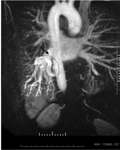
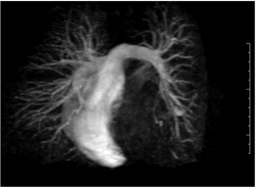
| A 30-year-old male non-smoker with good past health was noted to have a Grade 3 continuous heart murmur at the precordium during his pre-employment health check-up in the private sector in May 2011. An approximately 2.0 cm×4.0 cm shadow was also spotted at left retrocardiac region, with dilated tubular structures, in his preemployment chest radiograph (CXR) (Figure 1a). Respiratory physician suspected that it was a pulmonary Arterio-Venous Malformation (AVM); and subjected the patient to Magnetic Resonance (MR) study to measure the shunt ratio, as the consideration for embolization depended on the degree of Arterio-Venous (AV) shunting. However, MR images were not typical of pulmonary AVM. Magnetic Resonance Angiography (MRA) demonstrated serpiginous vascular tangle at left lower lobe, with systemic arterial blood supply and pulmonary venous drainage (Figure 1b). Cine phase contrast for flow volume showed Qp:Qs=1:1.1, indicative of no significant AV shunting. Magnetic Resonance (MR) pulmonary arteriogram showed attenuation of segmental branches of basal segments of Left Lower Lobe (LLL) (Figure 1c). Bronchopulmonary sequestration was suspected at that moment. |
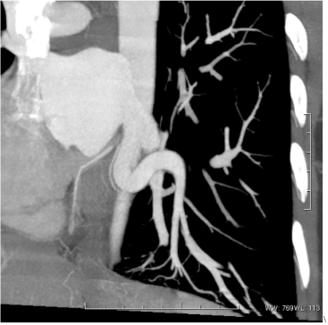
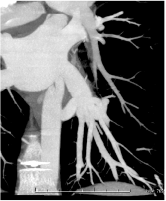
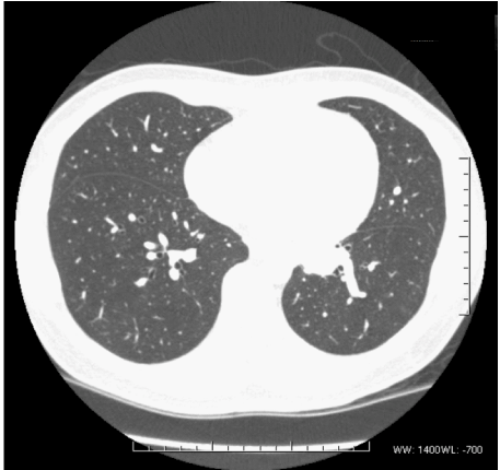
| A Computed Tomographic (CT) scan was subsequently performed and demonstrated a ~1.0 cm in caliber anomalous systemic artery from the descending aorta at T8 level to the basal segment of left lower lobe (Figure1d-1g). Dilated arteries were paralleling the normal bronchi. No direction communication with the pulmonary veins was evident. Tributaries of the Left Inferior Posterior Vein (LIPV) in left lower lobe were hypertrophies with variceal formation. Branches of Left Pulmonary Artery (LPA) supplying the basal segments, distal to origin of the apical segment branch, were however attenuated. Underlying lung parenchyma was normal, which made bronchopulmonary sequestration less likely. Overall findings were in favour of anomalous systemic arterial supply to normal lung parenchyma in the basal segments of left lower lobe, with associated Pulmonary Vein (PV) dilatation and attenuated Pulmonary Artery (PA) branches. Since our patient was asymptomatic, with no significant shunting, he was managed conservatively. |
| Case 2 |
| A 25-year-old young man, who was a chronic smoker and recreational drug abuser, was incidentally found to have a rough systolic murmur, loudest over the precordium, during a consultation for flu-like symptoms by a general practitioner in September 2011. He was referred to the cardiac clinic for further assessment and investigation. On further enquiry by the cardiologist, he volunteered a 2-month history of exertional dyspnea. Physical examination revealed a mildly engorged jugular vein and mild leg edema. Cardiac auscultation revealed a Grade 3/4 continuous murmur along the right sternal border. Transthoracic Echocardiography (TTE) revealed alternating jet-flows into the Right Atrium (RA) during different phases of the cardiac cycle; and structurally normal valves, with trivial Tricuspid Regurgitation (TR). |
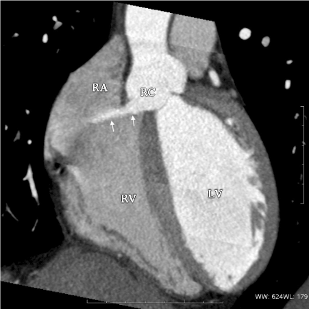
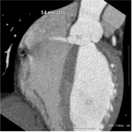
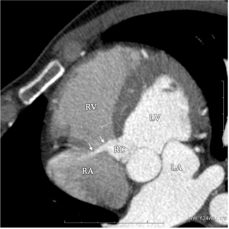
| Chest Radiographs (CXRs) revealed mild pulmonary congestion with cardiomegaly. (Figure 2a) The patient refused to undergo Transesophageal Echocardiography (TEE) even after a comprehensive description of the procedure was provided. Electric Cardiogram (ECG)-gated Computed Tomography (CT) was performed, and it revealed a tubular jet of contrast from the anterior base of the right sinus of Valsalva, into Right Atrium (RA), with a maximum diameter of ~5.8 mm (Figure 2b-2d). The hole of the right cusp was surgically repaired. The patient recovered uneventfully, with no long-term cardiac abnormality. |
| Discussion |
| Continuous heart murmur is a common clinical finding in asymptomatic patients and patients with various presentations, such as chest discomfort and shortness of breath. It is defined as a murmur that begins in systole, continues through the second heart sound and into part or all of diastole. There are a number of differential diagnoses for continuous heart murmur: Patent Ductus Arteriosus (PDA); cervical venous hum; hyperdynamic circulation of pregnancy; Arterio-Venous (AV) fistulas of coronary arteries; great vessel or haemodialysis; coarcation collaterals, truncus arteriosus; pseudo-truncus; total anomalous pulmonary venous connection; Cruveilhier-Baumgarten Syndrome; and post-operative status such as Blalock-Taussig shunt; Potts operation; and Waterston operation. Anomalous systemic arterial supply to the normal basal segments and rupture of sinus of Valsalva (RSOV) are two rare and vital causes of continuous heart murmur, as they are potentially fatal. |
| Radiological features of anomalous systemic arterial supply to the normal basal segments are often confused with pulmonary AVM or bronchopulmonary sequestration. In plain radiographs, the anomalous systemic artery can appear as dilated tubular structures or ill-defined nodular area of increased opacity in retrocardiac region, as in our cases. The anomalous systemic artery can silhouette out the outline of the lower thoracic descending aorta as focal obscuration. Anomalous systemic arteries are also well demonstrated on CT scans as vascular structures, with the same attenuation as the thoracic aorta in the left lower lobe. Moreover, CT can also provide information about the morphology of the bronchial tree and the pulmonary parenchyma, which are both normal in this condition. Another CT finding is the absence or attenuation of the interlobar artery distal to the origin of the superior segmental artery [1,2]. Bronchial tree and the pulmonary parenchyma are normal in this condition. Although MRI can demonstrate an anomalous systemic artery, yet it would not be easy to determine confidently whether the lung tissue supplied by a systemic artery is normal. Therefore, CT is a useful technique for the diagnosis of anomalous systemic arterial supply to the normal basal segments [3]. Patients having anomalous systemic arterial supply to the normal basal segments of left lower lobe are often asymptomatic, while occasional cases of hemoptysis are recognized. Without the presence of clinical symptoms such as hemoptysis and congestive heart failure, treatment is conservative; despite surgery is always imperative for the correction of left-to-right shunt. An anomalous systemic artery can be ligated if there is pulmonary supply to the involved segments of the lung. If, on the other hand, the aberrant artery represents the sole source of blood flow, lobectomy is required. In either case, contrast-enhanced CT is indispensable for correct diagnosis and proper treatment. |
| In patients suffering from RSOV, CXR may reveal cardiomegaly, right heart enlargement and pulmonary congestion. ECG-gated CT is superb in identifying the location of the rupture hole before surgical evaluation; providing early diagnosis of RSOV; delineating an aortocardiac shunt; and evaluating tricuspid valve involvement [4,5]. The high contrast resolution of CT makes it possible to delineate an aortocardiac shunt, if present, and to identify a ruptured aneurysm by depicting a jet of contrast material extending from the aneurysm into the adjacent cardiac chamber. The high contrast resolution of CT is also invaluable for evaluating Tricuspid Valve (TV) involvement. Tricuspid regurgitation is a serious consideration if there is TV involvement, and may increase the risk of endocarditis and the difficulty of surgical repair. If TV involvement is suspected, images with better quality focus around the TV can be obtained. Although RSOV may have potentially fatal complications, the prognosis is excellent after treatment. Thus, prompt and accurate diagnosis is crucial. |
| There are many different causes for continuous murmur and some of them may have potentially fatal complications. Anomalous systemic arterial supply to the normal basal segments and Rupture of Sinus of Valsalva (RSOV) are two rare causes of continuous heart murmur. These two entities are important, as they may have potentially fatal complications. Early diagnosis and prompt management are crucial. Nowadays, ECG-gated CT provides excellent anatomic depiction, and simultaneous evaluation of coronary arteries, great vessels and lungs. A combined approach to diagnoses contributed by both cardiologists and radiologists often reveals the nature of pathology better and aids successful management. |
References |
|
Figures at a glance
 |
 |
 |
 |
| Figure 1a | Figure 1b | Figure 1c | Figure 1d |
 |
 |
 |
 |
| Figure 1e | Figure 1f | Figure 1g | Figure 2a |
 |
 |
 |
|
| Figure 2b | Figure 2c | Figure 2d |
Relevant Topics
- Abdominal Radiology
- AI in Radiology
- Breast Imaging
- Cardiovascular Radiology
- Chest Radiology
- Clinical Radiology
- CT Imaging
- Diagnostic Radiology
- Emergency Radiology
- Fluoroscopy Radiology
- General Radiology
- Genitourinary Radiology
- Interventional Radiology Techniques
- Mammography
- Minimal Invasive surgery
- Musculoskeletal Radiology
- Neuroradiology
- Neuroradiology Advances
- Oral and Maxillofacial Radiology
- Radiography
- Radiology Imaging
- Surgical Radiology
- Tele Radiology
- Therapeutic Radiology
Recommended Journals
Article Tools
Article Usage
- Total views: 14073
- [From(publication date):
June-2013 - Jul 05, 2025] - Breakdown by view type
- HTML page views : 9529
- PDF downloads : 4544
