Case Report Open Access
Cone-Beam Computed Tomography: An Accurate Diagnostic Tool in Dental Practice for Evaluation of Anatomic Variations in Maxillary Bone Septa
| Maíra Fanha Souto1, Milena Bortolotto Felippe1, Thiago de Oliveira Gamba2, Isadora Luana Flores3*, Sérgio Lúcio Pereira de Castro Lopes4 and Luiz Roberto Coutinho Manhães Junior4 | |
| 1São Leopoldo Mandic Dental School, Campinas, São Paulo, Brazil | |
| 2Piracicaba Dental School, State University of Campinas, São Paulo, Brazil | |
| 3Pelotas Dental School, Federal University of Pelotas, Pelotas, Rio Grande do Sul, Brazil | |
| 4São José dos Campos Dental School, São Paulo State University, São José dos Campos, São Paulo, Brazil | |
| Corresponding Author : | Isadora Luana Flores Faculdade de Odontologia de Piracicaba – UNICAMP Departamento de Diagnóstico Oral – Semiologia Av. Limeira, 901 CEP 13.414-903 Piracicaba-São Paulo–Brazil Tel: +55 19 321065267 E-mail: isadoraluanaflores@gmail.com |
| Received April 24, 2015; Accepted June 15, 2015; Published June 18, 2015 | |
| Citation: Souto MF, Felippe MB, Gamba TO, Flores IL, de Castro Lopes SLP, et al. (2015) Cone-Beam Computed Tomography: An Accurate Diagnostic Tool in Dental Practice for Evaluation of Anatomic Variations in Maxillary Bone Septa. J Neuroinfect Dis S1:002. doi:10.4172/2314-7326.S1-002 | |
| Copyright: © 2015 Souto MF, et al. This is an open-access article distributed under the terms of the Creative Commons Attribution License, which permits unrestricted use, distribution, and reproduction in any medium, provided the original author and source are credited. | |
| Related article at Pubmed, Scholar Google | |
Visit for more related articles at Journal of Neuroinfectious Diseases
Abstract
Background: Advanced knowledge of the shape and length of the maxillary sinus and its internal septa is useful for treatment planning of extraction, implant and sinus lift procedures.
Purpose: This study evaluates the prevalence, morphology, orientation, location, height, and length of bone septa in the maxillary sinus using cone-beam computed tomography (CBCT) images, for completely dentate versus partially or fully edentulous patients. Also, since dental extractions may induce formation of sinus septa, this study assesses the correlation between edentulism and prevalence of the maxillary sinus septa.
Material and methods: Four hundred forty-three CBCT images were selected to evaluate the prevalence of a septum in the maxillary sinus to perform an anatomic study of septum morphology. All images were evaluated by one an expert in oral radiology. The χ2 test was used to verify the relationship between the presence of septa and sex, age, and edentulism status. Variance analysis and the Tukey test showed a relationship between partial or full edentulism and the height and length of the maxillary sinus septum.
Results: A maxillary sinus septum was found in 50.1% of the study sample size. 69.8% of patients with a septum were partially edentulous. Gender did not correlation with septum prevalence. The length of the septum in the transverse orientation, and the location of the septum in the medial aspect of the sinus, both correlated with tooth loss.
Conclusions: Maxillary sinus bone septa are more prevalent in partly or fully edentulous populations. Given the diversity of septa morphology among patients, a detailed evaluation of each patient’s septa using CBCT is useful for customizing bone graft and implant placement treatment planning for each patient.
| Keywords |
| Bone septum; Maxillary sinus; Cone-beam computed tomography; Anatomic variation |
| Introduction |
| The maxillary sinus is described universally as a pyramidal cavity in the maxilla [1] and it was mentioned for the first time in the literature by Underwood in 1910 [2,3]. The septum walls in humans are thin within the maxillary sinus [4] and the septum can be primary when it originates during maxillary development and tooth growth or secondary when it results from pneumatization of the maxillary sinus after tooth loss and resorption of the alveolar process [5-7]. Therefore, an antral septum is considered partly as an anatomic variation and partly an acquired structure in the maxillary sinus; prevalence in the population is variable [8]. The presence of insufficient alveolar bone height may render an implant site inappropriate for implantation and [2,8,9], in these cases, lifting the maxillary sinus floor is a technique commonly used for restorations in the posterior maxilla with a high success rate for placement of dental implants [9]. Consequently, the maxillary sinus septum should be considered as an anatomic structure of interest in planning surgery because the knowledge of its location can prevent perforating the septum. Moreover, it’s interesting know that the septum forms a compartment useful if the entire compartment was filled with bone grafting material [10]. |
| The septum has a size can vary between 2.5 and 12.7 mm in mean length [5] and spans from the medial wall to the lateral wall through the sinus floor [11]. Some clinical implications must be considered when approaching the sinus in the presence of a septum, such as access obstructed by the lateral wall of the maxillary sinus during the sinus elevation surgical procedure, the high risk of perforation due to the tight attachment of the membrane in the septum wall, and the difficult of inserting bone graft material when the lateral wall of the sinus is opened in only one area divided by septa [11,12]. |
| Thus, understanding of the sinus anatomy is required and conebeam computed tomography (CBCT), a widely used imaging tool in dental practice, can provide high-resolution images of hard tissues with no overlapping of adjacent anatomic structures allowing accurate surgical planning [12]. Thus, the aim of this study is to evaluate the prevalence, morphology, location, orientation, height, and length of bone septa in the maxillary sinus using CBCT images in dentate and edentulous patients. Moreover, the relationship between presence of a septum, age, sex, and edentulism was investigated. |
| Materials and Methods |
| After approval of this study by the Ethics Committee in Research of the Dental School of the São Leopoldo Mandic, Campinas, São Paulo, Brazil (Protocol Number: 2010/0297), 500 CBCT images were selected from a database of a private dental radiology clinic in Natal, RN, Brazil from January 2009 to April 2011 to investigate the anatomy of the maxillary sinus and bone septa. Jaw images were included when they covered the maxilla as a region of interest regardless of the FOV (Field of View), respectively, of 5.9 to 13 cm (height of the FOV). Images were excluded if some type of intervention, surgical or traumatic, had taken place; a total of 443 images remained for retrospective evaluation. |
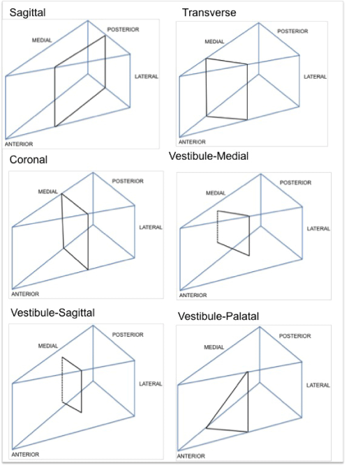
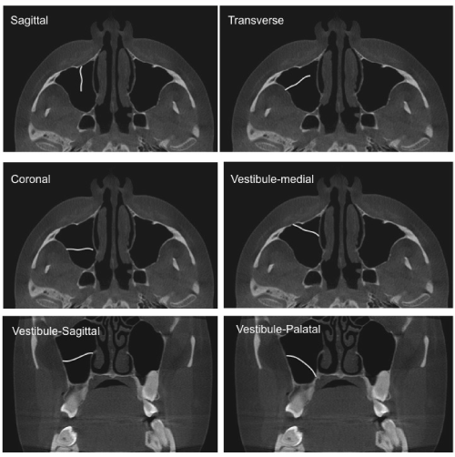
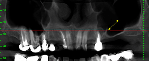
| CBCT images were obtained with an i-CAT scanner (Imaging Science, Hatfield, PA, USA) using the following parameters: 80 kVp, 4.8 mA, acquisition time of 40 s, reconstruction time of 62 s, and a voxel of 0.3 mm. All images were assessed by an expert oral radiologist using Xoran device software (Xoran Technologies, Imaging Science, Hatfield, PA, USA). Initially, demographic aspects, such as sex and age were taken from the patient records and the images were evaluated for the presence of a bone septum in the maxillary sinus. All images with a septum were subdivided into three groups: Fully dentate, partially edentulous and total edentulous. Fully dentate corresponds to presence of 28 teeth (no third molars) and partially edentulous means the absence of one or more of the superior-posterior teeth. Finally, total edentulous corresponds with no teeth. The images with a bone septum were analysed in detail by one expert in oral radiology experienced in computed tomography with regard to type, orientation, location, height, and length. An intra-observer test was realized before the image assessments to improve the study reliability. The type of the septum was categorized as complete (totally closed the sinus in its interior with a creation of secondary compartment of the maxillary sinus) or incomplete (not limits an entire sinus compartment) and the orientation was classified as sagittal, transverse, coronal, vestibule-medial, vestibule-sagittal, or vestibule- palatal. Figure 1 showed a schematic diagram of septum orientations. The axial CBCT slices showing septum orientation are presented in Figure 2. The septum location was categorized as premolar area, first/ second molar area, or tuberosity area for transverse orientation and the Figure 3 showed a premolar transverse septum inside the maxillary sinus in the panoramic reconstruction of the CBCT. For septa classified as sagittal and vestibule-sagittal, the location was categorized as premolar area, middle area (center of maxillary sinus), and tuberosity area or lateral, middle area and medial. For septa classified as vestibulemedial, vestibule-palatal and coronal, the location was categorized as inferior, middle, and superior or anterior, middle and posterior. The height of sinus septum (distance between lower and the higher point of the septum/inferior-superior direction) was measured in the coronal and sagittal depending on their orientations. The septa measuring less than 2 mm in height were not considered in order to exclude the alveolar recess within the floor by tooth loss or sinus pneumatization. The length of the sinus septum (distance between the more lateral and the medial point of the septum) was measured on axial slices. |
| The χ2 test was used to investigate the association between sex, age, type of edentulism, and septum presence in the maxillary sinus. Variance analysis and the Tukey test were applied to evaluate the relationship between height and length and the effect on each type of edentulism. P values less than 0.05 were considered statistically significant. |
| Results |
| A bone septum was found in 222 (50.1%) of 443 CBCT images evaluated and although the majority of patients selected in the study were female, the χ2 test revealed no significant relationship (χ2=0.20, P=0.655) between the presence of a bone septum and sex (male, 51.4%; female, 49.2%). No significant correlation was found between the presence of a septum and age (χ2=7.39, P=0.3894); nevertheless, the age group with the highest prevalence of a septum was between 49–68 years. |
| Of the 222 (50.1%) patients with a septum in the maxillary sinus, 9.9% were fully dentate, 69.8% were partially edentulous, and 20.3% were totally edentulous. The distribution of the number of septa, orientation, location, and edentulism is presented in Tables 1 and 2. Analysis of total axial CBCT scans showed complete septum in 54.8% of the sections and incomplete septum in 45.2%; however, the difference was not significant (χ2=5.19, P=0.0746). The most prevalent septum orientation, regardless of the type of edentulism, was transverse (Table 1), and in terms of location, the region of the first and second molars was the most common regardless of the type of edentulism (Table 2). |
| Analysis of variance revealed no difference in the height of the septum in dentate, partially edentulous, and totally edentulous patients (P=0.2210). However, the length of the septum varied significantly according to the type of tooth loss (P=0.0021) and was longer in partially edentulous patients than totally edentulous patients. The transverse orientation was found to have the greatest height and length (27.78 mm and 18.98 mm, respectively). The average height of the septum was 7.29 mm and the mean length was 9.33 mm. |
| Discussion |
| The maxillary sinus is the first of the paranasal sinuses to develop in the human fetus and is in close proximity to dental elements [2]. Bone septa are anatomic structures with an arched shape arising from the floor and the walls of the sinus and act as barriers of cortical bone, dividing the maxillary sinus into two or more cavities [5,6]. The prevalence of bone septa and their type, orientation, location, height, and length are heterogeneous among individuals and a careful analysis should be performed before surgical procedures to avoid rupture in one or more walls of the maxillary sinus during manipulation of this region [13]. |
| In the present study, after analysis of CBCT images of fully dentate, partially edentulous and totally edentulous patients, a bone septum was found in 50.1% of patients, a higher value compared with almost all articles in the English literature [5,14-28] Previous studies reported values of 14.7% to 47% [14-18,20-28]; only two studies reported higher values (58% and 70%) than ours [5,23]. Thus, the prevalence of a bone septum in the maxillary sinus is variable and the higher prevalence in some studies, including ours, may be due to the imaging tool used to investigate the bone septum, such as the thin slice interval of CBCT images (0.3 mm). Edentulism may also be an important factor associated with the variation in the prevalence of bone septa in different studies; tooth loss seems to be related to bone septum onset [29]. According to Jang et al. after the teeth extraction, the sinus septa start its formation between 6 and 12 months [29]. The CBCT images in the present study revealed prevalence of a bone septum of 69.8% and 20.3% in partially edentulous and totally edentulous patients, respectively, which is in agreement with most studies in the English literature. |
| The association between the prevalence of bone septum and sex and age showed no significant differences. Similar findings were found by others [5,6,17,18,20,22,23,27,28,30]. Therefore, these demographic aspects are probably not related to a bone septum and cannot be considered as relevant to the presence of this anatomic structure. The septum orientation in the maxillary sinus varies widely; transverse was the most frequent orientation found in this study. The transverse septum of the maxillary sinus tend to be more linear and regular this way, its trajectory can be bypassed, when the sinus lift/elevation surgical procedure is recommended, this is a great advantage on it [31]. However, other authors reported miscellaneous findings on bone septum orientation [6,22,24,27,28,30]. Thus, variation in bone septa orientation can be explained by the subjective rating for this factor, which may also vary from evaluator to evaluator as well as for different imaging methods. This study used CBCT images, providing high-resolution images of a delicate bone septum; conventional imaging methods may lead to misdiagnosis in 21.3% of cases due to the difficulty in differentiating overlapping anatomic structures [20,22,23,27]. Therefore, the three-dimensional structure of the maxillary sinus should be evaluated using three-dimensional imaging methods before implant surgery to avoid surgical complications. |
| The most frequent location of bone septa in the present study was the middle (region of the first and second molars) in agreement with several previous studies [5,6,8,15,19,20,22,26,27,30]. The location of a bone septum is directly related to its development, primary or secondary. In our study, 69.8% of the septa were discovered in images of partially edentulous individuals with a predominance of transverse orientation; the middle is the most likely location to find secondary septa. Another important feature of the bone septum is its type; however, analysis of variance showed no evidence of statistically significant differences between complete and incomplete type in the Brazilian population investigated in this study. On the contrary, incomplete predominance was found by some authors [14,17,27,32] suggesting that diverse outcomes are the result of population variability and differences in the tools, the evaluator, and data collection methods. |
| No statistical difference was found between septum height and tooth loss; higher septa were found in fully dentate patients. Corroborating our findings, a previous article reported that the height of the septum was greatest in dentate or nonatrophied segments [17]. However, the length of the septum was associated with edentulism, especially, in partially edentulous patients [17,24,28]. Reports relating height and length of bone septa and edentulism are scarce in the English literature and, despite the limitations of the findings, the height of the septum is probably not related to its length because higher septa were found in partial edentulous patients and larger septa were found in dentate patients. |
| Conclusions |
| According to our results, it is possible to conclude that the prevalence and the length of bone septa are related to loss tooth, particularly in partially edentulous patients. Nevertheless, variations in all anatomic aspects of a bone septum should always be evaluated and CBCT imaging is a useful tool in the planning for dental implants and bone grafts because it provide crucial information. |
| Conflicts of Interest |
| The authors declare there are no conflicts of interest. |
References
- Lawson W, Patel ZM, Lin FY (2008) The development and pathologic processes that influence maxillary sinus pneumatization. Anat Rec (Hoboken) 291: 1554-1563.
- Park YB,Jeon HS, Shim JS, Lee KW, Moon HS (2011) Analysis of the anatomy of the maxillary sinus septum using 3-dimensional computed tomography. J Oral MaxillofacSurg 69: 1070-1078.
- Underwood AS (1910) An Inquiry into the Anatomy and Pathology of the Maxillary Sinus. J AnatPhysiol 44: 354-369.
- González-Santana H,Peñarrocha-Diago M, Guarinos-Carbó J, Sorní-Bröker M (2007) A study of the septa in the maxillary sinuses and the subantral alveolar processes in 30 patients. J Oral Implantol 33: 340-343.
- Maestre-Ferrín L,Galán-Gil S, Rubio-Serrano M, Peñarrocha-Diago M, Peñarrocha-Oltra D (2010) Maxillary sinus septa: a systematic review. Med Oral Patol Oral Cir Bucal 15: e383-386.
- Rosano G,Taschieri S, Gaudy JF, Lesmes D, Del Fabbro M (2010) Maxillary sinus septa: a cadaveric study. J Oral MaxillofacSurg 68: 1360-1364.
- Rossi AC, Freire AR, Perussi MR, Caria PHF, Prado FB (2012) Use of homologous bone grafts in maxillary sinus lifting. Int JOdontostomat 6: 19-26.
- Kim MJ, Jung UW, Kim CS, Kim KD, Choi SH, et al. (2006) Maxillary sinus septa: prevalence, height, location, and morphology. A reformatted computed tomography scan analysis. J Periodontol 77: 903-908.
- vanZyl AW, van Heerden WF (2009) A retrospective analysis of maxillary sinus septa on reformatted computerised tomography scans. Clin Oral Implants Res 20: 1398-1401.
- Lehmann P,Bouaziz R, Page C, Warin M, Saliou G, et al. (2009) [Sinonasal cavities: CT imaging features of anatomical variants and surgical risk]. J Radiol 90: 21-29.
- OniÅŸor-Gligor F,Rotaru A, Juncar M, Bran S (2009) [Clinical study of sinus grafts and implants integration, in the posterior maxilla]. Rev Med ChirSoc Med Nat Iasi 113: 1141-1145.
- Bassiouny A, Newlands WJ, Ali H, Zaki Y (1982) Maxillary sinus hypoplasia and superior orbital fissure asymmetry. Laryngoscope 92: 441-448.
- van den Bergh JP, ten Bruggenkate CM, Disch FJ, Tuinzing DB (2000) Anatomical aspects of sinus floor elevations. Clin Oral Implants Res 11: 256-265.
- Ella B, Noble Rda C, Lauverjat Y, Sédarat C, Zwetyenga N, et al. (2008) Septa within the sinus: effect on elevation of the sinus floor. Br J Oral MaxillofacSurg 46: 464-467.
- Gosau M, Rink D, Driemel O, Draenert FG (2009) Maxillary sinus anatomy: a cadaveric study with clinical implications. Anat Rec (Hoboken) 292: 352-354.
- Koymen R,Gocmen-Mas N, Karacayli U, Ortakoglu K, Ozen T, et al. (2009) Anatomic evaluation of maxillary sinus septa: surgery and radiology. ClinAnat 22: 563-570.
- Krennmair G, Ulm C, Lugmayr H (1997) Maxillary sinus septa: incidence, morphology and clinical implications. J CraniomaxillofacSurg 25: 261-265.
- Krennmair G, Ulm CW, Lugmayr H, Solar P (1999) The incidence, location, and height of maxillary sinus septa in the edentulous and dentate maxilla. J Oral MaxillofacSurg 57: 667-671.
- Lee WJ, Lee SJ, Kim HS (2010) Analysis of location and prevalence of maxillary sinus septa. J Periodontal Implant Sci 40: 56-60.
- Li J, Zhou ZX, Yuan ZY, Yuan H, Sun C, et al. (2013) [An anatomical study of maxillary sinus septum of Han population in Jiangsu region using cone-beam CT]. Shanghai Kou Qiang Yi Xue 22: 52-57.
- Naitoh M,Suenaga Y, Kondo S, Gotoh K, Ariji E (2009) Assessment of maxillary sinus septa using cone-beam computed tomography: etiological consideration. Clin Implant Dent Relat Res 11 Suppl 1: e52-58.
- Neugebauer J, Ritter L, Mischkowski RA, Dreiseidler T, Scherer P, et al. (2010) Evaluation of maxillary sinus anatomy by cone-beam CT prior to sinus floor elevation. Int J Oral Maxillofac Implants 25: 258-265.
- Orhan K,KusakciSeker B, Aksoy S, Bayindir H, BerberoÄŸlu A, et al. (2013) Cone beam CT evaluation of maxillary sinus septa prevalence, height, location and morphology in children and an adult population. Med PrincPract 22: 47-53.
- Pommer B, Ulm C, Lorenzoni M, Palmer R, Watzek G, et al. (2012) Prevalence, location and morphology of maxillary sinus septa: systematic review and meta-analysis. J ClinPeriodontol 39: 769-773.
- Rysz M,Bakoń L (2009) Maxillary sinus anatomy variation and nasal cavity width: structural computed tomography imaging. Folia Morphol (Warsz) 68: 260-264.
- Shen EC, Fu E, Chiu TJ, Chang V, Chiang CY, et al. (2012) Prevalence and location of maxillary sinus septa in the Taiwanese population and relationship to the absence of molars. Clin Oral Implants Res 23: 741-745.
- Shibli JA,Faveri M, Ferrari DS, Melo L, Garcia RV, et al. (2007) Prevalence of maxillary sinus septa in 1024 subjects with edentulous upper jaws: a retrospective study. J Oral Implantol 33: 293-296.
- Ulm CW, Solar P, Krennmair G, Matejka M, Watzek G (1995) Incidence and suggested surgical management of septa in sinus-lift procedures. Int J Oral Maxillofac Implants 10: 462-465.
- Jang SY, Chung K, Jung S, Park HJ, Oh HK, et al. (2014) Comparative study of the sinus septa between dentulous and edentulous patients by cone beam computed tomography. Implant Dent 23: 477-481.
- Velásquez-Plata D, Hovey LR, Peach CC, Alder ME (2002) Maxillary sinus septa: a 3-dimensional computerized tomographic scan analysis. Int J Oral Maxillofac Implants 17: 854-860.
- Pignataro L,Mantovani M, Torretta S, Felisati G, Sambataro G (2008) ENT assessment in the integrated management of candidate for (maxillary) sinus lift. ActaOtorhinolaryngolItal 28: 110-119.
- Yang HM,Bae HE, Won SY, Hu KS, Song WC, et al. (2009) Thebuccofacial wall of maxillary sinus: an anatomical consideration for sinus augmentation. Clin Implant Dent Relat Res 11 Suppl 1: e2-6.
Tables and Figures at a glance
| Table 1 | Table 2 |
Figures at a glance
 |
 |
 |
| Figure 1 | Figure 2 | Figure 3 |
Relevant Topics
- Bacteria Induced Neuropathies
- Blood-brain barrier
- Brain Infection
- Cerebral Spinal Fluid
- Encephalitis
- Fungal Infection
- Infectious Disease in Children
- Neuro-HIV and Bacterial Infection
- Neuro-Infections Induced Autoimmune Disorders
- Neurocystercercosis
- Neurocysticercosis
- Neuroepidemiology
- Neuroinfectious Agents
- Neuroinflammation
- Neurosyphilis
- Neurotropic viruses
- Neurovirology
- Rare Infectious Disease
- Toxoplasmosis
- Viral Infection
Recommended Journals
Article Tools
Article Usage
- Total views: 14752
- [From(publication date):
specialissue-2015 - Apr 05, 2025] - Breakdown by view type
- HTML page views : 10034
- PDF downloads : 4718
