Make the best use of Scientific Research and information from our 700+ peer reviewed, Open Access Journals that operates with the help of 50,000+ Editorial Board Members and esteemed reviewers and 1000+ Scientific associations in Medical, Clinical, Pharmaceutical, Engineering, Technology and Management Fields.
Meet Inspiring Speakers and Experts at our 3000+ Global Conferenceseries Events with over 600+ Conferences, 1200+ Symposiums and 1200+ Workshops on Medical, Pharma, Engineering, Science, Technology and Business
Case Report Open Access
Cone Beam Computed Tomography: A Useful Tool in Diagnosis of Bone Island and Implant Insertion Guidance
| Chen Tie, Zhou Zhi-ying and Lai Ren-fa* | |
| The Medical Centre of Stomatology, the 1st Affiliated Hospital of Jinan University, Guangzhou 510630, Chinaru | |
| Corresponding Author : | Lai Ren-fa The Medical Centre of Stomatology the 1st Affiliated Hospital of Jinan University Guangzhou 510630, China E-mail: Prof.Dr.Lai@163.com |
| Received December 20, 2011; Accepted January 10, 2012; Published January 14, 2012 | |
| Citation: Tie C, Zhi-ying Z, Ren-fa L (2012) Cone Beam Computed Tomography: A Useful Tool in Diagnosis of Bone Island and Implant Insertion Guidance. OMICS J Radiology. 1:101. doi: 10.4172/2167-7964.1000101 | |
| Copyright: © 2012 Tie C et al. This is an open-access article distributed under the terms of the Creative Commons Attribution License, which permits unrestricted use, distribution, and reproduction in any medium, provided the original author and source are credited. | |
Visit for more related articles at Journal of Radiology
Abstract
The aim of this paper is to make surgeons aware of the use of cone beam computed tomography (CBCT) within the field of implantology. The paper describes one case illustrating the improved diagnostic yield using CBCT over conventional radiography thus facilitating the appropriate insertion guidance of implants.
| Keywords |
| Cone beam CT; Implants; Insertion; Imaging; Guidance |
| Introduction |
| Cone beam computed tomography (CBCT) has been used in dentistry since the mid 1990s. As the name implies, it uses a cone shaped X-ray beam which rotates around the patient to acquire a volumetric data set of the region of interest with a single rotation of the patient. The CBCT volumetric data set comprises a three-dimensional (3D) block of cuboid structures known as voxels, where each voxel represents a specific degree of X-ray beam absorption. Image reconstruction is achieved using computer algorithms ultimately producing 3D images at high resolution. The main advantage of CBCT is that the radiation dosage is considerably less than conventional CT scanning. In addition with most units the patient is scanned in the upright position, and so there is less distortion of the soft tissues in comparison to conventional CT where the patient is supine. This is particularly useful if the facial soft tissues are reconstructed. The literature is replete in clinical applications of CBCT within the oral medicine specialty the clinical applications include imaging of impacted teeth and dental abnormalities, assessment of alveolar bone heights and bone volume, investigation of the temporomandibular joint and so on. The use of CBCT in the field of endodontics has also been described as it is useful for diagnosing canal morphology, assessing root and alveolar fractures, analysis of resorptive lesions and identification of pathology. |
| This paper reports on one case where conventional radiographs suggested the need for a CBCT which yielded additional diagnostic information to allow the clinician to make the diagnosis and to guide the implant insertion process. In this case, CBCT imaging was carried out using the Kodark 9000C-3D (Imaging Sciences International, Hatfield, PA, USA). The data was then exported into Simplant (Materialise, Leuven, Belgium) to carry out the 3D reconstructions. The exporting of data and 3D construction of images takes additional time and resources. |
| Clinical Case |
| General condition |
| A 48 years old Female patient was referred to the Oral & Maxillofacial department by her local general dental practitioner regarding the right mandible tumor, which exits since her childhood, but without any symptoms and uncomforted. Recently, she worries about the tumor, and want to have a diagnosis. The patient presented a hard mass with a clear boundary in the right mandible premolar region and the premolars are slightly oblique. The patient hasn’t any other uncomforted feeling. |
| A panoramic radiograph showed the a mass in the right mandible premolar area, according to the characteristic of the X-ray film, the diagnosis of odontoma or complex odontoma was made, further examination and treatment were required and moreover the panoramic presented a dense sclerosis lesion near the mental foramen, with a diameter of 1.5cm in the left mandible, and there isn’t any Periosteal reaction surrounding this dense sclerosis lesion (Figure 1). |
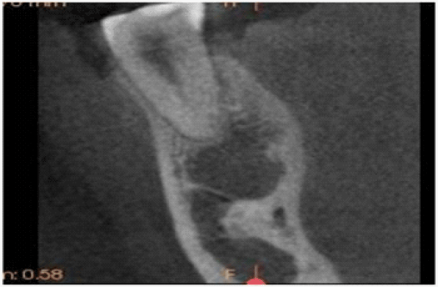
| CBCT ( Kodark 9000C - 3D ) scan was taken around the dense sclerosis lesion near the mental foramen in the left mandible premolar area. CBCT showed a high-density lesion, a homogeneously dense sclerotic focus in the cancellous bone, similar to the surrounding cortical bone, prominent inward from the from the lateral cortical, with the diameter of 1.5cm, the semi-circular shape, clear boundary (Figure 2). |
| Discussion |
| Bone Island, bone spot, also known as enotosis, is of different patterns of cortical bone nodules within the cancellous bone [1]. This benign lesion is probably congenital or developmental in origin and reflects failure of resorption during endochondral ossification, and the incidence is about 1/1,000,000 [2]. Typical asymptomatic, the lesion is usually an incidental finding by the occasional X-ray check such as by the other illness reasons [1,2]. The location of the bone island mostly occurs in trunk and limbs [3], but also occasionally occurs in mandible [4]. In this case, the female patient has a tumor in the right mandible, and the panoramic radiograph showed a dense sclerosis in the left mandible. |
| Diagnostic information is essential in influencing clinical decision making. Accurate imaging leads to better treatment planning decisions and potentially more predictable outcomes. CBCT is an emerging imaging modality that can offer the clinician information above that obtained from conventional radiographs [5]. |
| The advantages of CBCT in maximizing diagnostic yield and reduced radiation exposure have been well documented. It is essential that these images are requested appropriately and in order to maximize diagnostic yield for the patient all images should be analyzed and reported on. Currently there are no published referral criteria for CBCT. Some scholars emphasize that the CBCT should be used with caution and the surgeon should always ask whether the question for which the imaging is requested could be answered by conventional radiography [5,6]. There are currently no formalized selection criteria for CBCT in implantology, and that more evidence based research is necessary in this area [7,8]. |
| In recent years, Oral Implantology develops rapidly, in order to perform the implant operation well, the panoramic X-ray film was carried out routinely so as to get enough information about the alveolar bone height, width, density, and the adjacent teeth and so on. Therefore it gives us more possibility to find the existence of the bone island in the mandible. In case of finding a homogeneously dense, sclerotic focus in mandible by plain radiography, further CBCT scan can determine its exact location. And the radiologic features remind the oral surgeons to avoid the location of the bone island for the implant insertion, or to make some special care by injecting more cooling saline in order to avoid producing excessive heat during the implant insertion process leading to implant failure [8,9]. |
| Conclusions |
| Bone Island occasionally occurs in the mandible and it will influence the oral surgeon to perform the implant insertion if it exits in the implant insertion location in the mandible. CBCT allows the surgeons to have an accurate 3D picture of the position of areas of interest which facilitates diagnosis of Bone Island and guidance for implant insertion operation. |
References |
|
Figures at a glance
 |
 |
| Figure 1 | Figure 2 |
Post your comment
Relevant Topics
- Abdominal Radiology
- AI in Radiology
- Breast Imaging
- Cardiovascular Radiology
- Chest Radiology
- Clinical Radiology
- CT Imaging
- Diagnostic Radiology
- Emergency Radiology
- Fluoroscopy Radiology
- General Radiology
- Genitourinary Radiology
- Interventional Radiology Techniques
- Mammography
- Minimal Invasive surgery
- Musculoskeletal Radiology
- Neuroradiology
- Neuroradiology Advances
- Oral and Maxillofacial Radiology
- Radiography
- Radiology Imaging
- Surgical Radiology
- Tele Radiology
- Therapeutic Radiology
Recommended Journals
Article Tools
Article Usage
- Total views: 8627
- [From(publication date):
April-2012 - Mar 29, 2025] - Breakdown by view type
- HTML page views : 3996
- PDF downloads : 4631
