Case Report Open Access
Compromised Object Pattern Separation Performance in Parkinsons Disease Suggests Dentate Gyrus Neurogenesis may be Compromised in the Condition
Keith A Wesnes1* and David J Burn2
1Bracket, Goring on Thames, RG8 9RD, Centre for Human Psychopharmacology, Swinburne University, Melbourne, Australia, UK
2Institute for Ageing and Health, Newcastle University, Campus for Ageing and Vitality, Newcastle upon Tyne, NE4 5PL, UK
- Corresponding Author:
- Keith A Wesnes
Practice Leader, Bracket
Gatehampton Road, Goring-on-Thames
RG8 0EN, Adjunct Professor
Centre for Human Psychopharmacology
Swinburne University, Melbourne, Australia, UK
Tel: +44 (0) 1491 878 702
E-mail: keith.wesnes@BracketGlobal.com
Received date: August 31, 2013; Accepted date: November 15, 2013; Published date: November 22, 2013
Citation: Wesnes KA, Burn DJ (2013) Compromised Object Pattern Separation Performance in Parkinson’s Disease Suggests Dentate Gyrus Neurogenesis may be Compromised in the Condition. J Alzheimers Dis Parkinsonism 3:131. doi:10.4172/2161-0460.1000131
Copyright: © 2013 Wesnes KA, et al. This is an open-access article distributed under the terms of the Creative Commons Attribution License, which permits unrestricted use, distribution, and reproduction in any medium, provided the original author and source are credited.
Visit for more related articles at Journal of Alzheimers Disease & Parkinsonism
Abstract
Background: Destruction of dopaminergic neurones decreases hippocampal Dentate Gyrus (DG) neurogenesis in rodents and primates. Post-mortem work in Parkinson’s Disease (PD) patients identified evidence of compromised hippocampal neurogenesis. In both animals and man, difficult discriminations in tests which require discrimination between previously seen objects and closely similar ones (object pattern separation tasks) reflect DG activity and thus potentially neurogenesis. The object of this study was to use such a task in PD patients to seek evidence of compromised DG activity.
Methods: The CDR System picture recognition task has been validated as an object pattern separation task. In the task pictures of objects and scenes are presented which later must be discriminated from closely similar pictures. fMRI work in man has identified that difficult discriminations in object pattern separation tasks (i.e deciding whether or not a closely similar object was previously presented) to selectively result in increased DG activity. Data from this task in 72 patients with Parkinson’s disease were compared with 62 age and gender matched controls.
Results: The PD patients showed selective, marked and highly significant deficits to the DG-sensitive measure which was unrelated to dopaminergic medication. Such a pattern was not seen on a word recognition paradigm, which is consistent with fMRI work showing verbal tasks are not related to DG activation.
Conclusions: This is the first behavioural demonstration of compromised OPS in Parkinson’s patients, supporting work with rats and primates. This finding is consistent with impairment to DG function and thus potentially compromised neurogenesis. Implications for current and novel PD therapies will be discussed, in relation to compounds such as rasagiline which promote neurogenesis.
Keywords
Dentate Gyrus (DG); Neurogenesis; Parkinson’s disease; Object pattern separation
Introduction
The discovery that the human Dentate Gyrus (DG) retains its ability to generate neurons throughout life, has led numerous groups to study whether adult neurogenesis is compromised in various neurodegenerative disorders [1]. In Parkinson’s disease for example, speculation is growing that that impaired or failed neurogenesis may contribute to cognitive decline in the disorder [2]. There is accumulating evidence from animal models of Parkinson’s disease and human postmortem studies that adult neurogenesis is impaired in the condition. For example, the over expression of alpha-synuclein in transgenic mice had a negative impact on adult neurogenesis, and reduced levels of adult neurogenesis were seen in the hippocampal dentate gyrus [3]. The mechanism by which alpha-synuclein promotes neurotoxicity has been shown to be the inhibition of histone acetylation, and the neurotoxicity can be reversed by histone deacetylase inhibitors [4]. Considerable evidence has now accumulated that histone deacetylase inhibitors promote adult neurogenesis in the hippocampal dentate gyrus; leading to predictions that this may be a therapeutic target for Parkinson’s disease [5]. Evidence has just appeared firmly linking RbAp48 (a histone-binding protein that modifies histone acetylation) with dentate gyrus neurogenesis and age-related memory loss [6]. Overall, this body of data clearly links alpha-synuclein, histone acetylation in the hippocampal dentate gyrus, and cognitive dysfunction in Parkinson’s disease. Finally, a study of post-mortem brain autopsy tissue of PD patients identified a reduced number of neural precursor cells in the dentate gyrus, suggesting that the generation of neural precursor cells is impaired in PD as a consequence of dopaminergic denervation [7]. However, in vivo evidence of reduced neurogenesis in PD is lacking.
The human hippocampus supports the formation of episodic memory without confusing new memories with old ones [8]. To accomplish this, the brain must disambiguate memories and this is now widely recognised as a key role of the dentate gyrus (DG). Schmidt et al. write: “Over the course of the last 20 years, the overwhelming majority of data have supported the notion of the DG as a critical mediator of pattern separation within the hippocampal formation” (page 57) [8]. Human evidence from fMRI work has accumulated over the last few years linking the DG to performance on Object Pattern Separation (OPS) tasks. In OPS tasks, a series of pictures is presented and subjects have to decide whether or not each subsequent picture has been presented earlier. The consistent finding from different groups using OPS tasks has been that fMRI DG measured activity is highest when closely similar pictures are presented, and lowest when the original pictures or completely different ones are presented [9-11]. Thus in man OPS tasks can provide an index of DG activity by assessing the ability of subjects to correctly reject closely similar pictures, ie to perform difficult pattern separations. The CDR System picture recognition task has been validated as an OPS, and the DG sensitive measure has been found to selectively decline in normal aging, which is consistent with known age-related changes to hippocampal DG neurogenesis [12,13].
If hippocampal dentate gyrus activity is compromised in Parkinson’s disease, the prediction would be that performance on difficult OPS would be selectively impaired in the patients. The purpose of this analysis is to determine the pattern of performance of PD patients on an OPS task as well as a test of verbal recognition.
Methods
Subjects
72 Parkinson’s disease patients (27 female, 45 male), mean age 70.1 years (SD 7.8) and mean MMSE 27.4 (SD 2.4) participated in this study. 62 controls were also recruited (25 female, 37 male), mean age 71.3 years (SD 6.7) and mean MMSE 28.5 (SD 1.4). For the patients, the average duration of illness was 7.5 years (SD 5.1) and age of onset was 62.9 years (SD 9.5). The UPDRS II mean score for the patients was 13.7 (SD 6.5), UPDRS III 27.3 (SD 12.5) and Hoehn and Yahr score 2.4 (SD 0.7). All patients provided signed, written informed consent. The study was conducted according to the provisions of the Helsinki Declaration and was approved by the Northumberland Healthcare Trust Ethics Committee.
Methods
Patients were tested on the CDR System tests of delayed word and picture recognition, the testing being conducted by a trained administrator [14].
The CDR system: The CDR System is a set of automated tests of cognitive function which has been used previously in Parkinson’s disease (e.g. 15). Tests of attention, working memory and episodic memory were administered, the data from two tasks being reported in this analysis:
CDR System Picture Recognition test: A series of 20 pictures of everyday scenes and objects was presented on the screen at the rate of 1 every 3 s for the patient to remember. Around 12 minutes later the 20 pictures were re-presented mixed with 20 novel pictures which were individually matched to the original pictures to be closely similar. For each picture the patient was required to decide whether it was an originally presented picture or a novel picture by pressing the YES or NO button as appropriate as quickly as possible. Both the accuracy and speed of each response was recorded.
CDR System Word Recognition test: A series of 15 words was presented on the screen at the rate of 1 every 2 s for the patient to remember. Around 12 minutes later the 12 words were re-presented mixed with 12 novel words. For each word the patient was required to decide whether it was an originally presented word by pressing the YES or NO button as appropriate as quickly as possible. Both the accuracy and speed of each response was recorded.
Results and Discussion
CDR System data were available for all 72 patients and 62 controls. The CDR System picture recognition task has been validated as an object pattern separation (OPS) task [12] and is directly comparable to the task used in fMRI studies [10,11] in which Dentate Gyrus (DG) activity has been found to be specifically high during trials when patients are presented with a closely similar picture to one of the original pictures; and asked to determine whether or not it was the original. In contrast original pictures when re-presented are associated with considerably less DG activity. If the DG is not functioning optimally for the patient’s age, the ability to correctly reject the closely similar pictures should be impaired to a greater degree than controls. Further, while patients may be less accurate in recognising the original pictures than controls, the difference between these two aspects of task performance should be greater if DG activity is impaired. As a control for the performance of patients in forced-choice recognition tasks, the data for the picture task were contrasted to those from word recognition. Brickman et al. have confirmed that difficult discriminations in OPS tasks are associated with increased DG activity, but that not in verbal tasks; which instead increase activity in the entorhinal cortex [9].
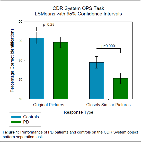
The patients had significantly lower MMSE scores than controls [27.4 vs. 28.6, F(1,131)=11.4, p<0.001], and to control for this, plus the small but not significant disparity in average age, both MMSE and age were fitted as covariates to the analyses. The SAS procedure MIXED was used to conduct a 2-factor ANCOVA of the accuracy scores from the picture and word recognition tasks. In each analysis: group (control vs. PD), type of response (original items vs. novel items) and the interaction between them were fitted as fixed terms; subject being fitted as a random term. For the pictures there were significant main effects of group and type of response (both p<0.001), and also a significant interaction between them [F(1,132)=4, p<0.05]. These results are illustrated graphically in Figure 1. There was no difference between the groups on the non-DG sensitive measure, the ability to correctly recognise the original pictures (p=0.57), but there was a highly significant difference on the DG sensitive difficult OPS separations (p=0.0001). The Cohen’s d effect size of this impairment was 0.68, a medium to large sized effect, which exceeds the 0.5 criterion of a clinically relevant effect [15,16]. To examine whether the dopaminergic treatment was related to the pattern of results, the patients were divided into two groups, one which was not on medication (n=7) and the other on medication (n=65). Repeating the analysis with the PD population divided into these 2 groups had no effect on the outcome, the interaction term between group and type of response remaining significant [F(2,131)=3.15, p<0.05]. There were significant differences for the novel pictures between controls and each of the two PD groups, but no difference between the PD groups on this measure. Thus it appears that the finding is not related to whether or not the patients in this sample were receiving dopaminergic medication.
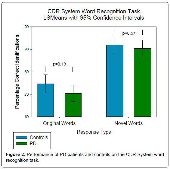
For the words, there was no main effect of group (p=0.121) or interaction (p=0.52), but a significant difference between the overall responses to original and novel words (p<0.0001). Figure 2 highlights the difference in patterns between the two tasks, patients and controls finding it more difficult to reject the closely similar pictures than to identify the original ones; whereas the pattern is reversed for word recognition, patients finding it easier to reject the novel words than correctly identify the original ones.
Overall, the PD patients showed a selective, large and statistically significant decline on difficult discriminations on the OPS task. As this measure has been associated with dentate gyrus activity from fMRI studies [9-11], the effect seen in this study is consistent with disrupted activity in this region in PD patients. The dentate gyrus is widely acknowledged to be the area where hippocampal neurogenesis occurs, and the findings are therefore consistent with preclinical PD models and post-mortem PD autopsy brain tissue findings of reduced neurogenesis in the dentate gyrus [3,7]. These are, to our knowledge, the first data from an OPS task in PD.
As enhanced neurogenesis has been shown to improve pattern separation, compounds which promote neurogenesis may help reverse these deficits in PD [17]. O’Sullivan et al. have shown L-Dopa to promote neurogenesis in post-mortem PD brain tissue [18]. The present study had too few patients who were not treated with dopaminergic agonists (n=7) to properly examine this hypothesis, but future work could examine this possibility. Newer treatments such as rasagiline have been shown to promote neurogenesis, and also to reverse neurogenesis related olfactory deficits in a mouse model of PD [19,20]. Future work with OPS could assess whether this mechanism is effective in PD patients. A research project funded by the Michael J Fox foundation is also underway entitled: “Increasing Endogenous Neurogenesis Using Neurosteroids: A Novel Therapeutic Strategy to Treat Parkinson’s disease” [21].
In clinical studies in PD of compounds targeted to improve neurogenesis, OPS tasks will be useful as they will serve, firstly, as non-invasive biomarkers which can provide ‘proof of mechanism’, and secondly, as efficacy endpoints.
References
- Eriksson PS, Perfilieva E, Björk-Eriksson T, Alborn AM, Nordborg C, et al. (1998) Neurogenesis in the adult human hippocampus. Nat Med 4: 1313-1317.
- Geraerts M, Krylyshkina O, Debyser Z, Baekelandt V (2007) Concise review: therapeutic strategies for Parkinson disease based on the modulation of adult neurogenesis. Stem Cells 25: 263-270.
- Winner B, Lie DC, Rockenstein E, Aigner R, Aigner L, et al. (2004) Human wild-type alpha-synuclein impairs neurogenesis. J Neuropathol Exp Neurol 63: 1155-1166.
- Kontopoulos E, Parvin JD, Feany MB (2006) Alpha-synuclein acts in the nucleus to inhibit histone acetylation and promote neurotoxicity. Hum Mol Genet 15: 3012-3023.
- Contestabile A, Sintoni S, Monti B (2012) Histone Deacetylase (HDAC) Inhibitors as Potential Drugs to Target Memory and Adult Hippocampal Neurogenesis. Current Psychopharmacology 1: 14-28.
- Pavlopoulos E, Jones S, Kosmidis S, Close M, Kim C, et al. (2013) Molecular mechanism for age-related memory loss: the histone-binding protein RbAp48. Sci Transl Med 5: 200ra115.
- Höglinger GU, Rizk P, Muriel MP, Duyckaerts C, Oertel WH, et al. (2004) Dopamine depletion impairs precursor cell proliferation in Parkinson disease. Nat Neurosci 7: 726-735.
- Schmidt B, Marrone DF, Markus EJ (2012) Disambiguating the similar: the dentate gyrus and pattern separation. Behav Brain Res 226: 56-65.
- Brickman AM, Stern Y, Small SA (2011) Hippocampal subregions differentially associate with standardized memory tests. Hippocampus 21: 923-928.
- Kirwan CB, Stark CE (2007) Overcoming interference: an fMRI investigation of pattern separation in the medial temporal lobe. Learn Mem 14: 625-633.
- Yassa MA, Stark CE (2011) Pattern separation in the hippocampus. Trends Neurosci 34: 515-525.
- Wesnes KA (2010) Visual object pattern separation: A paradigm for studying the role of the dentate gyrus in memory disorders. Alzheimer's & Dementia 6: e45.
- Villeda SA, Luo J, Mosher KI, Zou B, Britschgi M, et al. (2011) The ageing systemic milieu negatively regulates neurogenesis and cognitive function. Nature 477: 90-94.
- Simpson PM, Surmon DJ, Wesnes KA, Wilcock GR (1991) The Cognitive Drug Research computerised assessment system for demented patients: A validation study. International Journal of Geriatric Psychiatry 6: 95-102.
- Hudson G, Stutt A, Eccles M, Robinson L, Allcock LM, et al. (2010) Genetic variation of CHRNA4 does not modulate attention in Parkinson's disease. Neurosci Lett 479: 123-125.
- Harvey PD (2012) Clinical applications of neuropsychological assessment. Dialogues Clin Neurosci 14: 91-99.
- Sahay A, Scobie KN, Hill AS, O'Carroll CM, Kheirbek MA, et al. (2011) Increasing adult hippocampal neurogenesis is sufficient to improve pattern separation. Nature 472: 466-470.
- O'Sullivan SS, Johnson M, Williams DR, Revesz T, Holton JL, et al. (2011) The effect of drug treatment on neurogenesis in Parkinson's disease. Mov Disord 26: 45-50.
- Mandel SA, Sagi Y, Amit T (2007) Rasagiline promotes regeneration of substantia nigra dopaminergic neurons in post-MPTP-induced Parkinsonism via activation of tyrosine kinase receptor signaling pathway. Neurochem Res 32: 1694-1699.
- Petit GH, Berkovich E, Hickery M, Kallunki P, Fog K, et al. (2013) Rasagiline ameliorates olfactory deficits in an alpha-synuclein mouse model of Parkinson's disease. PLoS One 8: e60691.
- Adeosun S, Hou X, Jiao Y, Zheng B, Kyle B, et al. (2012) Increasing endogenous neurogenesis using neurosteroids: A novel therapeutic strategy to treat Parkinson's disease. Parkinsonism & Related Disorders 18: S214.
Relevant Topics
- Advanced Parkinson Treatment
- Advances in Alzheimers Therapy
- Alzheimers Medicine
- Alzheimers Products & Market Analysis
- Alzheimers Symptoms
- Degenerative Disorders
- Diagnostic Alzheimer
- Parkinson
- Parkinsonism Diagnosis
- Parkinsonism Gene Therapy
- Parkinsonism Stages and Treatment
- Stem cell Treatment Parkinson
Recommended Journals
Article Tools
Article Usage
- Total views: 14402
- [From(publication date):
February-2014 - Jul 04, 2025] - Breakdown by view type
- HTML page views : 9830
- PDF downloads : 4572


