Case Report Open Access
Complicative Extraction Lead of an Infected Pacemaker
| Clara Bonanad*, Maite Izquierdo, Angel Ferrero, Angel Martínez, Juan Miguel Sánchez and Ricardo Ruiz-Granell | |
| Department of Cardiology, Hospital Clinico y Universitario de Valencia, Universidad de Valencia, ICLIVA Blasco Ibanez 1746010, Valencia, Spain | |
| Corresponding Author : | Clara Bonanad Department of Cardiology Hospital Clinico y Universitario de Valencia Universidad de Valencia ICLIVA Blasco Ibanez 1746010–Valencia, Spain Tel: +34 96 3862658 Fax: +34 96 3862658 E-mail: clarabonanad@gmail.com |
| Received August 28, 2013; Accepted September 26, 2013; Published September 30, 2013 | |
| Citation: Bonanad C, Izquierdo M, Ferrero A, Martínez A, Sánchez JM (2013) Complicative Extraction Lead of an Infected Pacemaker. OMICS J Radiology 2:146 doi: 10.4172/2167-7964.1000146 | |
| Copyright: © 2013 Bonanad C, et al. This is an open-access article distributed under the terms of the Creative Commons Attribution License, which permits unrestricted use, distribution, and reproduction in any medium, provided the original author and source are credited. | |
Visit for more related articles at Journal of Radiology
Abstract
Endocarditis in Cardiac Devices (CD), either permanent pacemakers or implantable cardioverter defibrillators, is a severe disease associated with high mortality. The increasing number of patients with implanted cardiac devices explains the rising frequency of endocarditis. The treatment includes medical therapy with complete removal of CD either by surgery or percutaneous extraction depending on the size of vegetation, but there are no clear guidelines about the management of patient with intermediate size vegetations and significant associated comorbidity. We present a case of a 69 year-old man with significant comorbidity and dilated ischemic cardiomyopathy with moderately depressed Left Ventricular Ejection Fraction (LVEF), with a single pass VDD pacemaker presenting with adhered vegetation in the auricular surface of the lead.Endocarditis in Cardiac Devices (CD), either permanent pacemakers or implantable cardioverter defibrillators, is a severe disease associated with high mortality. The increasing number of patients with implanted cardiac devices explains the rising frequency of endocarditis. The treatment includes medical therapy with complete removal of CD either by surgery or percutaneous extraction depending on the size of vegetation, but there are no clear guidelines about the management of patient with intermediate size vegetations and significant associated comorbidity. We present a case of a 69 year-old man with significant comorbidity and dilated ischemic cardiomyopathy with moderately depressed Left Ventricular Ejection Fraction (LVEF), with a single pass VDD pacemaker presenting with adhered vegetation in the auricular surface of the lead.
| Keywords |
| Endocarditis; Pacemaker; Percutaneous extraction |
| Introduction |
| Endocarditis in Cardiac Devices (CD), either permanent pacemakers or implantable cardioverter defibrillators, is a severe disease associated with high mortality. In adición to patient factors, procedural characteristics may also play an important role in the development of CD infection. Despite the greater ease of device implantation with pectoral rather than other routes and increasing experience with implantation, the rate of CD infection has been increasing [1]. |
| Another factors associated with a greater risk of CD infection have been described, including the following: (1) Immunosuppression (renal dysfunction and corticosteroid use); (2) oral anticoagulation use; (3) patient coexisting illnesses; (4) periprocedural factors, including the failure to administer perioperative antimicrobial prophylaxis; (5) device revision/replacement; (6) the amount of indwelling hardware; (7) operator experience; and (8) the microbiology of bloodstream infection in patients with indwelling CDs [2]. |
| Among other causes, the increasing number of patients with implanted cardiac devices and important comorbidities explains the rising frequency of endocarditis [1-6]. The treatment includes medical therapy with CD complete removal in all cases [1,4]. |
| Case Report |
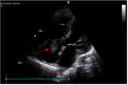
| We present a case of a 69 year-old hypertensive man with renal insufficiency, Chronic Pulmonary Obstructive Disease (CPOD) and ischemic cardiomyopathy with moderate depressed left ventricular ejection fraction (LVEF). Ten years back, he was implanted with a permanent transvenous single lead VDD pacemaker because of a complete atrio-ventricular block. The generator was electively replaced nine years later. After the intervention the wound showed signs of infection and was treated with antibiotic therapy. Six months later, the patient was admitted to the hospital because of high fever. Transthoracic echocardiography demonstrated a vegetation of 40×30 mm in the auricular surface of the single lead near the tricuspid valve (Figure 1), with moderate tricuspid regurgitation. Cultures of 3 blood samples were positive for coagulase-negative staphylococci. Four weeks after the completion of antibiotic therapy (Vancomycine, Rifampicin and Gentamicin), the patient remained free of symptoms, but tranesophageal echocardiography demonstrated no change in the size of the vegetation. Guidelines state that surgical treatment of vegetations larger than 25 mm is warranted. However, this patient was considered a high-risk candidate for surgery, due to the risk of the intervention and the important comorbidities associated (calculated Euroscore of 32%). Therefore, it was decided to perform percutaneous lead extraction. |
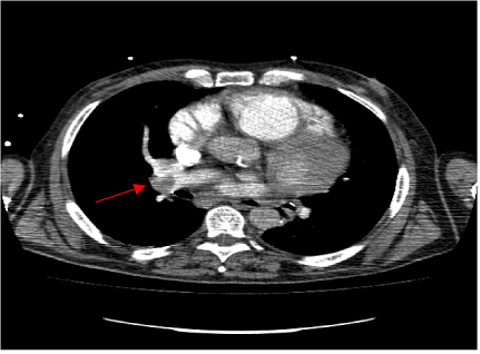
| Using mechanical dilator sheaths, the lead was removed, but the tip was fractured and migrated to the accessory hemiazygos vein. The patient was hemodynamically stable during the procedure. With a new provisional pacemaker, he was admitted to the Intensive Care Unit for monitoring. One hour later, he developed severe hypotension and desaturation with respiratory failure that required mechanical ventilation. Computed Tomography (CT) was carried out, showing an embolism in the right pulmonary artery due to the migration of the big vegetation (Figure 2). Prophylactic anticoagulation, antibiotic treatment, hemodynamic and vital support measures were established. The patient improved clinically over the next 24 hours and eventually recovered completely. One week later, a new single lead VDD pacemaker was implanted and the patient was discharged. |
| Discussion |
| The reported incidence after permanent endocardial pacemaker implantation varies in the literature from 1% to 7% [1-3]. CD extraction can be performed percutaneously without need for surgical intervention in the majority of patients. Guidelines on the prevention, diagnosis, and treatment of infective endocarditis (new version 2009) of the European Society of Cardiology recommend surgical extraction in patients with very large vegetations (>25 mm), owing to the high risk of septic pulmonary embolism resulting from vegetation displacement during percutaneous extraction [3-5]. However, these episodes are said to be commonly asymptomatic, and percutaneous extraction remains the recommended method even in cases of large vegetations in many publications [4-13]. Following that line of thought and taking into account that mortality associated with surgical removal is higher [6-13] especially in elderly patients with associated comorbidities, we decided a percutaneous lead extraction. |
| Treatment of a septic embolism includes antimicrobial therapy that has to range from 4 to 8 weeks, embolectomy if indicated and managing the possible complications associated such as haemoptysis. Trying an interventional solution like embolectomy was proposed but conservative measures were finally decided. In a few hours the patient was fully recovered which indicates that the vegetation splitted up; some authors have postulated that emboli from lead vegetations are of minimal consequence because they are friable, as compared with the solid form of venous trombi [5,10,11]. |
| In conclusion, percutaneous removal of infected pacemaker leads is an alternative to cardiac surgery even in larger vegetations (>15 mm in largest diameter). An application of this technique in large vegetations carries the risk of embolism, which highlights the importance of selecting the cases, individualizing risk-benefit ratio compared with other alternatives. Literature offers evidence that this complication is rare, usually asymptomatic and even when the episode presentation is symptomatic, the long-term prognosis of these patients after removal of pacemaker and antibiotic therapy could be excellent [12,13]. |
References |
|
Figures at a glance
 |
 |
| Figure 1 | Figure 2 |
Relevant Topics
- Abdominal Radiology
- AI in Radiology
- Breast Imaging
- Cardiovascular Radiology
- Chest Radiology
- Clinical Radiology
- CT Imaging
- Diagnostic Radiology
- Emergency Radiology
- Fluoroscopy Radiology
- General Radiology
- Genitourinary Radiology
- Interventional Radiology Techniques
- Mammography
- Minimal Invasive surgery
- Musculoskeletal Radiology
- Neuroradiology
- Neuroradiology Advances
- Oral and Maxillofacial Radiology
- Radiography
- Radiology Imaging
- Surgical Radiology
- Tele Radiology
- Therapeutic Radiology
Recommended Journals
Article Tools
Article Usage
- Total views: 13771
- [From(publication date):
October-2013 - Jul 18, 2025] - Breakdown by view type
- HTML page views : 9222
- PDF downloads : 4549
