Case Report Open Access
Complicated Pott’s Puffy Tumour Involving both Frontal and Parietal Bones: A Case Report
| Jude-Kennedy C Emejulu* and Ofodile C Ekweogwu | ||
| NnamdiAzikiwe University/Teaching Hospital, Nnewi, Anambra State, Nigeria | ||
| Corresponding Author : | Jude-Kennedy C Emejulu Nnamdi, Azikiwe University/Teaching Hospital Nnewi, Anambra State, Nigeria Tel: +234-803-328-3 E-mail: judekenny2003@yahoo.com |
|
| Received August 04, 2014; Accepted September 9, 2014; Published September 16, 2014 | ||
| Citation: Emejulu JC, Ekweogwu OC (2014) Complicated Pott’s Puffy Tumour Involving both Frontal and Parietal Bones: A Case Report. J Infect Dis Ther 2:166. doi:10.4172/2332-0877.1000166 | ||
| Copyright: © 2014 Emejulu JC, et al. This is an open-access article distributed under the terms of the Creative Commons Attribution License, which permits unrestricted use, distribution, and reproduction in any medium, provided the original author and source are credited. | ||
Related article at Pubmed Pubmed  Scholar Google Scholar Google |
||
Visit for more related articles at Journal of Infectious Diseases & Therapy
Abstract
Pott’s puffy tumour is a rare clinical condition, especially in this antibiotic era. It presents as frontal skull osteomyelitis with resultant frontal subperiosteal abscess, arising usually from frontal sinusitis. Other extracranial regions may also be unusually involved in addition to intracranial, periorbital and intra-orbital extensions, with various clinical manifestations, including neurological deficits, and potentially lethal complications. Its potential to cause significant morbidity emphasizes the need for a high index of suspicion in the setting of frontal scalp swellings in order to facilitate optimal outcome. This report, which is the case of a 7-year old male sickle cell anaemia patient with Pott’s puffy tumour and unusual involvement of both parietal bones, in addition to the usual frontal bone affectation, serves to highlight the need for a thorough evaluation of patients with hemoglobinopathies and skull bosselations, in order not to miss this potentially lethal disease.
| Keywords |
| Antibiotics; Osteomyelitis; Sickle cell anaemia; Subperiosteal abscess; Surgery |
| Introduction |
| Pott’s puffy tumour is characterized by frontal skull osteomyelitis with associated subperiosteal abscess [1-6]. Unusually, other extracranial regions may be involved [7,8]. It can also spread to the intracranial, periorbital and intraorbital regions [1-4,6-12]. Sir Percival Pott first described this lesion in 1760 as a “puffy, circumscribed, indolent tumour of the scalp and the spontaneous separation of the pericranium from the scalp under such tumour” with associated frontal bone osteomyelitis following head injury. Subsequently, in 1775, he described a second case resulting from frontal sinusitis [2,5-8,11]. |
| This condition is now rare with the advent of antibiotics [2,4-6,8,10,13-16]. Common aetiological factors include direct spread of frontal sinusitis and frontal trauma. Rarer causes such as mastoiditis, insect and animal bites, frontal tumour, and intranasal cocaine use have also been implicated [2,7,14]. With an intracranial extension, the clinical manifestations progressively became severe with attendant neurological deficits. |
| Cranial computed tomographic scanning is the radiological investigation of choice because it delineates extracranial and intracranial soft tissue lesions in addition to the associated bony lesions. Cranial magnetic resonance imaging can further elucidate intracranial lesions, and technetium-99m scan, and gallium-67 scan can also be employed [6,15]. |
| Treatment of Pott’s puffy tumour is an urgent surgical intervention with a long period of adjuvant antibiotic therapy. Surgery involves incision and drainage of the abscess with debridement of the osteomyelitic bone and devitalized soft tissues. Associated intracranial collections are also evacuated and debrided. Endoscopic approaches have also been described [2,6,15,17]. Antibiotics are concurrently administered parenterally in high doses and on a long term basis, usually for 4-6 weeks. |
| Pott’s puffy tumour is associated with significant morbidity and complications such as intracranial sepsis, cerebral vein thrombosis, seizures, focal neurological deficits, chronic calvarial osteomyelitis. Mortality has significantly reduced from as high as 60% in the preantibiotic era to as low as 0-3.5% in the antibiotic era [1]. |
| We, hereby, report a case of a 7-year old male sickle cell anaemia patient with a rare presentation of Pott’s puffy tumour involving both the frontal and parietal bones bilaterally, with intracranial and neurological involvement, as well. He was managed operatively with adjuvant antibiotic therapy and recovered fully without neurological deficits. |
| Case Report |
| The patient was a 7-year old right handed male, known sickle cell anaemia patient, referred to our service in March 2014, with complaints of fever of 5 weeks’ duration, multiple scalp swellings of 4 weeks’ duration, and right sided body weakness of 3 weeks’ duration. There were associated headaches and vomiting, but no visual disturbance, seizure or sphincteric dysfunction. The history was suggestive of a preceding upper respiratory tract infection, but none of head trauma, ear infection, intranasal medications, head surgery or malignancy in any part of his body. Prior to his presentation at our hospital, he was treated with oral herbal medications, and following the worsening clinical condition he was brought to our paediatric service which subsequently referred him to our neurosurgery unit. |
| On clinical examination, his vital signs were normal, his Glasgow consciousness score (GCS) was 15 and mental status was grossly intact. The pupils were 4 mm bilaterally and briskly reactive to light, but there was a right supranuclear facial paresis and right abducens palsy, but no sign of meningeal irritation. The muscle bulk was normal but tone was increased globally with a right hemiparesis. There was bilateral flexor plantar response and tendon stretch reflexes were exaggerated globally. Sensory, cerebellar and autonomic functions were normal. On the scalp there were bilateral frontal and parietal non-tender, fluctuant scalp swellings with no differential warmth, left frontal punctate defect with exudation of pus and bilateral upper posterior cervical (non-tender) lymphadenopathies (Figure 1). |
| A clinical diagnosis of Pott’s puffy tumour, with possibly, left intracranial abscess was made. |
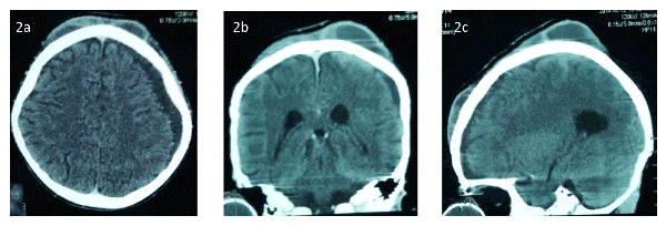
| Cranial computed tomographic (CT) scan showed three extracranial soft tissue cystic collections with peripheral contrast enhancement (bifrontal and biparietal in location), left fronto-parietal subdural collection with no midline shift, but with subtle enlargement of the ventricles (Figure 2). |
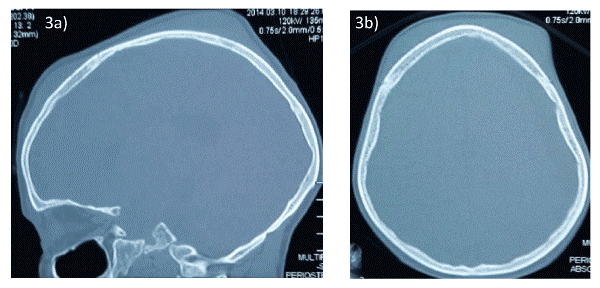
| The bone windows of the CT scan showed only mild bi-frontal cranial bony involvement with no bony destruction; and the paranasal sinuses appeared not to be well developed, possibly, due to the young age of the patient and the sickle cell haemoglobin status (Figure 3). |
| The HIV I and II screening was non-reactive, haemoglobin concentration was 8.3 g/dl, packed cell volume was 23.9%, total white blood cell count was 8.6x10³/µL with 48% lymphocytosis, and other haematological and biochemical laboratory reports were normal. |
| He underwent incision and drainage of the scalp abscesses, bilateral frontal and parietal craniectomy and debridement, and evacuation of the left fronto-parietal subdural empyema. |
| Intraoperative findings included |
| •Multiple bifrontal and biparietal scalp bosselations with a pointing left frontal sinus |
| •Associated subpericranial abscesses/soft tissue inflammation |
| •Chronic bilateral calvarial osteomyelitis with cloacae, sequestrum and involucrum |
| •Deep amber left fronto-parietal subdural collection under marked pressure |
| The scalp incisions were tagged with prolene 3-0 sutures and the wound beds were packed with honey-soaked gauze (Figures 4a-4c). Subsequent daily honey-soaked gauze wound packing and dressings were done and intravenous broad spectrum antibiotics (ceftriaxone, gentamycin and metronidazole), were administered. |
| Culture of the left parietal soft tissue specimen yielded Staphylococcus aureus sensitive to ceftriaxone and gentamycin which the patient was already receiving. Cultures of other soft tissue specimen and pus were sterile. The right hemiparesis progressively and completely resolved. |
| On the 9th day postoperatively, delayed primary wound closure of the bilateral frontal and parietal scalp incisions were done. Alternate wound sutures and later, all the sutures were removed within the week with good wound healing, while the patient was continued on intravenous antibiotics for a total of 4 weeks and he was discharged in good health without residual deficits. His out-patient review was 4 weeks later, and he still had no residual neurological deficits. |
| Discussion |
| Pott’s puffy tumour is a rare clinical condition, especially with the advent of antibiotics [2,4-6,8,10,13-16]. The classical lesions of skull osteomyelitis and subperiosteal abscess usually involve the frontal region, but on rare occasions there may be involvement of other extracranial regions and/or associated intracranial and intraorbital extensions [1-4,6-12]. The index patient also had bilateral parietal and left fronto-parietal subdural involvements in addition to the bilateral frontal lesion though the intraorbital cavities and structures were spared. The biparietal occurrence is a very rare event which has not been reported before from Nigeria, and rarely from elsewhere [8]. |
| Pott’s puffy tumour affects all age groups and is more common in males of all ages (range of 2-83 years in literature) and a male: female ratio of 9:1 reported in literature, though, it has a predilection for preteen and teenage males, the age group of the index patient [15]. |
| Frontal sinusitis and trauma are the most common aetiological factors and our patient had a history suggestive of upper respiratory tract infection (URTI) which may have predisposed him to Pott’s puffy tumour. But, beyond the URTI, which could well be a red herring, his sickle cell anaemia status, offered a predilection for infections, not least, osteomyelitis of the skull, and a combination of these two conditions could more readily explain the origin of this present case. Spread of the infection could also result directly from the bony involvement or through the venous drainage of the frontal sinus to both contiguous and distant regions. |
| The cranial computed tomographic scan findings were diagnostic in this case and also assisted in identifying the subdural empyema in order to facilitate a comprehensive definitive treatment. Intravenous antibiotic treatment for 4-6 weeks is the recommended adjuvant treatment after surgical excision and evacuation. Also, a culture of the specimen from the patient yielded growths of Staphylococcus aureus in keeping with literature reports [6-8,17]. Previous antibiotic use may be responsible for the sterile cultures of some of the specimens, though gram-negative anaerobic cultures could have supported this contention if they were done. |
| We could not readily access any facilities for anaerobic culture in our services. More so, in the Nigerian health care system, run on “cash-and-carry” basis, the patient’s parents, who were small scale traders of nuts and screws for motorcycle spare parts, could barely afford the cost of the surgery and four weeks of postoperative intravenous antibiotic treatment, and so, we decided to withhold a further chase for anaerobic culture. |
| At the time the patient was discharged from the hospital, there were still a few spots of underlying cranial defects at the sites of the craniectomy, but no gross neurological deficits. At the age of 7 years, we decided to follow up on the cranial defects in the coming years, reserving a decision on cranioplasty till later in his follow up, if the need ever arises. |
| Conclusion |
| This was a rare form of Pott’s puffy tumour with a biparietal bone involvement in association with the more usual frontal bony location. On a background of sickle cell anaemia, this report makes a call for a higher index of suspicion by clinicians who manage patients with haemoglobinopathies, because skull bone bosselations may not ordinarily be reckoned with the possibility of harbouring an underlying bacterial inoculation and osteomyelitis. |
References
- Skomro R, McClean KL (1998) Frontal osteomyelitis (Pott's puffy tumour) associated with Pasteurellamultocida-A case report and review of the literature. Can J Infect Dis 9: 115-121.
- Suwan PT, Mogal S, Chaudhary S (2012) Pott's Puffy Tumor: An Uncommon Clinical Entity. Case Rep Pediatr 2012: 386104.
- Feder HM Jr, Cates KL, Cementina AM (1987) Pott puffy tumor: a serious occult infection. Pediatrics 79: 625-629.
- Masterson L, Leong P (2009) Pott's puffy tumour: a forgotten complication of frontal sinus disease. Oral MaxillofacSurg 13: 115-117.
- McDermott C, O'Sullivan R, McMahon G (2007) An unusual cause of headache: Pott's puffy tumour. Eur J Emerg Med 14: 170-173.
- Jung J, Lee HC, Park IH, Lee HM (2012) Endoscopic Endonasal Treatment of a Pott's Puffy Tumor. ClinExpOtorhinolaryngol 5: 112-115.
- Schwartz RH, Zingariello C, Levorson R, Myceros JS, Kamat R, et al. (2013)Pott puffy tumour in an unusual location. OJPed3: 147-150.
- Evliyaoglu C
- , Bademci G, Yucel E, Keskil S (2005) Pott's puffy tumor of the vertex years after trauma in a diabetic patient: case report. Neurocirugia (Astur) 16: 54-57.
- Domville-Lewis C, Friedland PL, Santa Maria PL (2013)Pott’s puffy tumour and intracranial complications of frontal sinusitis in pregnancy. J LaryngolOtol 1: S35-S38.
- Karaman E, Hacizade Y, Isildak H, Kaytaz A (2008) Pott's puffy tumor. J CraniofacSurg 19: 1694-1697.
- Morley AM (2009) Pott's puffy tumour: a rare but sinister cause of periorbital oedema in a child. Eye (Lond) 23: 990-991.
- Durur-Subasi I, Kantarci M, Karakaya A, Orbak Z, Ogul H, et al. (2008) Pott's puffy tumor: multidetector computed tomography findings. J CraniofacSurg 19: 1697-1699.
- Emejulu JKC, Iloabachie IBC (2010)Pott’s puffy tumour – Report of a grotesque case. Annals of Neurosurgery 10:1-4.
- Khan MM, Khwaja S, Bhatt YM, Karagama Y (2014) Pott’s Puffy Tumour Complicating Frontal Sinus Osteoma – A Case for Combined Approach Surgery. Otolaryngology 4: 152-154.
- Shehu BB, Mahmud MR (2008) Pott's puffy tumour: a case report. Ann Afr Med 7: 138-140.
- Monteiro M, Castillo R, Andre C, Santos M, Antunes L, et al. (2007)Pott’s puffy tumour, a rare complication of frontal sinusitis. Fr ORL 93: 350-352.
Figures at a glance
 |
 |
 |
 |
|||
| Figure 1 | Figure 2 | Figure 3 | Figure 4 |
Relevant Topics
- Advanced Therapies
- Chicken Pox
- Ciprofloxacin
- Colon Infection
- Conjunctivitis
- Herpes Virus
- HIV and AIDS Research
- Human Papilloma Virus
- Infection
- Infection in Blood
- Infections Prevention
- Infectious Diseases in Children
- Influenza
- Liver Diseases
- Respiratory Tract Infections
- T Cell Lymphomatic Virus
- Treatment for Infectious Diseases
- Viral Encephalitis
- Yeast Infection
Recommended Journals
Article Tools
Article Usage
- Total views: 15736
- [From(publication date):
October-2014 - Apr 05, 2025] - Breakdown by view type
- HTML page views : 11156
- PDF downloads : 4580
