Comparison between the Singular Action and the Synergistic Action of Therapeutic Resources in the Treatment of Knee Osteoarthrosis in Women: A Blind and Randomized Study
Received: 22-Apr-2019 / Accepted Date: 01-May-2019 / Published Date: 07-May-2019 DOI: 10.4172/2165-7025.1000411
Abstract
Osteoarthritis is a degenerative joint disease that promotes chronic and joint decrease, which is prevalent in the elderly. There are several therapeutic approaches for the treatment of pain, including therapeutic ultrasound and laser therapy, therapeutic resources are not included in the treatment of this disease. The aim of this study was to distinguish between a single application of the laser therapy/therapeutic ultrasound resources in relation to the effect of the synergistic applications of the same resources in patients with knee osteoarthrosis. Thirty patients were selected, being 10 patients per group, randomly distributed. In addition, patients did not have access to the form of a course, characterizing a blind study. Patients were assessed by Western Ontario and McMaster University Osteoarthritis Index (WOMAC) for osteoarthritis of the knee. All patients completed the Visual Analogue Scale. Analogue scale, fossil of greater abnormality, larger or smaller wall, conjugated according to natural resources. A greater percentage difference was observed in the comparison between laser versus ultralaser (50%) and therapeutic ultrasound versus ultralaser (130%), when compared to functionality (WOMAC). When comparing the Visual Analogue Scale, a greater reduction of pain was observed by the combined use of resources in relation to the resources singularly, for both the right and left knees. The results showed that synergic therapy was more efficient in the treatment of knee osteoarthrosis in relation to the use of the therapies alone, in relation to both joint function and pain. The use of new technologies provides a new treatment approach, without the use of drugs and non-invasively, allowing a better quality of life of patients with this chronic and degenerative pathology.
Keywords: Osteoarthritis; Laser therapy; Therapeutic ultrasound; Ultralaser
Introduction
Osteoarthritis is an irreversible degenerative joint disease, which leads to pain and loss of joint function. It can be defined as a condition characterized by focal areas of loss of articular cartilage in the synovial joints and affects about 20% of the world population. Between the age group of 40 to 60 years a linear increase of the degeneration happens, and with the increasing life expectancy, will result in an increase of osteoarthrosis; worldwide estimates of diseases show that by 2030 the prevalence for symptomatic osteoarthritis is 30%. The knee joint is among the three most commonly affected joints, along with hand and hip joints, being more common in females [1,2].
Treatments for osteoarthritis include pharmacological therapies such as analgesics and anti-inflammatories, as well as nonpharmacological therapies such as stretching and muscle strengthening, improving physical fitness and reducing pain, thereby increasing quality of life. They also include as conventional treatments the use of laser therapy and therapeutic ultrasound for the purpose of reducing pain due to its analgesic and anti-inflammatory effect [1-3].
In recent studies carried out by the research group of the Institute of Physics of São Carlos, University of São Paulo, it was observed that the prototype of the device that simultaneously emits ultrasonic waves and laser of low intensity, allows the potentialization of the effects of produced cavitation by the therapeutic ultrasound, as well as the enzymatic and mitochondrial modulation produced by the low power laser, obtaining a greater anti-inflammatory effect, analgesic of both resources and thus allowing the stabilization of cartilage degeneration, improving the quality of life of the patient [3-5].
The purpose of the study was to evaluate the differences in the different treatments of laser therapy, therapeutic ultrasound and ultralaser (therapeutic ultrasound conjugated to laser therapy) in patients with osteoarthritis of the knee, evaluated by the functionality by Western Ontario and McMaster Universities Osteoarthritis Index (WOMAC) and pain level by Visual Analogue Scale (VAS).
Materials and Methods
It was used a prototype of laser and ultrasound, where the device had a project and support carried out by the Laboratory of Technological Support of the Institute of Physics of São Carlos, University of São Paulo. This prototype allows the emission of laser and ultrasound simultaneously, conditioning the overlap of both fields, ultrasonic and luminous, and contributing to the synergistic therapeutic effect.
Patients and evaluation
For the study, 30 female patients, average age of 45 years old (45 ± 4.5) and diagnosed with osteoarthrosis of both knees, were selected. As exclusion factors for the project were considered pre-existence of other degenerative, autoimmune diseases and fibromyalgia. Infiltration treatment was also considered as exclusion factors in a period of three months prior to the beginning of the project. Patients were assessed using the Western Ontario and McMaster Universities Osteoarthritis Index (WOMAC) Questionnaire for knee osteoarthritis. All patients were evaluated before and after each session. The Visual Analogue Scale (VAS) was used as a way of measuring the level of pain.
Intervention protocol
The intervention protocol was applied twice a week, totaling 8 sessions. The groups were randomly divided into Laser (n=10), ultrasound (n=10) and Ultralaser (n=10). The Laser group used the parameters: wavelength 660 nm, continuous, 100 mW and power density of 60 mW/cm², application time of 8 minutes for each knee. The Ultrasound group uses the parameters: ultrasound pulsed, frequency 1 MHZ, 100 Hz, 50% of the work cycle and average spatial mean time (SATA) of 0.5 W/cm², application time of 8 minutes for each knee. The Ultralaser group used the parameters: ultrasound: pulsed, frequency 1 MHz, 100 Hz, 50% of the work cycle and average spatial mean time (SATA) of 0.5 W/cm²; Laser: wavelength 660 nm, 100 mW and a power density of 60 mW/cm². Application time: 8 minutes for each knee.
To characterize the blind study, the patients were not informed about their intervention group.
Statistical Analysis
Statistical analysis was performed using Instat 3.0 software for Windows 7 (Graph Pad, USA, 1998). All data were expressed as mean and standard deviation. The level of significance was set at p<0.05. The Kolmogorov-Smirnov test was used to analyze the normality of the data. Subsequently, a one-way ANOVA with a post-test was performed, using Student-Newman-Keuls for parametric data in comparison between protocols and post-hoc-Tukey-Kramer for comparison between therapeutic resources.
Results
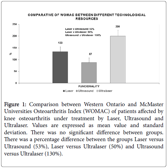
The results obtained through the evaluation of WOMAC (Figure 1), show the comparison between different technological resources used in the study. It is possible to observe the comparisons between them, but there is no significant difference found. The observed comparison between the laser and X-ray groups showed greater functionality obtained by the patient of the laser group in relation to the ultrasound group, in 53%. When compared to the laser group versus the ultralaser group, the ultrasonic laser and ultrasound treatment performed by the ultralaser provided greater functionality in this group of patients compared to the laser group in 50%. Furthermore, when comparing the ultrasound and ultralaser groups, the functionality achieved by the patients with laser and ultrasound treatment in a conjugated way reached 130% greater than the ultrasound group.
Figure 1: Comparison between Western Ontario and McMaster Universities Osteoarthritis Index (WOMAC) of patients affected by knee osteoarthritis under treatment by Laser, Ultrasound and Ultralaser. Values are expressed as mean value and standard deviation. There was no significant difference between groups. There was a percentage difference between the groups Laser versus Ultrasound (53%), Laser versus Ultralaser (50%) and Ultrasound versus Ultralaser (130%).
Table 1 shows the visual analogue pain scale observed in initial and final evaluations, in the right and left knees. It was observed that the actions of both technologies were efficient for the reduction of patients' pain, according to the analogous visual scale, indicating a significant difference. However, the observed action of the ultralaser (p<0.001) was better than the action observed by the laser action (p<0.006) and the ultrasound (p<0.006) in relation to the right knee in the comparison between initial and final values.
| Right Knee | Left Knee | |||||
|---|---|---|---|---|---|---|
| Pre-Treatment | Post-Treatment | p | Pre-Treatment | Post-Treatment | p | |
| Laser | 5.75 | 2.5 | 0.006 | 5 | 2 | 0.05 |
| Ultrasound | 6.4 | 2.6 | 0.006 | 5 | 2.4 | 0.04 |
| Ultra-laser | 8.6 | 2 | 0.001 | 8.3 | 4 | 0.003 |
Table 1: Comparison between the initial and final values of the Visual Analogue Scale (EVA) of patients affected by osteoarthritis of the knee under treatment by Laser, Ultrasound and Ultralaser. Values are expressed as mean value and standard deviation. There was a significant difference for the Laser (p<0.001), Ultrasound (p<0.006) and Ultralaser groups (p<0.001), when the directional effort was observed in relation to the initial versus final. We found significant devaluation for the Laser (p<0.05), Ultrasound (p<0.04) and Ultralaser groups (p<0.003), when the initial and final values were found.
Likewise, the action of the therapeutic resources on the patients' left knee was more efficient in the ultralaser group (p<0.003) than in the laser (p<0.05) and ultrasound (p<0.04) groups. Representative table of initial and final pain values visual analogue scale
Discussion
Osteoarthrosis of the knee is an inflammatory and degenerative disease that results in the destruction of the articular cartilage and may lead to a deformity of the joint. The etiology of the degenerative process is complex, it begins with aging, however during life several factors that can cause natural degeneration may arise, as well as inflammatory, infectious diseases, and traumas that degenerate the cartilage. The structure of cartilage and the inflammatory aspects of the degenerative process have been studied and recent advances have shown that the resolution of knee osteoarthrosis could be by therapeutic resources and by surgical procedures [1,6].
Among the therapeutic technologies is the ultrasound. This technology is a mechanical waveform that, when used in the pulsed mode, provides several therapeutic effects, such as analgesia, stimulation of the anti-inflammatory response, chondrocyte proliferation and cartilage growth, as well as increasing vascularization and collagen synthesis leading tissue repair. Still as therapeutic technology is the low intensity laser. Photobiomodulation is characterized by electromagnetic waves that induce the occurrence of modulatory and stimulating actions, contributing to increased blood flow, reduction of edema and increased supply of tissue oxygen, resulting in pain relief and tissue regeneration [6,7] . Previous studies conducted by this research group show that the synergistic application of ultrasound and laser offers the potential of these benefits, thus showing a new therapeutic approach for osteoarthrosis, significantly improving patients' quality of life [8,5].
In the present study, we observed the potentialization of these results observed in Figure 1, where the action of the ultralaser was superior to the isolated action of the ultrasound and laser therapeutic resources. The potentiated action of these resources, through the cavitational action of the ultrasound in synergy with the laser light action, provided greater analgesia and anti-inflammatory action. In this way, it is possible, in Figure 1, to verify the great increase in the functionality of the knee joint in a patient affected by osteoarthrosis. Also, in relation to the Visual Analogous Scale (VAS), Table 1, we observed that the action of the ultralaser was more positive for pain reduction in both knees, in relation to the groups of patients treated with the technologies in isolation.
In theory, the synergistic effect of ultrasound and low-intensity laser was evidenced as the best approach for analgesic and antiinflammatory modulation in relation to the use of individual therapy. The stimulation of cartilage growth and the proliferation of chondrocytes that occurs by the mechanical effect of ultrasound, causes analgesia and anti-inflammatory action, with no significant heat production. The extensibility of collagen tendons, ligaments and contracture of the joint capsule and connective tissue are structural effects that can occur [3].
Ultrasonic waves in pulsed mode produce longitudinal vibration that result in negative pressure on the cells, causing cellular and biomolecular changes bringing as a benefit increased cellular metabolism and greater fluidity of blood in the blood vessels. Consequently, there is an increase in temperature and supply of cellular oxygenation. The additional heating may convert to electrical and mechanical energies inside the cells, adding the molecular movement. The physical effects promote the permeability of the cell, ion exchange through the cell membrane and the synthesis of proteins resulting in increased tissue repair. Such alterations contribute to the analgesic action regulating the thermodynamic mechanism related to mechanoreceptors and pain [3].
The effect of photo-biomodulation stimulates the proliferation of chondrocytes promoting cell regeneration and the extracellular matrix performs synthesis and secretion, favoring increased blood flow, decreasing edema and improving tissue oxygenation, aiding in physiological mechanisms that depend on photochemical actions at the cellular level, increasing the release of neurotransmitters such as serotonin that are involved in pain modulation [3].
In this way, the present study showed the efficiency of the synergic performance of the ultrasound and low intensity laser therapeutic resources, increasing the joint function and consequently reducing pain. The sum of these factors enabled not only numerically but also visually the improvement of the patient's quality of life.
Conclusion
Based on the evidences found in the present study, it is explicit that the use of therapeutical ultrasound and laser therapy resources, when applied in a conjugated way significantly potentiates the effects when compared to the use of the individual therapy, greatly reducing the inflammatory picture and pain relief, increasing mobility and functionality, providing a significant increase in quality of life.
References
- Thomas AC, Hubbard-Turner T, Wikstrom EA, Palmieri-Smith RM (2017) Epidemiology of Posttraumatic Osteoarthritis. J Athl Train 52: 491-496.
- Pereira D, Peleteiro B, Araújo J, Branco J, Santos RA, et al. (2011) The effect of osteoarthritis definition on prevalence and incidence estimates: a systematic review. Osteoarthritis Cartilage 19: 1270-1285.
- De Souza Simão ML, Fernando AC, Cesarino RL, Zanchini AL, Ciol H, et al. (2018) Sinergic Effect of Therapeutic Ultrasound and Low-Level Laser Therapy in the Treatment of Hands and Knees Ostheoarthritis. J Arthritis 7: 277.
- Jorge AES, Simão MLd, Fernades AC, Chiari A, de Aquino Jr AE, et al. (2018) Ultrasound conjugated with Laser Ðerapy in treatment of osteoarthritis: A case study. J Sports Med Ðer 3: 024-027.
- Paolillo AR, Paolillo FR, João JP, João HA, Bagnato VS (2015) Synergic effect of ultrasound and laser on the pain relief in women with hand osteoarthritis. Lasers Med Sci 30: 279-286
- Camanho GL (2011) Treatment of knee osteoarthritis. Revista Brasieleira de Ortopedia 36: 135-140.
- Vascocelos Y (2015) Two in one: device uses ultrasound and laser simultaneously to rehabilitate patients with arthrosis (Testimony to Yuri Vasconcelos). Pesquisa FAPESP 229: 76-77.
- Paolillo FR, Paolillo AR, João JP, Frascá D, Duchêne M, et al. (2018) Ultrasound plus low-level laser therapy for knee osteoarthritis rehabilitation: a randomized, placebo-controlled trial. Rheumatol Int 38: 785-793.
Citation: de Souza Simão ML, Fernandes AC, Ferreira KR, de Oliveira LS, Mário EG, et al. (2019) Comparison between the Singular Action and the Synergistic Action of Therapeutic Resources in the Treatment of Knee Osteoarthrosis in Women: A Blind and Randomized Study. J Nov Physiother 9: 411. DOI: 10.4172/2165-7025.1000411
Copyright: © 2019 de Souza Simão ML, et al. This is an open-access article distributed under the terms of the Creative Commons Attribution License, which permits unrestricted use, distribution, and reproduction in any medium, provided the original author and source are credited.
Share This Article
Recommended Journals
Open Access Journals
Article Tools
Article Usage
- Total views: 3276
- [From(publication date): 0-2019 - Apr 02, 2025]
- Breakdown by view type
- HTML page views: 2411
- PDF downloads: 865

