CNS Abnormalities at 11-14 Weeks
Received: 26-Oct-2017 / Accepted Date: 21-Nov-2017 / Published Date: 29-Nov-2017 DOI: 10.4172/2572-4983.1000141
Introduction
The implementation of the routine ultrasound assessment in obstetric practice both as a diagnostic as well as a screening tool in the management of the antenatal care, has proved its benefits in the detection of fetal anomalies with great medical, socio-economical and psychological impact. The two components of the fetal imaging: screening and diagnosis need to be very well defined as the latter require a higher level of expertise and appropriate sonographic equipment, especially for cases difficult to imagine, as the first trimester malformations [1,2]. Nevertheless, these two terms should live in a perfect symbiosis, as the development of one of them causes the progress of the other, in favour of the patient and of a healthy society. The late improvements in technology has enabled the health providers to lower the timing of the fetal morphologic assessment during pregnancy and made possible the detection of the fetal disorders earlier than before [1,2]. Its utility in counselling the couple is incontestable, and offers them the possibility to legally terminate the pregnancy in cases with severe fetal anomalies or scheduling the delivery in a centre that fulfils the needs of their pathology in curable cases, with good outcomes and significant expenditure decrease.
Importance
Central nervous system (CNS) malformations are important, because they are the largest group of fetal abnormalities, more prevalent than trisomy 21 and similar to congenital heart diseases, accounting for more than 10 cases in 1000 births [3,4]. They also represent an important factor of morbidity and mortality among neonates and children, as some of these disorders become symptomatic only in early infancy. Their detection is important because of the high degree of gravity with modest postnatal treatment and possibilities of recovery and due to their disabling evolution with medical, social and economic implications.
Limitations of the First Trimester Scan
Although the ultrasound screening for brain anomalies is worldwide performed at 19-22 weeks of gestational age because of the continuous development of brain structures by mid-pregnancy, further attempts are made to decrease the detection age for some CNS malformations as much as possible and even to the 11-13 weeks, when routinely, a morpho-genetic scan is recommended. Still there are important limitations in the FT evaluation. First, most of the CNS abnormalities are undetectable or associate only subtle findings in early gestation, as the brain continues to develop during pregnancy and after birth. There are a small number of brain structures that can be assessed at this gestational age, as the appearance of the brain is much different in later stages of pregnancy, due to its later development and differentiation during the second trimester. Therefore, a skilled sonographer needs a thorough knowledge of the embryological development of the fetus. Some disorders such as neural migration, proliferation and organization, as well as acquired lesions like haemorrhage and tumors occur in the late second and even in the third trimester, and these anomalies cannot be suspected during the previous fetal evaluations [5,6]. Agenesis of corpus callosum, microcephaly, hydrocephaly, lissencephaly, cysts, posterior fossa abnormalities usually are apparent only in late stages. Some other abnormalities represent the consequence of acquired prenatal or perinatal insults: infections, hemorrhage or hypoxia.
A second limitation is related to the fact that the extensive assessment of the fetal anatomy at the FT scan necessitates appropriate training, equipment and increased examination time, which means financial resources, that health care systems are not yet ready to provide [4,7,8].
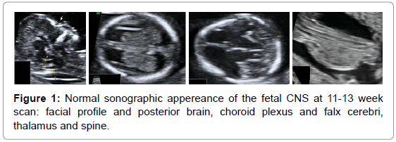
What is the usefulness and effectiveness of the 11-13 weeks’ scan in CNS abnormalities’ detection, what can we detect and how? There are strong arguments in favour for CNS early assessment. We have this opportunity/obligation to examine the fetus at the end of the FT since there is strong evidence toinvert the pyramid of prenatal care and to look for major fetal abnormalities at the end of the first trimester [9]. There is no need for further investments in training, equipment and time, because the CNS parameters are easily assessed during the standard examination, with no additional scanning time (Figure 1).
We should not forget the advantages of an earlier detection, which is safer, with less emotional stress and less economic costs [10]. Indeed, most of the CNS MA are indeed undetectable, but the most important CNS congenital anomalies concerning prevalence and severity are usually detectable in the first trimester: holoprosencephaly and neural tube defects (Table 1) [11-22].
| Fetal abnormality | First trimester detection [nr, %] | |
|---|---|---|
| Neural tube | 81/116 | 69.82% |
| Acrania/iniencephaly | 63/64 | 98.44% |
| Encephalocele | 3/3 | 100.00% |
| Open spina bifida | 12/42 | 28.57% |
| Hemivertabrae | 2/4 | 50.00% |
| Sacrococcygeal teratoma | 1/3 | 33.33% |
| Brain | 25/83 | 30.12% |
| Microcephaly | 0/2 | 0.00% |
| Craniosynostosis | 0/2 | 0.00% |
| Corpus callosum agenesis | 0/11 | 0.00% |
| Ventriculomegaly | 8/36 | 15.15% |
| Holoprosencephaly | 11/13 | 84.61% [alobar 100%] |
| Cerebellar hypoplasia | 5/13 | 38.46% |
| Vermian agenesis | 1/5 | 20.00% |
| Porencephaly | 0/1 | 0.00% |
Table 1: Detection of CNS abnormalities. Analysis after Hernadi, et al. [11], Bilardo, et al. [12], D’Ottavio, et al. [13], Whitlow, et al. [14], Chen, et al. [15], Taipale, et al. [16], Cedergren, et al. [17], Dane, et al. [18], Chen, et al. [19], Oztekin, et al. [20], Syngelaki, et al. [21], Iliescu, et al. [22].
Due to the early stages of calvarium ossification, fetal brain structures are easily seen and a certain number of CNS anomalies can be detected during this period [11-13]. Anomalies like anencephaly and holoprosencephaly can be confidently diagnosed at the end of the first trimester (Table 1), because the respective defective structures, as calvaria and falx cerebri are already well seen.
Studies show a detection rate between 66 and 84% for major CNS abnormalities in the first trimester (Table 2).These figures makes early CNS scan efficient, because ISUOG guidelines state that the most optimistic 2nd trimester detection rates report 80% of the major abnormalities detected [4,19,22,23].
| Study | Year | Patients | First trimester detection |
|---|---|---|---|
| D’Ottavio et al. [13] | 1997 | 3.514 | 4/6 (66%) |
| Whitlow et al. [14] | 1999 | 6.443 | 16/19 (84.2%) |
| Chen et al. [15] | 2008 | 7.642 | 7/9 (77.8%) |
| Iliescu et al. [22] | 2013 | 5.472 | 16/23 (68.6%) |
Table 2: Detection rates for CNS abnormalities at 11-13 weeks scan [13,14,19,22].
Why this small difference? Perhaps the answer is that the most prevalent major abnormalities are detectable early in pregnancy, while the FT undetectable MA are often missed during the second trimester anomaly scan. Only 50% of the agenesis of corpus callosum and posterior fossa abnormalities are detected at the morphologic scan. However, this hypothesis should be verified in large population studies [24-26].
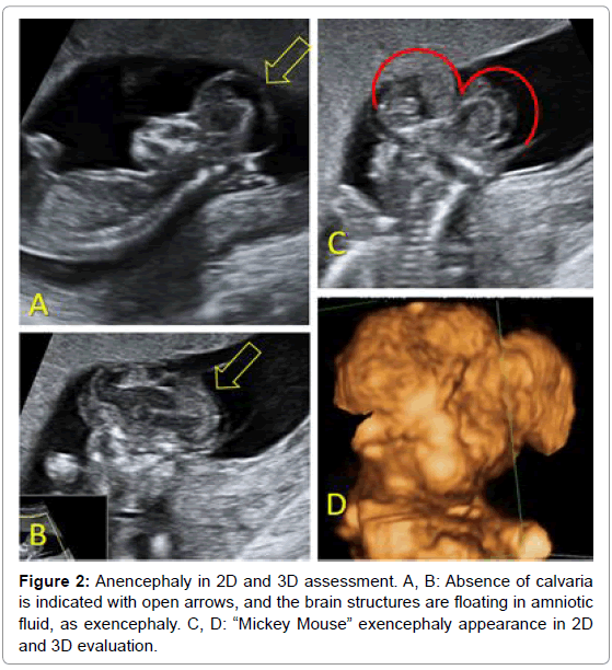
Anencephaly can be easily recognized at 11-13 weeks scan, because of the absence of calvaria and brain abnormalities. Usually exencephaly is evident, with the cerebral hemispheres still present but in contact with the destructive amniotic fluid and giving the Mikey Mouse appearance [27], described sixteen years ago (Figure 2). The absence of calvaria must be differentiated from skeletal dysplasia like achondrogenesis, osteogenesis imperfecta type II and hypophosphatasia, where due to the severe hypomineralisation of the calvarium, the skull cannot be visualised [1].
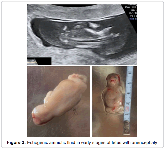
A high percentage (89%) of fetuses with acrania has echogenic amniotic fluid and this finding could potentially be used as a marker of fetal acrania in the first trimester (Figure 3) [28]. This also supports the hypothesis of the transition from acrania to anencephaly, with the unprotected brain undergoing progressive destruction. The prognosis of anencephaly is gloomy, as this disorder is incompatible with life, therefore termination of pregnancy is recommended and feasible at this gestational age.
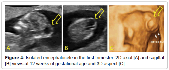
Encephaloceleis easily detectable because of the occipital herniation mass. Because of the similar unfavourable prognosis, termination should be offered (Figure 4).
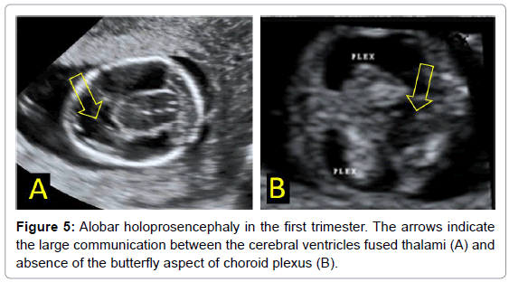
Another detectable malformation that can be detected early is holoprosencephaly, a CNS disorder that affects around 0.4% of all conceptuses [6]. The earliest limit for diagnosis is 10 weeks of gestation, as the development of the telencephalon into the two halves of the cerebrum is accomplished at the beginning of the 10th week of gestation. The abnormal midline is easily recognized in axial planes, with the presence of a large fluid collection in the fetal head and the fusion of the thalami. A biparietal diameter (BPD) below the 5th centile was found in 32.4% of pathologic cases and below the 50th centile in 67.6% [29-32].The ‘butterfly’ sign, representing the normal appearance of the choroid plexus has a higher sensitivity (100%), higher than BPD (40%), implying that microcephaly is not a prominent FT feature [33]. These findings, in association with the fetal facial abnormalities such as facial asymmetry, hypotelorism, central clefts and abnormal orbits, support the diagnosis of holoprosencephaly [1]. Since most of the affected fetuses die shortly after birth, and the surviving ones are severely mentally retarded, termination of the pregnancy is offered (Figure 5) [6].
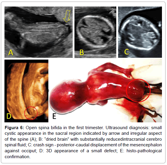
At the 11-13 week scan the diagnosis of open spina bifida (OSB) cannot be relied upon the well-known indirect cranian markers from the second trimester, as the lemon and banana signs [34]. Also, a cystic mass is rarely observed in the FT (Figure 6). But similar to the second trimester markers, early cranian features were initially proposed: reduced BPD, abnormal spine shape, retraction of the frontal bones and parallelism of cerebral peduncles. However, in the last decade, many studies highlighted the possibility of an efficient early OSB detection by assessing parameters of the posterior brain region. In such abnormal cases the sonographer may encounter a thickened brainstem, an increase ratio between brainstem diameter and brainstem-occipital bone distance to more than 1 (normal <0,9), a shortened cisterna magna and fourth ventricle [also called intracranial translucency], which is not visible or has a less amount of fluid than normal fetuses. The studies emphasized the high specificity of a normal posterior brain for OSB exclusion which is compulsory for an efficient screening test [30,31]. The optimal plane used to assess the posterior brain is the mid-sagittal view of the fetal face, which is routinely investigated at this gestational age to proper evaluate the genetic markers: nuchal translucency, nasal bone and fronto-maxillary facial angle. However, the posterior brain may be also assessed by experienced sonographers in the axial plane, more confidently by transvaginal approach [34].
Figure 6: Open spina bifida in the first trimester. Ultrasound diagnosis: small cystic appearance in the sacral region indicated by arrow and irregular aspect of the spine (A); B: “dried brain” with substantially reducedintracranial cerebro spinal fluid; C: crash sign - posterior-caudal displacement of the mesencephalon against occiput; D: 3D appearance of a small defect; E: histo-pathological confirmation.
What is more? There is a continuous progress in early CNS investigation. Several new markers for OSB were added in the last years to the multitude of early features of this pathology. It seems that the BPD/transverse abdominal diameter ratio improves considerably the diagnostic performance of using BPD measurement alone, to 76.9% [29,35]. Also, combining alpha fetoprotein and BPD with free β-hCG as part of first trimester aneuploidy screening, would allow early detection of about two-thirds of cases [36]. Another early feature of OSB foetuses is that the intracranial collection of cerebrospinal fluid is substantially reduced; giving the aspect of a “dried brain”, therefore, the roof of the third ventricle, aqueduct of Sylvius and fourth ventricle cannot be properly visualized [37]. Another marker recently proposed is the “crash” of the thalamus against the occiput, meaning the posterior-caudal displacement of the mesencephalon against occiput, with 92.3% detection rate [38].
Several years ago, we suggested the potential of the posterior brain morphometry to highlight conditions other than OSB: posterior fossa abnormalities, hydrocephaly, holoprosencephaly, other neural tube defects and chromosomal abnormalities. Our findings were later confirmed by other researches [39,40], regarding Dandy-Walker syndrome, vermian hypoplasia, Blake’s pouch cyst, and trisomies [13,18] and triploid fetuses that have measurable abnormalities in the posterior brain [40-43].
Nowadays we are heading toward the unthinkable. Until recently, we could not imagine identifying agenesis of corpus callosum in the first trimester, and now we have a marker - midbrain diameter-to-falx diameter ratio that seems to correctly identify 87% of the cases [44]. And we are bringing Kaneth score in the early stages of fetal development as the3/4D sonography enables precise study of embryonic and fetal activity and behaviour [45-47].
Challenges
Despite the impressive progress in early fetal diagnosis, we still must face an important challenge. Pathology is not easy to perform because of the small fetal dimensions and the brain damage that is also commonly seen at later gestational ages [48,49]. And as always, we expect great things from genetic investigation, with better characterization of CNS abnormalities [50].
Is 11-13 weeks’ scan reliable fora precise diagnosis and can it solely represent the indication of termination of pregnancy? In most of the cases, further investigations as genetic tests should be undertaken to correlate the sonographic with genetical findings to obtain a complete diagnosis. Generally, the brain anomalies detected in the first trimester are disorders with poor prognosis and outcome, with disabling or even lethal postpartum evolution.
Yet, the 11-13 weeks scan is not exempted of false-positive conditions. For instance, due to the incomplete development of some structures such as the cerebellar vermis, a false diagnosis of Dandy Walker variants can be made by an unexperienced sonographer, if it is not taken into consideration the cerebellum embryological development. Therefore, a wide communication between the fourth ventricle and cisterna magna is physiological at 11-13 weeks and should not be considered a pathological finding, unless an evident cystic posterior fossa is detected [1]. In case of anomalies with dynamic evolution pattern such as ventriculomegaly, the sonographer may suspect at the 11-13 weeks scan the diagnosis, but usually without certainty and further serial re-evaluation is mandatory [40]. In such cases the counselling of the couple is troublesome, because of the uncertainty of the diagnosis and the necessity of second trimester confirmation [51]. Hence, ventriculomegaly may be considered only by an experienced fetal medicine specialist. Along the subjective impression, the objective assessment of the ratio between choroid plexus and lateral ventricle lengths, or the ratio between the choroid plexus and lateral ventricle areas have proved their prognostic value and good reproducibility among observers [52].
Conclusions
Nowadays, we should be able to detect at a routine first trimester ultrasound scan most major CNS abnormality: neural tube defects, holoprosencephaly and iniencephaly. What is more, the pyramid of prenatal care is more and more reversed, as we now aim early in pregnancy for diagnosis traditionally reserved for second and third trimester, regarding posterior fossa abnormalities, agenesis of corpus callosum or neurobehavioral scoring. We still must face several important challenges, regarding the false positive results, pathology confirmation and genetic correlations. And even first trimester scan gives us great information regarding CNS, we should not forget to counsel patients that first trimester anomaly scan is not a replacement for second trimester morphologic sonography, because a lot of brain MA may develop or become apparent later in pregnancy.
References
- Paladini D, Volpe P (2007) Ultrasound of congenital fetal anomalies, diferential diagnosis and prognostic indicators. Informa Healthcare. CRP Press.
- Schoonen HMHJD, Essink-Bot ML, Van AGT HME, Wildschut HI, Steegers EAP, et al. (2011) Informed decision-making about the fetal anomaly scan: what knowledge is relevant?. Ultrasound Obstet Gynecol 37: 649-657.
- Pilu G, Hobbins JC (2002) Sonography of fetal cerebrospinal anomalies. Prenat Diagn 22: 321-330.
- Manegold-Brauer G, Oseledchyk A, Floeck A, Berg C, Gembruch U, et al. (2016) Approach to the sonographic evaluation of fetal ventriculomegaly at 11 to 14 weeks gestation. BMC Pregnancy and Childbirth 16: 3.
- Wong HS, Lam YH, Tang MHY, Cheung LWK, Ng LKL, et al. (1999) First-trimester ultrasound diagnosis of holoprosencephaly: three case reports. Ultrasound Obstet Gynecol 13: 356-359.
- Katorza E, Achiron R (2012) Early pregnancy scanning for fetal anomalies--the new standard? Clin Obstet Gynecol 55:199-216.
- Timor-Tritsch IE (2012) Evolving applications of first-trimester ultrasound. OBG Manag 24: 36-45.
- Nicolaides KH (2011) A model for a new pyramid of prenatal care based on the 11 to 13 weeks' assessment. Prenat Diagn 31: 3-6.
- Davies V, Gledhill J, McFadyen A, Whitlow B, Economides D (2005) Psychological outcome in women undergoing termination of pregnancy for ultrasound-detected fetal anomaly in the first and second trimesters: a pilot study. Ultrasound Obstet Gynecol 25: 389-392.
- Hernadi L, Torocsik M (1997) Screening for fetal anomalies in the 12th week of pregnancy by transvaginal sonography in an unselected population. Prenat Diagn 17: 753-759.
- Bilardo CM, Pajkrt E, de Graaf I, Mol BW, Bleker OP (1998) Outcome of fetuses with enlarged nuchal translucency and normal karyotype. Ultrasound Obstet Gynecol 11: 401-406.
- D’Ottavio G, Mandruzzato G, Meir YJ, Rustico MA, Fischer-Tamaro L, et al. (1998) Comparison of first trimester and second trimester screening for fetal anomalies. Ann NY Acad Sci 847: 200-209.
- Whitlow BJ, Chatzipapas IK, Lazanakis ML, Kadir RA, Economides DL (1999) The value of sonography in early pregnancy for the detection of fetal abnormalities in an unselected population. BJOG 106: 929-936.
- Chen M, Lam YH, Lee CP, Tang MH (2004) Ultrasound screening of fetal structural abnormalities at 12 to 14 weeks in Hong Kong. Prenat Diagn 24: 92-97.
- Taipale P, Ammala M, Salonen R, Hilesmaa V (2004) Two-stage ultrasonography in screening for fetal anomalies at 13-14 and 18-22 weeks of gestation. Acta Obstet Gynecol Scand 83: 1141-1146.
- Cedergren M, Selbing A (2006) Detection of fetal structural abnormalities by an 11-14-week ultrasound dating scan in an unselected Swedish population. Acta Obstet Gynecol Scand 85: 912-915.
- Dane B, Dane C, Sivri D, Kiray M, Cetin A (2007) Ultrasound screening for fetal major abnormalities at 11-14 weeks. Acta Obstet Gynecol Scand 86: 666-670.
- Chen M, Lee CP, Lam YH, Tang RYK, Chan BCP, et al. (2008) Comparison of nuchal and detailed morphology ultrasound examinations in early pregnancy for fetal structural abnormality screening: a randomized controlled trial. Ultrasound Obstet Gynecol 31: 136-146.
- Oztekin O, Oztekin D, Tınar S, Adıbelli Z. (2009) Ultrasonographic diagnosis of fetal structural abnormalities in prenatal screening at 11-14 weeks. Diagn Interv Radiol 15: 221-225.
- Syngelaki A, Chelemen T, Dagklis T, Allan L, Nicolaides KH (2011) Challenges in the diagnosis of fetal non-chromosomal abnormalities at 11-13 weeks. Prenat Diagn 31: 90-102.
- Iliescu D, Tudorache S, Comanescu A, Antsaklis P, Cotarcea S, et al. (2013) Improved detection rate of structural abnormalities in the first trimester using an extended examination protocol. Ultrasound Obstet Gynecol 42: 300-309.
- Whitlow BJ, Chatzipapas IK, Lazanakis ML, Kadir RA, Economides DL (1999) The value of sonography in early pregnancy for the detection of fetal abnormalities in an unselected population. BJOG: An International Journal of Obstetrics & Gynaecology 106: 929-936.
- Santo S, D'Antonio F, Homfray T, Rich P, Pilu G, et al. (2012) Counseling in fetal medicine: agenesis of the corpus callosum. Ultrasound Obstet Gynecol 40: 513-521.
- Wald M, Lawrenz K, Deutinger J, Weninger M (2004) Verification of anomalies of the central nervous system detected by prenatal ultrasound. Ultraschall in der Medizin 25: 214-217.
- Carroll SGM, Porter H, Abdel-Fattah S, Kyle PM, Soothill PW (2000) Correlation of prenatal ultrasound diagnosis and pathologic findings in fetal brain abnormalities. Ultrasound Obstet Gynecol 16: 149-153.
- Chatzipapas IK, Whitlow BJ, Economides DL (1999) The ‘Mickey Mouse’ sign and the diagnosis of anencephaly in early pregnancy. Ultrasound Obstet Gynecol 13: 196-199.
- Cafici, Daniel, Sepulveda, Waldo (2003) First-Trimester Echogenic Amniotic Fluid in the Acrania-Anencephaly Sequence. Journal of ultrasound in medicine : official journal of the American Institute of Ultrasound in Medicine 22: 1075-1079.
- Bernard J, Cuckle H, Stirnemann J, Salomon LJ, Ville Y (2012) OC05.02: Screening for fetal spina bifida by ultrasound examination in the first trimester of pregnancy at 11-14 weeks' using fetal biparietal measurement diameter. Ultrasound Obstet Gynecol 40: 10.
- Mangione R, Dhombres F, Lelong N, Amat S, Atoub F, et al. (2013) Screening for fetal spina bifida at the 11-13 week scan using three hallmarks of the posterior brain anatomy. Ultrasound in obstetrics & gynecology : the official journal of the International Society of Ultrasound in Obstetrics and Gynecology 42: 416-420.
- Acuna JG, Lau G, Rad S, Burk C, Gornbein J, et al. (2012) OP14.02: Posterior brain in fetuses with open spina bifida at 11 to 13 weeks gestation. Ultrasound Obstet Gynecol 40: 96-97.
- Khalil A, Papageorghiou A, Bhide A, Akolekar R, Thilaganathan B (2014) Biparietal diameter at 11 to 13 weeks' gestation in fetuses with holoprosencephaly. Prenat Diagn 34: 134-138.
- Sepulveda W, Wong AE (2013) First trimester screening for holoprosencephaly with choroid plexus morphology (‘butterfly’ sign) and biparietal diameter. Prenat Diagn 33: 1233-1237.
- Chaoui R, Nicolaides KH (2011) Detecting open spina bifida at the 11–13-week scan by assessing intracranial translucency and the posterior brain region: mid-sagittal or axial plane?. Ultrasound Obstet Gynecol 38: 609-612.
- Simon EG, Arthuis CJ, Haddad G, Bertrand P, Perrotin F (2015) Biparietal/transverse abdominal diameter ratio ≤ 1: potential marker for open spina bifida at 11–13-week scan. Ultrasound Obstet Gynecol 45: 267-272.
- Bernard JP, Cuckle H, Bernard M, Brochet C, Salomon L, Ville Y (2013) Combined screening for open spina bifida at 11-13 weeks using fetal biparietal diameter and maternal serum markers. American journal of obstetrics and gynecology 209: 223e1-223e5.
- Loureiro T, Ushakov F, Montenegro N, Gielchinsky Y, Nicolaides KH (2012) Cerebral ventricular system in fetuses with open spina bifida at 11–13 weeks' gestation. Ultrasound Obstet Gynecol 39: 620-624.
- Ushakov F, Fernandez M, Lesmes Heredia C, Pandya P (2014) OP06.08: “Crash signâ€: displacement and deformation of mesencephalon against occipital bone in the diagnosis of spina bifida at 11-13 weeks. Ultrasound Obstet Gynecol 44: 80.
- Iliescu DA, Comănescu P, Antsaklis, Tudorache S, Ghiluşi M, et al. (2011) Cernea Neuroimaging parameters in early open spina bifida detection. Further benefit in first trimester screening? Rom J Morphol Embryol 2011 52: 809-817.
- Lee MY, Won HS, Hyun MK, Lee HY, Shim JY, et al. (2012) One case of increased intracranial translucency during the first trimester associated with the Dandy-Walker variant. Prenatal diagnosis 32: 602-603.
- Bornstein E, RodrÃguez JLG, Pavón ECÃ, Quiroga HD, Divon MY (2013) First-Trimester Sonographic Findings Associated With a Dandy-Walker Malformation and Inferior Vermian Hypoplasia. Journal of Ultrasound in Medicine 32: 1863-1868.
- Garcia-Posada R, Eixarch E, Sanz M, Puerto B, Figueras F, et al. (2013) Cisterna magna width at 11–13 weeks in the detection of posterior fossa anomalies. Ultrasound Obstet Gynecol 41: 515-520.
- Ferreira AFA, Syngelaki A, Smolin A, Vayna AM, Nicolaides KH (2012) Posterior brain in fetuses with trisomy 18, trisomy 13 and triploidy at 11 to 13 weeks' gestation. Prenat Diagn 32: 854-858.
- Lachmann R, Sodre D, Barmpas M, Akolekar R, Nicolaides KH (2012) OC11.05: Midbrain and falx in fetuses with absent corpus callosum at 11–13 weeks. Ultrasound Obstet Gynecol 40: 23.
- Annemarie BL, Hadders AM, Van Kan CM, Johanna De Vries IP (2008) Fetal Onset Of General Movements Pediatric Research 63: 2.
- Kurjack A, Azumendi G, Andonotopo W, Aida S (2007) Three- and four-dimensional ultrasonography of the fetal face. Am J Obstet Gynecol 196: 16-28.
- Kurjak A, Miskovic B, Stanojevic M, Amiel-Tison C, Ahmed B, et al. (2008) New scoring system for fetal neurobehavior assessed by three- and four-dimensional sonography. Journal of perinatal medicine 36: 73-81.
- Isaksen CV, Eik-Nes SH, Blaas HG, Torp SH (1998) Comparison of prenatal ultrasound and postmortem findings in fetuses and infants with central nervous system anomalies. Ultrasound Obstet Gynecol 11: 246-253.
- Becher JC, Bell J, Keeling J, Liston W, McIntosh N, et al. (2006) The Scottish Perinatal Neuropathology Study—clinicopathological correlation in stillbirths. BJOG: An International Journal of Obstetrics & Gynaecology 113: 310-317.
- Huang JWYM, Pooh R, Wai CK (2012). Molecular genetics in fetal neurology. Seminars in fetal & neonatal medicine. 17: 341-346.
- Carvalho MHB, Brizot ML, Lopes LM, Chiba CH, Miyadahira S, et al. (2002) Detection of fetal structural abnormalities at the 11-14 week ultrasound scan. Prenat Diagn 22: 1-4.
- Hernádi L, Töröcsik M (1997) Screening for Fetal Anomalies in the 12th week of Pregnancy by Transvaginal Sonography in an Unselected Population. Prenat Diagn 17: 753-759.
Citation: Iliescu DG, Comanescu C (2017) CNS Abnormalities at 11-14 Weeks. Neonat Pediatr Med 3: 141. DOI: 10.4172/2572-4983.1000141
Copyright: © 2017 Iliescu DG, et al. This is an open-access article distributed under the terms of the Creative Commons Attribution License, which permits unrestricted use, distribution, and reproduction in any medium, provided the original author and source are credited.
Select your language of interest to view the total content in your interested language
Share This Article
Recommended Journals
Open Access Journals
Article Tools
Article Usage
- Total views: 28212
- [From(publication date): 0-2017 - Nov 06, 2025]
- Breakdown by view type
- HTML page views: 26953
- PDF downloads: 1259

