Conference Proceeding Open Access
Clinical and Radiographic Evaluation of Two Newly Pulp Medicaments Used in Primary Molars Pulpotomy
El-Morsy Badran MM1*, Awad SM2 and Shalan HM11Faculty of Dentistry, Pediatric Dentistry and Dental Public Health Department, Mansoura University, Egypt
2Chairman of Pediatric Dentistry and Public Health Department, Mansoura University, Egypt
- *Corresponding Author:
- Badran El-Morsy MM
Faculty of Dentistry
Pediatric Dentistry and Dental Public Health Department
Mansoura University, Egypt
Tel: 002-01228066974
E-mail: mahmoudbadran2010@mans.edu.eg
Received Date: February 10, 2017; Accepted Date: March 15, 2017; Published Date: March 25, 2017
Citation: El-Morsy Badran MM, Awad SM, Shalan HM (2017) Clinical and Radiographic Evaluation of Two Newly Pulp Medicaments Used in Primary Molars Pulpotomy. Pediatr Dent Care 2:135.
Copyright: © 2017 El-Morsy Badran MM, et al. This is an open-access article distributed under the terms of the Creative Commons Attribution License, which permits unrestricted use, distribution, and reproduction in any medium, provided the original author and source are credited.
Visit for more related articles at Neonatal and Pediatric Medicine
Abstract
Primary molars pulpotomy is a very common therapy for primary molars with extensive caries. Many agents including formaldehydebased materials, electro surgery, lasers, glutaraldehyde, haemostatic medicaments, zinc oxide eugenol, bone morphogenic protein (BMP), collagen and calcium involving, Dentin Bridge inducing materials have been developed. However, the ideal pulpotomy treatment still needs to be improved, so this research was done.
Introduction
Primary molars pulpotomy is a very common therapy for primary molars with extensive caries. Many agents including formaldehydebased materials, electro surgery, lasers, glutaraldehyde, haemostatic medicaments, zinc oxide eugenol, bone morphogenic protein (BMP), collagen and calcium involving, Dentin Bridge inducing materials have been developed. However, the ideal pulpotomy treatment still needs to be improved, so this research was done.
Aim
To evaluate clinically and radiographically the effect of (Biodentine and Mineral Trioxide Aggregate) as pulpotomy medicament agents vs. Formocresol in primary molars.
Design
The design groups are divided according to Split Mouth design so, the number of control group was 30 primary molar and the two experimental groups (Bio dentine, MTA) was 15 primary molar for each. The 3 groups were divided randomly without any bias and written content was taken from their parents for participating acceptance. All teeth were examined both clinically and radio graphically according to Coll and Sadrian Criteria for 3,6,9 months expect 2 cases did not come the last follow up.
Criteria of Coll and Sadrian
Clinical criteria
No pain on percussion on recall checkup.
No gingival swelling or sinous tract 6 months postoperatively.
No purulent exudate expressed from the gingival margin.
No abnormal mobility of tooth.
Radiographic criteria
No pathologic root resorption
A furcation radiolucency resolved 6-12 months postoperatively
No periapical radiolucency formation postoperatively
Sixty carious primary molars, followed pulpotomy indications, for 17 child were used in this study. The teeth were divided into 3 groups
• Group I (Control group ) 30 molar treated by Formocresol
• Group II (Experimental group) 15 molar treated by Biodentine
• Group III (Experimental group)15 molar treated by MTA
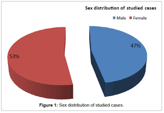
Patients preparation, profound local anesthesia, isolation by rubber dam was done then the whole caries was removed, all of undermined enamel was removed, the whole coronal pulp was amputated by sharp spoon excavator, initial stabilized clot was established [1-3], then the various pulp medicaments were applied over the pulp stump, so the pulp was treated by group I (formocresol), group II (Bio dentine), group III (MTA). Final restoration was performed with composite [4-6]. Then, both clinical and radiographic evaluation was done for all teeth at 3,6,9 months according to Coll and Sadrian Criteria. The data were analyzed to obtain Descriptive statistics and Analytical statistics to test the significance of difference between groups (Figure 1 and Table 1).
| n=17 | % | |
|---|---|---|
| Age (years) | ||
| Mean ± SD | 5.53 ± 1.07 | |
| Sex | ||
| Male | 8 | 47.1 |
| Female | 9 | 52.9 |
Table 1: Demographic characters of studied groups.
Results
The results of this study showed that no significant difference in between Biodentine and MTA in the three periods of follow up (P>0.05) on the other hand there was a statistically significant difference between biodentine and its control group (P<0.05) and between MTA group and its control group( P<0.05).
Statistical results: These two tables show the comparison between the 3 groups both clinically and radiographically at 3,6,9 months follow up (Tables 2 and 3).
| Period of follow up | Clinical assessment | Groups | Chi-square test | |||||
|---|---|---|---|---|---|---|---|---|
| Biodentine group | Control | MTA | ||||||
| n (%) | group | group | ||||||
| n (%) | n (%) | |||||||
| 3 months | pain | 0 (0.0) | 1 (3.3) | 0 (0.0) | p=0.6 | |||
| Gingival Swelling | 0 (0.0) | 1 (3.3) | 0 (0.0) | p=0.6 | ||||
| Purulent exudates | 0 (0.0) | 1 (3.3) | 0 (0.0) | p=0.6 | ||||
| Abnormal mobility | 0 (0.0) | 1 (3.3) | 0 (0.0) | p=0.6 | ||||
| 6 | pain | 0 (0.0) | 5 (16.7) | 2 (13.3) | p=0.25 | |||
| months | Gingival Swelling | 0 (0.0) | 3 (10.0) | 1 (6.7) | p=0.45 | |||
| Purulent exudates | 0 (0.0) | 3 (10.0) | 1 (6.7) | p=0.45 | ||||
| Abnormal mobility | 0 (0.0) | 2 (6.7) | 0 (0.0) | p=0.36 | ||||
| 9 | pain | 2 (14.3) | 6 (21.4) | 2 (14.3) | p=0.78 | |||
| months | Gingival Swelling | 0 (0.0) | 3 (10.7) | 1 (7.1) | p=0.56 | |||
| Purulent exudates | 1 (7.1) | 4 (14.3) | 1 (7.1) | p=0.75 | ||||
| Abnormal mobility | 0 (0.0) | 2 (7.1) | 0 (0.0) | p=0.36 | ||||
Table 2: Three groups at 3,6,9 months follow up clinical assessment.
| Period of follow up | Radiographic evaluation | Groups | Chi-square test | ||
|---|---|---|---|---|---|
| Biodentine group | Control | MTA | |||
| n (%) | group n (%) | group n (%) | |||
| 3 months | Pathological Root Resorption | 0 (0.0) | 1 (3.3) | 0 (0.0) | p=0.6 χ2=1.02 |
| Furcation radiolucency resolved6-12 months postoperatively | 0 (0.0) | 1 (3.3) | 0 (0.0) | p=0.6 χ2=1.02 |
|
| Periapical radiolucency formation | 0 (0.0) | 1 (3.3) | 0 (0.0) | p=0.6 χ2=1.02 |
|
| 6 months | Pathological Root Resorption | 0 (0.0) | 3 (10.0) | 1 (6.7) | χ2=1.02 p=0.4 |
| Furcation radiolucency resolved6-12 months postoperatively | 1 (6.7) | 4 (13.3) | 1 (6.7) | p=0.69 χ2=0.7 |
|
| Periapical radiolucency formation | 0 (0.0) | 2 (6.7) | 0 (0.0) | p=0.35 χ2=0.7 |
|
| 9 months | Pathological Root Resorption | 2 (14.3) | 5 (17.9) | 2 (14.3) | p=0.94 χ2=0.13 |
| Furcation radiolucency resolved6-12 months postoperatively | 0 (0.0) | 3 (10.7) | 1 (7.1) | p=0.45 χ2=1.6 |
|
| Periapical radiolucency formation | 0 (0.0) | 3 (10.7) | 0 (0.0) | p=0.2 χ2=3.17 |
|
χ2=Chi-square test.
p value significant if <0.05.
Table 3: Three groups at 3,6,9 months follow up Radiographic evaluation.
Conclusion
Both MTA and Biodentine can be considered as a great substitute to Formocresol as pulp medicaments after pulpotomy.
Future Aspects
The clue for future about primary molars treatment after surgery. I really recommend the use of MTA and Biodentine as a great substitute to Formocresol as many researches proved that it is mutagenic and carcinogenic.
References
- Mohammad SG, Raheel SA, Baroudi K (2014) Clinical and Radiographic Evaluation of Allium sativum Oil as a New Medicament for Vital Pulp Treatment of Primary Teeth. Journal of International Oral Health 6: 32.
- Rodd HD, Waterhouse PJ, Fuks AB, Fayle SA, Moffat MA (2006) Pulp therapy for primary molars. International Journal of Paediatric Dentistry 16: 15-23.
- Markovic D, Zivojinovic V, Vucetic M (2005) Evaluation of three pulpotomy medicaments in primary teeth. European Journal of Paediatric Dentistry 6: 133.
- Avram DC, Pulver F (1988) Pulpotomy medicaments for vital primary teeth. Surveys to determine use and attitudes in pediatric dental practice and in dental schools throughout the world. ASDC Journal of Dentistry for Children, 56: 426-434.
- Waterhouse PJ (1995) Formocresol and alternative primary molar pulpotomy medicaments: a review. Dental Traumatology 11: 157-162.
- Waterhouse PJ, Nunn JH, Whitworth JM (2000) Paediatric dentistry: An investigation of the relative efficacy of Buckley's Formocresol and calcium hydroxide in primary molar vital pulp therapy. British Dental Journal 188: 32-36.
Relevant Topics
- About the Journal
- Birth Complications
- Breastfeeding
- Bronchopulmonary Dysplasia
- Feeding Disorders
- Gestational diabetes
- Neonatal Anemia
- Neonatal Breastfeeding
- Neonatal Care
- Neonatal Disease
- Neonatal Drugs
- Neonatal Health
- Neonatal Infections
- Neonatal Intensive Care
- Neonatal Seizure
- Neonatal Sepsis
- Neonatal Stroke
- Newborn Jaundice
- Newborns Screening
- Premature Infants
- Sepsis in Neonatal
- Vaccines and Immunity for Newborns
Recommended Journals
Article Tools
Article Usage
- Total views: 3372
- [From(publication date):
specialissue-2017 - Apr 02, 2025] - Breakdown by view type
- HTML page views : 2458
- PDF downloads : 914

