Research Article Open Access
Clinical and Histological Comparison of Pulp Anaesthesia and Local Diffusion after Periodontal Ligament Injection and Intrapapillary Infiltration Anaesthesia
| Peer W. Kämmerer1*, Victor Palarie2, Eik Schiegnitz1, Thomas Ziebart1, Bilal Al-Nawas1 and Monika Daubländer3 | |
| 1Department of Oral and Maxillofacial Surgery, University Medical Centre Mainz, Mainz, Germany | |
| 2Clinic for Oral & Maxillofacial Surgery, State Medical and Pharmaceutical University “Nicolae Testemitanu,” Chisinau, Moldova | |
| Corresponding Author : | Peer W. Kämmerer MD DMD Assistant Professor Department of Oral and Maxillofacial Surgery University Medical Centre Mainz Augustusplatz 2, 55131 Mainz, Germany Tel: 00496131-175086 Fax: 06131/17-6602 E-mail: peer.kaemmerer@unimedizin-mainz.de |
| Received April 10, 2012; Accepted June 11, 2012; Published June 18, 2012 | |
| Citation: Kämmerer PW, Palarie V, Schiegnitz E, Ziebart T, Al-Nawas B, et al. (2012) Clinical and Histological Comparison of Pulp Anaesthesia and Local Diffusion after Periodontal Ligament Injection and Intrapapillary Infiltration Anaesthesia. J Pain Relief 1:108 doi: 10.4172/2167-0846.1000108 | |
| Copyright: © 2012 Kämmerer PW, et al. This is an open-access article distributed under the terms of the Creative Commons Attribution License, which permits unrestricted use, distribution, and reproduction in any medium, provided the original author and source are credited. | |
Visit for more related articles at Journal of Pain & Relief
Abstract
Improvements in local anaesthetic techniques may minimize pain during dental treatment. Block techniques of the trigeminal branches (II and III) are considered to be the most used and trusted methods in dentistry . However, the incidence of inadequate anaesthesia and technical complications is reported to be between 15-30% of cases. Other possibilities to achieve local anaesthesia are infiltration, intra-pulpal injection, intraosseous , intraseptal and periodontal ligament injection (PDL). In the present time, PDL technique increases in popularity, especially because of specialized high pressure syringe
systems and computer controlled local anaesthesia devices. PDL has shown to provide sufficient anaesthesia of the pulp. It is less unpleasant and painful than inferior alveolar and palatinal injections.
Other advantages are well documented: single tooth anaesthesia, minimal anaesthetic agent dosage, rapid onset and safety in patients with bleeding problems as well as in medical compromised patients. Though, disadvantages like damage of periodontal tissue, damage of interdental septal crest, root resorption and severe bacteraemia are reported as well. Infection and/or inflammation may decrease
the anaesthetic efficacy and PDL may induce systemic reactions manifested in cardiovascular parameters changes Intraseptalintraosseous anaesthesia (ISA) is described to be an alternative to PDL in order to overcome inconveniences associated with PDL, especially in compromised periodontal tissues. Still, probably because of the enosseous pathway, ISA has shown to cause cardiovascular
changes similar to PDL. Intrapapillary infiltration anaesthesia (IPA) is an intermediate of PDL and ISA and consists of application of the local anaesthetic agent into the mesial and distal papillae without
touching the periodontal ligament and without application in the bone. Therefore, cardiovascular side effects may be reduced. Such as for ISA, potential side effects like damage of periodontal tissue and
bacteraemia are limited. For IPA, normal syringes as well as pressure delivery systems can be used [18]. In order to minimize all possible side effects in general practice, we tested the new IPA technique for pulp anaesthesia. To our knowledge, there are no published studies that compare PDL and IPA. Therefore, the aim of the present study was to evaluate pulp anaesthesia (clinical setting) as well as local diffusion of the anaesthetic agent (histological setting) in both methods.
| Keywords |
| Clinical study; Histological study; Intraligamentary injection; Intraosseous resorption; Intrapapillary injection; Periodontal ligament; Infiltration; Dental local anaesthesia |
| Introduction |
| Improvements in local anaesthetic techniques may minimize pain during dental treatment. Block techniques of the trigeminal branches (II and III) are considered to be the most used and trusted methods in dentistry [1]. However, the incidence of inadequate anaesthesia and technical complications is reported to be between 15-30% of cases [2,3]. Other possibilities to achieve local anaesthesia are infiltration, intra-pulpal injection, intraosseous [4], intraseptal [5] and periodontal ligament injection (PDL). In the present time, PDL technique increases in popularity, especially because of specialized high pressure syringe systems [6] and computer controlled local anaesthesia devices [7,8]. PDL has shown to provide sufficient anaesthesia of the pulp. It is less unpleasant and painful than inferior alveolar and palatinal injections. Other advantages are well documented: single tooth anaesthesia, minimal anaesthetic agent dosage, rapid onset and safety in patients with bleeding problems as well as in medical compromised patients [6, 9-11]. Though, disadvantages like damage of periodontal tissue, damage of interdental septal crest, root resorption and severe bacteraemia are reported as well [12-15,8]. Infection and/or inflammation may decrease the anaesthetic efficacy [16] and PDL may induce systemic reactions manifested in cardiovascular parameters changes [17]. Intraseptalintraosseous anaesthesia (ISA) is described to be an alternative to PDL in order to overcome inconveniences associated with PDL, especially in compromised periodontal tissues [16,5]. Still, probably because of the enosseous pathway, ISA has shown to cause cardiovascular changes similar to PDL [18]. Intrapapillary infiltration anaesthesia (IPA) is an intermediate of PDL and ISA and consists of application of the local anaesthetic agent into the mesial and distal papillae without touching the periodontal ligament and without application in the bone. Therefore, cardiovascular side effects may be reduced. Such as for ISA, potential side effects like damage of periodontal tissue and bacteraemia are limited. For IPA, normal syringes as well as pressure delivery systems can be used [18]. In order to minimize all possible side effects in general practice, we tested the new IPA technique for pulp anaesthesia. To our knowledge, there are no published studies that compare PDL and IPA. Therefore, the aim of the present study was to evaluate pulp anaesthesia (clinical setting) as well as local diffusion of the anaesthetic agent (histological setting) in both methods. |
| Materials and Methods |
| The experimental solution tested was 4% Articaine with epinephrine 1:200.000 (Ultracain DS®, Sanofi-Aventis GmbH, Berlin, Germany) in dental cartridges. For both methods of anaesthesia, a system composed from Varioject INTRA® syringe and single use plastic device VarioSafe® with a pressure limit at 70 N (both Pajunk® GmbH Medizintechnologie, Geisingen, Germany, Figure 1) was used. A 27-gauge short needle was employed. To achieve the objectives, the study was composed of a human clinical study and animal experiments of both techniques. |
| Clinical experiments |
| The clinical part was performed in 30 healthy volunteers, aged between 28 and 42 years in the Unit of Oral Surgery, Policlinic of Stomatology, Chisinau, Moldova. In accordance to the regulations of the local ethics committee, no patient-related data were recorded and the patients were pseudonymised. The local Ethic Committee Approvals were obtained prior initiating experiments. The written informed consent was obtained before injection. They were currently taking no medications and had never had an allergic or toxic reaction to any of the local anaesthetic solutions. Upon clinical examination, all teeth were vital, free of caries, restorations, and had no exposed dentin. Mobility was tested for each tooth. No tooth exhibited pathological mobility in any direction. There were no periodontal pathologies such as probing depths > 5mm. Before injection, the oral mucosa was decontaminated with Clorhexidine 0.12% solution (Paroex®, Sunstar GmbH, Kriftel, Germany). Each volunteer received both of the following methods of anaesthesia in the region of two mandibular premolars on each side. To achieve a blinding, volunteers were not informed which method was used (neither before nor while nor after injection). Via Visual Analogue Scale (VAS), injection pain was measured for both techniques after each injection. Unknown to the volunteers, in the right inferior jaws, PDL technique was always used. Injections were given with Varioject INTRA® syringe system and 30 gauge single use short needles. The needle was inserted in an up to 30° angle to the tooth axis through the gingival sulcus to a point of maximum penetration. The handle of the syringe was squeezed firmly until back-pressure was achieved; this pressure was then sustained for approximately 25 seconds 0.2 ml of anaesthetic solution was deposited during 20 seconds [19]. In the left inferior jaws, an IPA technique was used with the same injection system. The puncture was conducted in the vestibular middle of the interdental papilla. The needle was inserted until bone contact and then slightly withdrawn before injection of the local anaesthetic. The soft tissues near the bone mesial and distal of the tooth were infiltrated. The injection was performed at a rate of 0.2 ml during 20-30 seconds for each papilla [18] (total 0.4 ml in a total of 30 seconds). As the efficiency of infiltration was tested, no additional injection into the intraseptal bone [18] was conducted. |
| To assess the effect of each technique to pulp response, an electric pulp testing was performed by the dentist 1 min after injection using an electronic pulp vitality scanner (Kerr Vitality Scanner, Kerr dental, Orange, CA, USA). The teeth were isolated with cotton rolls, dried with air, and the probe tip placed on the buccal surface midway between the gingival margin and the occlusal edge. A dental gel (Elmex® gelée, GABA GmbH, Lörrach, Switzerland) was used as between pulp tester probe and tooth. Prior and after PDL and IPA injections, baseline readings were recorded for each experimental tooth by another person. Each subject was instructed to respond when a sensation was first felt within the tooth. Values of pulp vitality scanner are given in arbitrary units. Maximal stimulation is 80/80. The method is seen to be objective. The time of anaesthesia was also evaluated with the pulp tester. Every minute after injection, a measurement was done until the reading was the same as the first baseline reading. |
| Animal experiments |
| Two 4-month-old male sheep, weighting approximately 3 kg, were used in the present study. Their anatomy and physiology are suitable for the design of this experimental project [20]. In order to evaluate diffusion of the local agent towards teeth of the next dentition, injections were given to young animals in the first dentition [21]. All animals were treated in accordance with both policies and principles of laboratory animal care and with the European Union guidelines (86/609/EEC). The local Ethic Committee Approvals were obtained prior initiating experiments. Sheep were housed in individual cages in an animal room maintained at 22°C and 55% relative humidity with ventilation 18–20 times/h and a 12-h light–dark cycle. Animals were allowed free access to diet and water. Adaptation and observation period was 1 week before surgery. The experiments were performed under a general anaesthesia by intramuscular injections of a combination of a dose of 35 mg/kg Ketamine and a dose of 5 mg/kg Xylazine. |
| To observe anaesthetic solution spreading into the tissues, trypan blue 1% solution was used as marker. It was incorporated into the anaesthetic agent by replacing one third of the Articaine liquid of the cartridges. This method of mixture was previously reported and dose not influences the flow of the solution [21]. Each animal received both of the methods of anaesthesia described above in the region of two inferior incisors and one inferior premolar on each side (3 teeth per method per animal) [20]. The right part served for the PDL injections. In the left mandible, the IPA technique was used with the same injection system, in the same way as in the clinical setting. After 15 minutes of injection, animals were sacrificed with an intra muscular excess dose of Pentobarbitone at 100 mg/kg and specimens were obtained for the histology evaluation. |
| Three type of specimen were examined: macroscopic surfaces of non-decalcified bone parts, microscopic surfaces of the non-decalcified bone parts and histological sections with decalcified cut surfaces. The bone was cut with a commercial water-cooled saw (Exakt, Hamburg, Germany) for a direct examination of macroscopic surfaces. Afterwards, the slices were cut down to a thickness of 5 mm perpendicular to the axis of the teeth. Microscopic evaluation was conducted with a Leica DM8000 M microscope (Leica Microsystems, Heidelberg, Germany). For further histological analyses with decalcified cut surfaces, the modified Tagger decalcification method of bone blocks was performed [22,21]: fixed specimens were rinsed in distillate water and demineralised with 5% formic acid, then dehydrated by passing through ascending graded of alcohol and cleared in methyl salicylate. Histological examination on the non-stained samples was performed on sections obtained from both animals. The areas of interest were the site of injection of interdental papilla, interdental bone, periodontal ligament and gingival tissue. |
| Statistics |
| The nature of this experiment was seen to be explorative; therefore, neither prior sample size calculation nor power analysis was conducted. Student’s T-test was used to examine the statistical significance of the differences in injection pain, pulp vitality testing and length of pulpal anaesthesia. We report descriptive p-values of tests; p-values of p≤0.05 were termed to be significant. The further histological analyses were also descriptive and cannot be considered as significant at any level. The analyses were conducted using SPSS version 20.0 for Mac (IBM, Armonk, NY, USA). |
| Results |
| Clinical experiments |
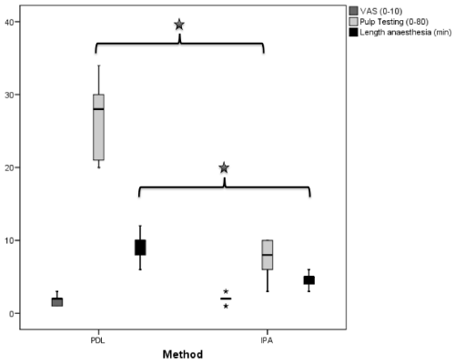
| VAS-measurements showed a mean score of 1.7 (standard deviation (SD): 0.7) for PDL and value of 1.9 (SD: 0.6) for IPA. The difference between the both groups was not significant (p=0.28). In PDL cases, a mean pulp testing value of 24.8 (SD: 5.6), in IPA, a mean value of 9.6 (SD: 2.6) was seen. This difference was statistically significant (p<0.0001). A significant difference was also seen in duration of pulp anaesthesia: whereas PDL had a mean duration of 9.2 min (SD: 2.1), IPA lasted 4.6 min (SD: 1.1; p<0.0001; Figure 2). In both groups, no tooth demonstrated an increased mobility when tested tactile after injection. At recall examination one day after treatment, mobility, the periodontium and vitality was within normal limits. No inflammation or necrotic events were observed. |
| Animal experiments |
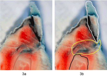
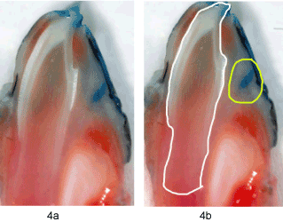
| For the cases of PDL injection, the evaluation of macroscopic surfaces of the non-decalcified, deep frozen bone parts could show a throughout diffusion of local anaesthetic solution into the periodontal ligament and deep into the surrounding bone (Figure 3a and Figure 3b). For cases of IPA injection, a local depot of anaesthetic without further diffusion was seen (Figure 4a and Figure 4b). |
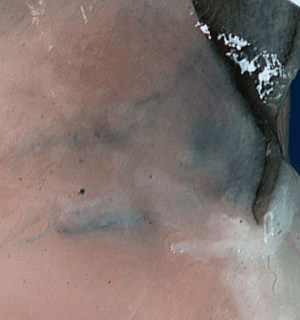
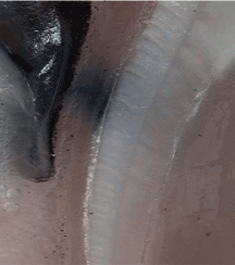
| The microscopic evaluation of the non-decalcified bone could verify these findings. For PDL, a diffusion of anaesthetic agent around the tooth and into the surrounding soft and hard tissue was seen (Figure 5). Microscopic evaluation of IPA injection could show local depots of stained anaesthetic agent only. Nevertheless, it could be seen that the anaesthetic solution was also reaching the tooth at some points (Figure 6). |
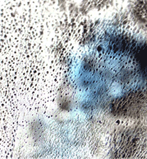
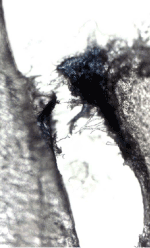
| The histological sections with decalcified cut surfaces could demonstrate the bone diffusion after PDL. The coloured solution was following vascular channels, spreading into bone cavities (Figure 7). A mechanical trauma of the needle and destruction of the periodontal ligament after PDL injection could be seen in one case, indicated by invasion of the blue agent from the puncture of the mucosa to the alveolar bone crest (Figure 8). |
| Discussion |
| The effect of PDL analgesia in patients of different age was discussed in numerous papers [23,11,24,18]. PDL injection has several advantages compared with block anaesthesia. It is more suitable in case of dental management of patients with haemophilia and other congenital bleeding disorders [25,26]. Also, it is more acceptable than direct painful palatinal technique and provides suitable anaesthesia for rubber dam, matrix band and crown placement in all teeth. It will give adequate analgesia for extraction of the primary incisors and canines [27]. However, it is recommended that PDL injection should not be used with infection or significant inflammation at the injection site [28]; in particular cases an intrusion of bacteria from the blood during injection may lead to serious complications [27,29]. Further negative side effects of PDL are reversible destruction of the periodontal ligament, and–possibly-damage to erupting teeth. ISA, the injection of anesthetic agent into the papillary soft tissue and interdental septum, has described to be a valid alternative to PDL, possibly without these negative side effects [18,30]. In order to minimize cardiovascular reactions, IPA, as non-osseous anesthesia was tested. As Articaine is suggested to be the anesthetic agent that diffuses best within soft and hard tissues [31], it was used in the study. Publications are concentrated on comparisons of the PDL versus classic infiltrative technique or nerve block [32]. IPA has to be discussed as a special part of infiltration technique. Accordingly, both techniques were used with different injection dosages and times. This disparity is clinically methodimmanent [10]. The animal sheep-mandible model is used for training purposes in periodontal surgery as well as for experimental surgery in oral and maxillofacial surgery [33,34]. The anterior mandible of the sheep was described to be an alternative to human mandible and has been used before to evaluate pressure by dental injections [20]. |
| Our results show that PDL injection archived more profound pulp anaesthesia than IPA. Based on electrical pulp testing, in our study, successful PDL was achieved in premolars in all cases. For IPA only superficial anaesthesia could be noted. Accordingly, the solely infiltrative technique of IPA is not equitable to PDL. The time of anaesthesia differed significantly between the two groups. PDL had a mean duration of anaesthesia of 9.2 min versus 4.6 min for IPA. The duration of pulp anaesthesia of PDL is in accordance to the literature [11,35]. To our knowledge, for IPA no analogical publications exist. PDL injections have shown to be more uncomfortable than infiltrations in the mandibular central incisor region [32]. In our study the pain response on mandibular premolars was the same in both groups of techniques. Kaufman’s study reported, that pain response during PDL is situated between the infiltration and mental nerve block techniques [26]. |
| The choice of marker was based on pervious reports on optimal for showing the effect of the local anaesthetics agents [36]. In contrary to PDL, in case of IPA a macroscopical spread of the coloured infiltration was found only into sub mucosal area. The histological data confirmed, that loosely attached tissues allow spread of solution because of the lower interstitial pressure and resistance. But, in microscopical specimens, a further spread of the solution after IPA could be seen as well. Therefore, in further studies, the soft tissue anaesthesia as well as the subjective patients’ pain while treated after IPA should be evaluated. Based on our findings, regarding the problem of possible influence of the PDL injection to permanent tooth buds, the risk of injury and toxicity appears to be minimal. The blue ink was not observed to diffuse into the bud and its attached membranes. These results are in accordance to the literature [21,35,37]. The histological findings showed a mechanical damage of the periodontal tissue after PDL injection. This damage was localized in the region of immediately adjacent to the injection site and caused by injury of the needle. The literature dates report that this is a reversible damage and these alterations subsequently show signs of rapid repair [38,8]. |
| Conclusion |
| According to histological and clinical data, in comparison to infiltrative IPA, intraosseous PDL seems to be favourable in achieving single tooth pulpal anaesthesia. It could be validated that the injection of the anesthetic agent into the bone (PDL, ISA) is a pivotal factor for pulp anesthesia; a sole intrapapillary infiltration, even with Articaine as anesthetic agent, does not seem to be sufficient. The effect of IPA on soft tissue anaesthesia has not been examined yet. |
References
- Blanton PL, Jeske AH (2003) Dental local anesthetics: alternative delivery methods. J Am Dent Assoc 134: 228-234.
- Meechan JG (1999) How to overcome failed local anesthesia. Br Dent J 186: 15-20.
- Kaufman E, Weinstein P, Milgrom P (1984) Difficulties in achieving local anesthesia. JADA 108: 205-208.
- Gow-Gates GA (1973) Mandibular conduction anesthesia: a new technique using extraoral landmarks. Oral Surg Oral Med Oral Pathol 36: 321-328.
- Woodmansey K (2005) Intraseptal anesthesia: a review of a relevant injection technique. Gen Dent 53: 418-420.
- Meechan JG (1992) Intraligamentary anaesthesia. J Dent 20: 325-332.
- Hochman MN (2007) Single-tooth anesthesia: pressure-sensing technology provides innovative advancement in the field of dental local anesthesia. Compend Contin Educ Dent 28: 186-188, 190, 192-183.
- Froum SJ, Tarnow D, Caiazzo A, Hochman MN (2000) Histologic response to intraligament injections using a computerized local anesthetic delivery system. A pilot study in mini-swine. J Periodontol 71: 1453-1459.
- Stoll P, Bührmann K (1983) [Intraligamental anesthesia for tooth extraction in patients with hemorrhagic diathesis]. ZWR 92: 545.
- Daubländer M, Kämmerer PW (2011) Lokalanästhesie in der Zahnmedizin. forum med dent. Sanofi, Berlin.
- Endo T, Gabka J, Taubenheim L (2008) Intraligamentary anesthesia: benefits and limitations. Quintessence Int 39: e15-25.
- Galili D, Kaufman E, Garfunkel AA, Michaeli Y (1984) Intraligamentary anesthesia--a histological study. Int J Oral Surg 13: 511-516.
- Garfunkel AA, Kaufman E, Marmary Y, Galili D (1983) Intraligamentary--intraosseous anesthesia. A radiographic demonstration. Int J Oral Surg 12: 334-339.
- Kaufman E, Galili D, Garfunkel AA (1983) Intraligamentary anesthesia: a clinical study. J Prosthet Dent 49 : 337-339.
- Roahen JO, Marshall FJ (1990) The effects of periodontal ligament injection on pulpal and periodontal tissues. J Endod 16: 28-33.
- Malamed SF (2004) Supplemental injection techniques. Handbook of local anesthesia, 5th edn. Mosby, St. Louis.
- Nusstein J, Berlin J, Reader A, Beck M, Weaver JM (2004) Comparison of injection pain, heart rate increase, and postinjection pain of articaine and lidocaine in a primary intraligamentary injection administered with a computer-controlled local anesthetic delivery system. Anesth prog 51: 126-133.
- Brkovic BM, Savic M, Andric M, Jurisic M, Todorovic L (2010) Intraseptal vs. periodontal ligament anaesthesia for maxillary tooth extraction: quality of local anaesthesia and haemodynamic response. Clin Oral Investig 14: 675-681.
- Meechan JG (2002) Supplementary routes to local anaesthesia. Int Endod J 35: 885-896.
- Whitworth JM, Ramlee RA, Meechan JG (2005) Pressures generated in vitro during Stabident intraosseous injections. Int Endod J 38: 291-296.
- Tagger E, Tagger M, Sarnat H, Mass E (1994) Periodontal ligament injection in the dog primary dentition: spread of local anaesthetic solution. Int J Paediatr Dent 4: 159-166.
- Tagger M (1976) Clearing of teeth for study and demonstration of pulp. J Dent Educ 40: 172-174.
- Naidu S, Loughlin P, Coldwell SE, Noonan CJ, Milgrom P (2004) A randomized controlled trial comparing mandibular local anesthesia techniques in children receiving nitrous oxide-oxygen sedation. Anesth Prog 51: 19-23.
- Forbes WC (2005) Twelve alternatives to the traditional inferior alveolar nerve block. J Mich Dent Assoc 87 : 52-56, 58, 75.
- Brewer AK, Roebuck EM, Donachie M, Hazard A, Gordon K, et al. (2003) The dental management of adult patients with hemophilia and other congenital bleeding disorders. Haemophilia 9: 673-677.
- Kaufman E, Epstein JB, Naveh E, Gorsky M, Gross A, et al. (2005) A survey of pain, pressure and discomfort induced by commonly used oral local anesthesia injections. Anesth Prog 52: 122-127.
- Zugal W, Taubenheim L, Schulz D (2005) Triade des Anästhesie-Erfolgs: Instrumente - Anästhetika - Methoden-Beherrschung. Stomatologie 1: 9-14.
- Reitz J, Reader A, Nist R, Beck M, Meyers WJ (1998) Anesthetic effiacy of the intraosseous injection of 0.9 ml of 2% lidocaine (1:100.000 epinephrine) to augment an inferior alveolar nerve block. Oral Surg Oral Med Oral Pathol Oral Radiol Endod 86: 516-523.
- Roberts GJ, Holzel HS, Sury MRJ, Simmons NA, Gardner P, et al. (1997) Dental bacteriemia in children. Pediatric Cadiology 18: 24-27.
- Saadoun AP, Malamed SF (1985) Intraseptal anesthesia in periodontal surgery. JADA 111: 249-256.
- Vree TB, Gielen MJ (2005) Clinical pharmacology and the use of articaine for local and regional anaesthesia. Best Pract Res Clin Anaesthesiol 19: 293-308.
- Meechan JG, Ledvinka JI (2002) Pulpal anesthesia for mandibular central incisor teeth: a comparison of infiltration and intraligamentary injections. Int Endod J 35: 629-634.
- Al-Qareer AH, Afsah MR, Muller HP (2004) A sheep cadaver model for demonstration and training periodontal surgical methods. Eur J Dent Educ 8: 78-83.
- Ayoub AF, Richardson W, Koppel D, Thompson H, Lucas M, et al. (2001) Segmental mandibular reconstruction by microincremental automatic distraction osteogenesis: an animal study. Br J Oral Maxillofac Surg 39: 356-364.
- Kaufman E, Solomon V, Rozen L, Peltz R (1994) Pulpal anesthesia efficacy of four lidocaine solutions injected with an intraligamentary syringe. Oral Surg Oral Med Oral Pathol 78: 17-21.
- Tateno K, Inoue K, Sato T, Fukayama H (2008) Differences in the degree of infiltration of local anesthesia according to the site of injection in rats. Oral Surg Oral Med Oral Pathol Oral Radiol Endod 106: e6-10.
- Hammarström L (1970) Specific uptake of some drugs in ameloblasts and developing enamel. Acta Odontol Scand 28: 187-195.
- Walton RE, Garnick JJ (1982) The periodontal ligament injection: histologic effects on the periodontium in monkeys. J Endod 8: 22-26.
Figures at a glance
 |
 |
 |
 |
 |
| Figure 1 | Figure 2 | Figure 3 | Figure 4 | Figure 5 |
 |
 |
 |
| Figure 6 | Figure 7 | Figure 8 |
Relevant Topics
- Acupuncture
- Acute Pain
- Analgesics
- Anesthesia
- Arthroscopy
- Chronic Back Pain
- Chronic Pain
- Hypnosis
- Low Back Pain
- Meditation
- Musculoskeletal pain
- Natural Pain Relievers
- Nociceptive Pain
- Opioid
- Orthopedics
- Pain and Mental Health
- Pain killer drugs
- Pain Mechanisms and Pathophysiology
- Pain Medication
- Pain Medicine
- Pain Relief and Traditional Medicine
- Pain Sensation
- Pain Tolerance
- Post-Operative Pain
- Reaction to Pain
Recommended Journals
Article Tools
Article Usage
- Total views: 15873
- [From(publication date):
October-2012 - Jul 13, 2025] - Breakdown by view type
- HTML page views : 11169
- PDF downloads : 4704
